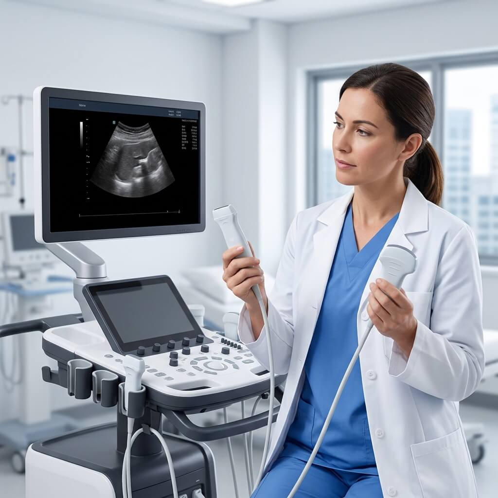
In modern medical imaging, the overall performance, resolution, and clinical accuracy of any ultrasound system depend heavily on its most critical component: the ultrasound transducer, commonly referred to as the ultrasound probe or ultra sound wand.
For hospital procurement managers, radiology department directors, and medical device distributors, selecting the right probe portfolio is as important as choosing the main imaging console or hand held ultrasound machine. Transducers directly determine acoustic wave frequency, penetration depth, field of view, and spatial resolution—ultimately dictating what clinical applications your facility can perform.
This comprehensive guide breaks down the core technology, mechanical types, clinical applications, maintenance considerations, and procurement strategies for ultrasound probes, while placing them within the broader context of hospital imaging suites and point-of-care equipment.
1. How Ultrasound Probes Work: Core Physics & Design Architecture
An ultrasound probe acts as both a transmitter and a receiver of high-frequency acoustic sound waves. Understanding its internal construction helps procurement teams evaluate transducer quality, longevity, and repairability.
                  [ Transducer Cross-Section Anatomy ]

  ┌─────────────────────────────────────────────────────────────────┐
  │ Acoustic Lens / Matching Layer  ──► Direct Contact & Focusing   │
  ├─────────────────────────────────────────────────────────────────┤
  │ Piezoelectric Crystal Array      ──► Electrical ◄► Acoustic     │
  ├─────────────────────────────────────────────────────────────────┤
  │ Backing Material                ──► Dampens Rear Reflections    │
  ├─────────────────────────────────────────────────────────────────┤
  │ Internal Circuitry & Strain Relief ──► Cable / Wireless Module   │
  └─────────────────────────────────────────────────────────────────┘
The Piezoelectric Effect
At the heart of every probe are piezoelectric crystals (traditionally Lead Zirconate Titanate, or PZT, and modern single-crystal materials). When an electrical current is applied, these crystals vibrate rapidly, producing acoustic sound waves ($1.0 \text{ MHz}$ to $22.0 \text{ MHz}$). As these sound waves bounce off tissue boundaries of varying acoustic impedance, the echoes return to the crystals, generating electrical signals that the system converts into real-time 2D, 3D, or Color Doppler images.
Frequency vs. Penetration Trade-Off
-
High Frequency ($7.0\text{–}18.0\text{ MHz}$): Short wavelengths deliver high spatial resolution but offer limited depth penetration. Best for superficial structures (vascular, small parts, musculoskeletal).
-
Low Frequency ($1.0\text{–}5.0\text{ MHz}$): Longer wavelengths penetrate deep into body tissues but sacrifice fine axial resolution. Best for deep abdominal and pelvic imaging.
2. Main Types of Ultrasound Probes and Their Clinical Applications
To build a versatile diagnostic suite, healthcare facilities must stock a balanced combination of specialized transducers. Below are the primary probe architectures used in hospitals today:
                      [ Major Probe Configurations ]

    Linear Array           Convex Array          Phased Array         Endocavitary
  ┌──────────────┐       ┌──────────────┐      ┌──────────────┐     ┌──────────────┐
  │ High Freq    │       │ Low Freq     │      │ Small Matrix │     │ Extended Field│
  │ Parallel Beam│       │ Curved Field │      │ Sector Beam  │     │ Intracavitary│
  │ Vascular/MSK │       │ Abdominal/OB │      │ Cardiac/ICU  │     │ Transvaginal │
  └──────────────┘       └──────────────┘      └──────────────┘     └──────────────┘
1. Linear Array Probes
-
Frequency Range: $5.0 \text{ MHz} – 18.0 \text{ MHz}$
-
Footprint: Flat, linear arrangement of piezo elements.
-
Clinical Applications: Vascular access, thyroid, breast, musculoskeletal (MSK), and superficial nerve blocks.
-
Key Strengths: Exceptional near-field resolution and accurate linear measurements.
2. Convex (Curved) Array Probes
-
Frequency Range: $2.0 \text{ MHz} – 6.0 \text{ MHz}$
-
Footprint: Curved element geometry providing a wide trapezoidal field of view.
-
Clinical Applications: General abdominal examinations, renal imaging, OB/GYN, and emergency FAST scans.
-
Key Strengths: Deep acoustic penetration with an expanded field of view at depth.
3. Phased Array (Cardiac) Probes
-
Frequency Range: $1.0 \text{ MHz} – 5.0 \text{ MHz}$
-
Footprint: Compact, small footprint designed to fit between intercostal spaces (ribs).
-
Clinical Applications: Adult and pediatric echocardiography, transcranial Doppler, and cardiac resuscitation monitoring.
-
Key Strengths: Electronically steered acoustic beams that allow deep imaging through tight acoustic windows.
4. Endocavitary (Transvaginal / Transrectal) Probes
-
Frequency Range: $5.0 \text{ MHz} – 10.0 \text{ MHz}$
-
Footprint: Long shaft with a wide-angle curved tip ($130^\circ – 180^\circ$ FOV).
-
Clinical Applications: Early pregnancy ultrasound confirmation, pelvic organ assessment, prostate biopsy guidance, and monitoring early gestational milestones such as a week 13 sonogram.
-
Key Strengths: Close proximity to target pelvic anatomy, avoiding beam attenuation through abdominal wall fat and bowel gas.
5. Specialized 3D/4D Volumetric & TEE Probes
-
Volumetric Probes: Contain mechanically sweeping or matrix-array crystals to capture real-time 3D volume rendering (widely used in obstetric imaging and complex cardiac valve evaluation).
-
Transesophageal Echocardiography (TEE): Endoscopic cardiac probes that provide high-resolution images of heart valves and septal defects via the esophagus.
3. Clinical Capabilities: What Diseases Can Be Detected by Ultrasound?
Procurement teams must ensure that their probe configuration directly matches the clinical services their institution offers. A common query from clinical directors is: what diseases can be detected by ultrasound?
                  [ Ultrasound Diagnostic Applications ]

      Abdominal & GI                   Obstetrics & GYN              Cardiology & Vascular
  ┌──────────────────────┐        ┌──────────────────────┐      ┌──────────────────────┐
  │ Gallstones / Sludge  │        │ Fetal Viability      │      │ DVT / Arterial Plaque│
  │ Hepatic Steatosis    │        │ Gestational Age      │      │ Valve Regurgitation  │
  │ Renal Cysts/Calculi  │        │ Placental Position   │      │ Pericardial Effusion │
  └──────────────────────┘        └──────────────────────┘      └──────────────────────┘
-
Abdominal Pathology: Fatty liver disease (steatosis), cirrhosis, gallstones, cholecystitis, renal stones, pancreatic cysts, and splenomegaly. Proper clinical workflow requires strict liver ultrasound prep (such as $6\text{–}8$ hours of fasting) to clear bowel gas. Recognizing non-optimal imaging parameters helps clinicians fix signs of a bad abdominal ultrasound—such as acoustic attenuation, improper gain control, or selecting an incorrect low-frequency probe.
-
Obstetrics & Gynecology: Confirming intrauterine pregnancy, tracking fetal anatomical growth, checking amniotic fluid index, and ruling out ectopic pregnancy during early triage or a week 13 sonogram.
-
Cardiology & Vascular: Deep Vein Thrombosis (DVT), carotid artery stenosis, cardiac valve regurgitation, pericardial effusion, and abdominal aortic aneurysms (AAA).
-
Emergency & MSK: Internal free fluid (FAST protocol), joint effusions, tendon ruptures, and soft tissue abscesses.
Diagnostic Turnaround & Patient Expectation
Administrators frequently address operational timelines: how long do ultrasound results take?
-
Scan Duration: Standard ultrasound scans take 15 to 30 minutes.
-
Immediate Triage: Real-time imaging allows emergency clinicians or sonographers to spot critical conditions (such as aortic rupture or ectopic pregnancy) instantly at the bedside.
-
Formal Reporting: Official written reports in PACS are usually finalized by radiologists within 1 to 4 hours.
4. Console vs. Handheld Ultrasound: The Rise of POCUS
The rapid evolution of miniaturized electronics has split ultrasound hardware into traditional cart-based systems and ultra-compact point-of-care ultrasound (POCUS) solutions.
             [ Cart-Based Console vs. Handheld POCUS ]

    Cart-Based Systems                  Wireless Handheld Systems
  ┌────────────────────────┐          ┌────────────────────────┐
  │ Multi-Port Probe Hub   │          │ Single / Dual-Head Probe│
  │ Advanced Processing    │    vs    │ iOS / Android Connect  │
  │ High Capital Cost      │          │ Pocket Portability     │
  │ Dedicated Suite        │          │ Emergency / Ambulance  │
  └────────────────────────┘          └────────────────────────┘
Traditional Cart Systems
Cart-based consoles offer multi-transducer active connectors, integrated gel warmers, large touchscreens, and advanced signal processing features like shear-wave elastography and spatial compound imaging. They remain the core workhorse for radiology and cardiology departments.
Wireless & Handheld Transducers
Miniaturized hand held ultrasound machine options and wireless probes—such as the GE Vscan Air or Vscan series—have revolutionized point-of-care clinical workflows.
-
Dual-Probe Versatility: Modern handheld units feature a dual-head transducer (combining a Linear probe on one end and a Convex probe on the other) connected via Wi-Fi/Bluetooth to a mobile phone or tablet.
-
Clinical Utility: Ideal for emergency departments, disaster response, rural clinics, home health visits, and ICU rounds where moving a full console cart is impractical.
5. Comparative Imaging Modalities: Ultrasound, CT, and MRI
When building an imaging department, healthcare organizations must balance ultrasound acquisition against complementary diagnostic modalities like Computed Tomography (CT) and Magnetic Resonance Imaging (MRI).
                      [ Diagnostic Modality Spectrum ]

   Modality         Ionizing Radiation        Portability           Cost / Exam
  ─────────────────────────────────────────────────────────────────────────────
   Ultrasound       None (0 mSv)             High (Cart / POCUS)   Low
   CT Scan          Moderate-High (2-10 mSv) Low-Medium (Mobile CT) Medium
   MRI              None (0 mSv)             Stationary            High
Modality Trade-Off Matrix
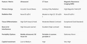
When analyzing ct scan vs ultrasound or ultrasound vs mri, ultrasound offers unmatched point-of-care mobility, real-time dynamic imaging, zero radiation hazard, and low ongoing maintenance costs. However, CT remains critical for complex acute trauma, multi-slice lung scans, and bone fractures, while MRI is superior for brain and spinal tissue details.
6. Comprehensive Hospital Procurement & Fleet Integration
Medical equipment purchasing departments rarely buy ultrasound probes in isolation. A complete hospital equipment procurement plan integrates imaging tools with peripheral patient monitoring and diagnostic equipment.
                 [ Integrated Hospital Diagnostic Suite ]

      Bedside Triage                 Imaging Suite                Vital Monitoring
  ┌────────────────────┐        ┌────────────────────┐        ┌────────────────────┐
  │ Portable X-Ray     │        │ Mobile Ultrasound  │        │ Vital Signs Equip. │
  │ Mobile X-Ray       │ ──────►│ Multi-Probe Array  │ ──────►│ ECG EKG Machines   │
  │ Portable CT        │        │ Handheld POCUS     │        │ Defibrillators     │
  └────────────────────┘        └────────────────────┘        └────────────────────┘
Complementary Equipment Procurement Checklist
-
General & Emergency Radiology: Combine cart-based ultrasound with mobile x-ray machine units and portable x-ray machines for immediate bedside chest, orthopedic, and abdominal screening.
-
Critical Care & Neurological ICUs: Pair mobile ultrasound with specialized portable ct scanner or portable ct units to perform rapid cranial CT scans without transferring unstable patients out of the ICU.
-
Cardiology & Emergency Response: Ensure cardiac ultrasound suites are supported by high-precision ECG EKG machines for cardiac gating and multi-parameter vital signs equipment to track pulse, blood pressure, and oxygen saturation during invasive procedures.
7. Cost Analysis & Buying Guide: Ultrasound Probes & Systems
Navigating ultrasound machines for sale and calculating total ultrasonography machine cost requires evaluating both the central console and individual transducer prices.
                    [ Procurement Cost Structure ]

  Component / System               Price Range (USD)        Key Cost Drivers
  ─────────────────────────────────────────────────────────────────────────────
  Single Replacement Probe         $1,500 - $8,000          Crystal Type, Matrix
  3D/4D or TEE Probe               $6,000 - $18,000         Mechanical Servo, Matrix
  Handheld System (Vscan Air)      $2,500 - $7,500          Wireless, App License
  Mid-Range Cart Ultrasound        $15,000 - $45,000        2-3 Probes Included
  High-End Radiology Console       $50,000 - $110,000+      Matrix Probes, Elastography
Key Procurement Factors for Transducers
-
Connector Compatibility: Ensure new probes use standard pin or pinless connectors compatible with your existing console hardware fleet.
-
Strain Relief & Cable Durability: Probe failure often occurs at the cable neck or connector junction. Inspect strain relief flex testing data before purchasing.
-
Disinfection Compatibility: Confirm that the probe material withstands your hospital’s standard high-level disinfection (HLD) chemical wipes or automated UV-C sterilization systems (especially for endocavitary and intraoperative probes).
-
Warranty & Service Contracts: Always negotiate multi-year warranty coverage, probe replacement discounts, and loaner probe availability to prevent clinical downtime.
Summary: Optimizing Your Hospital’s Imaging Fleet
Choosing the right ultrasound probes is fundamental to maximizing the diagnostic versatility of your facility’s imaging equipment. By combining high-frequency linear array transducers, deep-penetration convex probes, cardiac phased arrays, and modern wireless point-of-care tools like the GE Vscan Air, healthcare providers can deliver rapid, high-accuracy diagnostic care across every department.
Supported by secondary diagnostic tools—including portable x-ray machines, vital signs equipment, and ECG EKG machines—your facility can establish a resilient, high-throughput diagnostic infrastructure tailored to modern clinical demands.