The Time and Cost Cliff: Rebuilding the Chemistry of Life
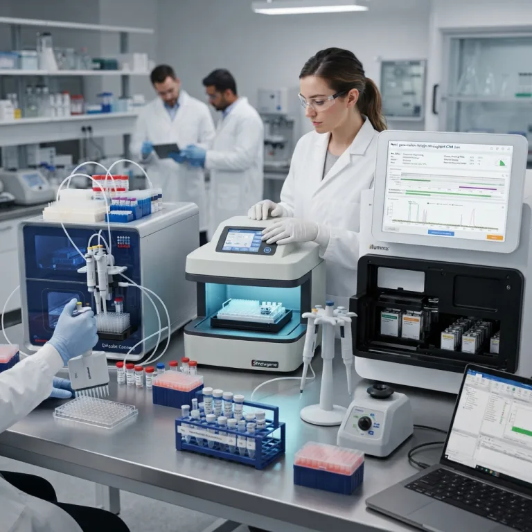
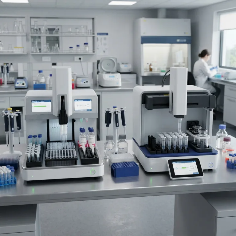
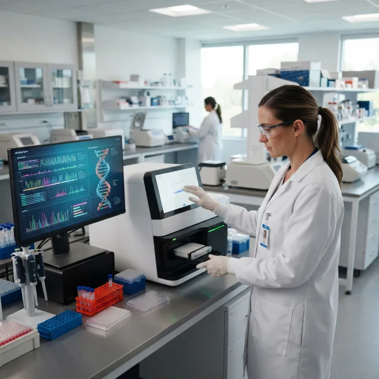
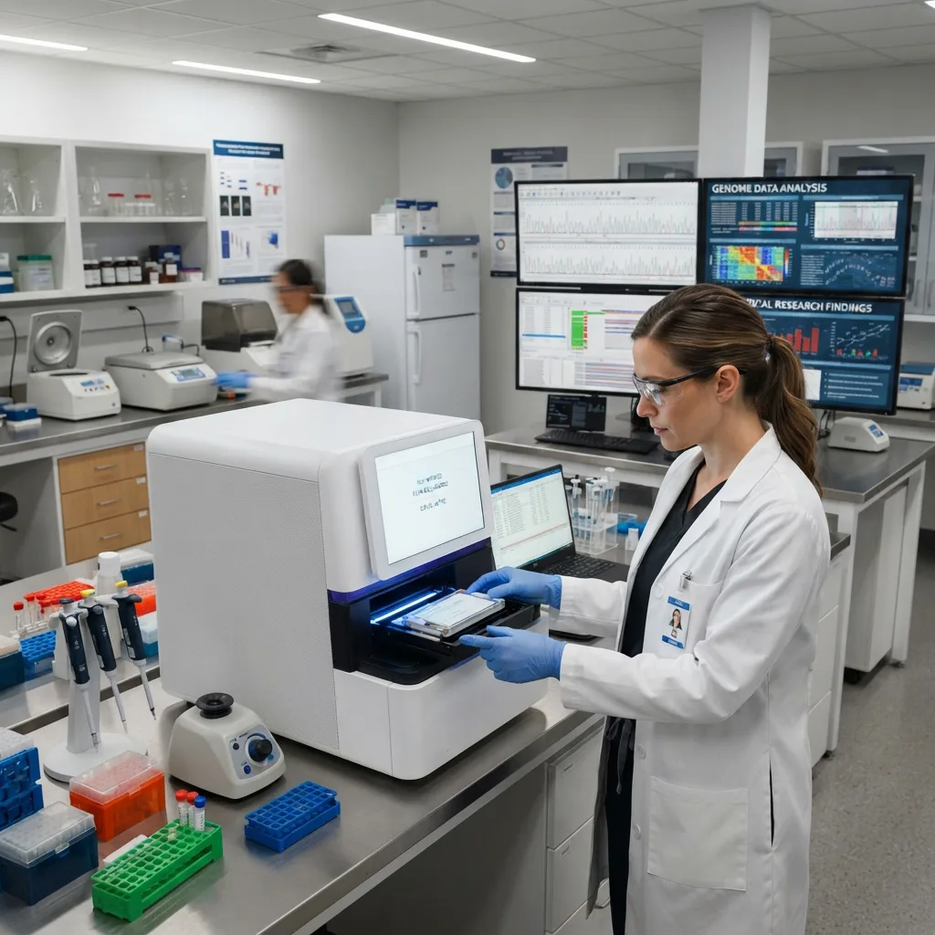
The original Human Genome Project required 13 years, an international consortium of thousands of scientists, and approximately 3 billion dollars to read a single human genetic blueprint. Today, a compact desktop sequencing machine sitting in a hospital laboratory can decode a patient’s entire genome in less than 24 hours for a few hundred dollars.
This is not simply a matter of computers getting faster. This breathtaking collapse in time and cost—often cited as outpacing Moore’s Law of computing—was driven by a fundamental tear-down and rebuild of sequencing chemistry.
To understand how modern clinical genetics, personalized oncology, and viral surveillance operate today, one must understand how laboratory hardware evolved from reading one DNA strand at a time in a glass tube to reading billions of strands simultaneously on a microscopic glass chip.
1. First-Generation: Sanger Sequencing and Capillary Electrophoresis
Developed by Frederick Sanger in 1977 (earning him a Nobel Prize), the Sanger method is the foundation of modern genomics. Despite its age, it remains a critical technology in every major diagnostic laboratory.
The Chemistry: Chain Termination
DNA polymerase is the enzyme that copies DNA by attaching free-floating nucleotides (dNTPs) to a growing strand. Sanger sequencing introduces a sabotaged version of these building blocks: dideoxynucleotides (ddNTPs). These sabotaged nucleotides lack a crucial chemical hook (the 3′-OH group). When the polymerase enzyme accidentally grabs a ddNTP and attaches it, the chain cannot grow any further. It is permanently terminated.
Each of the four sabotaged bases (A, T, C, G) is tagged with a different colored fluorescent dye. If you run this reaction millions of times in a test tube, you generate millions of DNA fragments of every possible length, each ending with a glowing color that represents the final letter of that specific fragment.
The Hardware: Capillary Electrophoresis (CE)
To read these colors in order, the liquid is injected into a Capillary Electrophoresis machine.
-
The sample is pulled through microscopic, polymer-filled glass capillaries using a high-voltage electrical field.
-
Because DNA is negatively charged, it moves toward the positive electrode. Shorter DNA fragments move faster than longer, heavier fragments.
-
As the fragments pass through a detection window one by one (from shortest to longest), an argon laser excites the fluorescent dyes.
-
An optical sensor records the sequence of colors (e.g., Red, Blue, Green, Green, Yellow), directly translating them into the genetic sequence (e.g., T, C, A, A, G).
Clinical Application: The Gold Standard
Sanger sequencing is slow. A standard CE machine might only process 8 to 96 capillaries at a time, reading about 800 to 1,000 base pairs per capillary. However, its accuracy is an unparalleled 99.99%. Today, clinical laboratories use Sanger sequencing not to discover new genomes, but to precisely verify specific, known disease mutations (such as confirming a Cystic Fibrosis mutation) or to cross-check ambiguous results generated by newer, high-throughput technologies.
2. Second-Generation: Next-Generation Sequencing (NGS)
If Sanger sequencing is like reading a book line by line with a magnifying glass, Next-Generation Sequencing (NGS) is like tearing a book into millions of pieces, handing those pieces to a million different people to read simultaneously, and using a supercomputer to stitch the story back together.
This concept is known as Massively Parallel Sequencing. The dominant technology driving NGS is Sequencing by Synthesis (SBS) paired with optical flow cells.
                  THE NGS SEQUENCING BY SYNTHESIS PIPELINE
+-------------------------------------------------------------------------+
| 1. LIBRARY PREPARATION & CLUSTERING                                     |
|    - Patient DNA is chopped into short fragments (150 - 300 base pairs).|
|    - Fragments are washed over a glass "Flow Cell" and bind to it.      |
|    - PCR amplification creates localized clusters of identical DNA clones|
|      (boosting the optical signal so a camera can see them).            |
|                                                                         |
| 2. REVERSIBLE TERMINATION                                               |
|    - Fluorescently tagged nucleotides wash over the flow cell.          |
|    - A polymerase adds ONE base to every cluster simultaneously.        |
|    - The nucleotides have a chemical block preventing a second addition.|
|                                                                         |
| 3. OPTICAL IMAGING                                                      |
|    - A laser fires. A high-resolution CMOS camera photographs the entire|
|      flow cell, recording millions of colored glowing dots.             |
|                                                                         |
| 4. CLEAVAGE & REPETITION                                                |
|    - A chemical wash removes the fluorescent dye and the chemical block.|
|    - The cycle repeats 150 to 300 times to spell out the sequences.     |
+-------------------------------------------------------------------------+
Hardware: The Optical Flow Cell
The flow cell is a highly engineered glass slide riddled with microscopic fluidic channels. Modern patterned flow cells contain billions of nano-wells spaced perfectly apart. A high-throughput NGS machine uses advanced microfluidic pumps to flush expensive reagents back and forth across this chip while an immense internal microscope photographs the chip frame by frame.
Clinical Applications of NGS
The sheer data output of NGS (ranging from gigabases to terabases per run) unlocks diagnostics that were previously impossible:
-
Non-Invasive Prenatal Testing (NIPT): A pregnant mother’s blood contains trace amounts of cell-free fetal DNA. NGS can sequence millions of these fragments to detect fetal chromosomal abnormalities (like Trisomy 21 / Down Syndrome) without risking a dangerous amniocentesis needle biopsy.
-
Oncology Tumor Profiling: Cancers mutate rapidly. NGS allows oncologists to sequence a biopsy sample against a panel of 500+ known cancer genes simultaneously, identifying the exact actionable mutations driving the tumor so they can prescribe highly targeted chemotherapy.
-
Whole Exome Sequencing (WES): Sequencing only the 1% to 2% of the genome that codes for proteins (the exome) to diagnose rare pediatric genetic diseases rapidly.
3. Third-Generation: Long-Read Sequencing Technologies
While NGS revolutionized throughput, it suffers from a major mathematical weakness: it can only read very short fragments of DNA (typically 150 to 300 base pairs).
Imagine trying to put together a puzzle of a blue sky composed of 10,000 identical blue pieces. Because the NGS fragments are so short, bioinformatic algorithms struggle to reassemble highly repetitive regions of the human genome. Third-Generation Sequencing solves this by reading massive, continuous strands of DNA (10,000 to 100,000+ base pairs) in real-time.
Method A: Single-Molecule Real-Time (SMRT) Optical Sequencing
Instead of amplifying DNA into clusters, this technology places a single DNA polymerase enzyme at the bottom of a microscopic well (a Zero-Mode Waveguide). As the enzyme pulls a long strand of DNA through itself, it incorporates fluorescent bases. An optical sensor at the bottom of the well records the flashes of light in real-time as the enzyme works.
Method B: Biological Nanopore Sequencing
This technology abandons optics entirely in favor of electrical physics.
-
An engineered protein pore (a nanopore) is embedded in an electrically resistant synthetic membrane.
-
A voltage is applied across the membrane, causing an ionic current to flow through the hollow pore.
-
A motor protein unwinds the DNA double helix and feeds a single strand through the nanopore.
-
Because each of the four bases (A, T, C, G) has a different physical shape and size, they block the flow of ions differently as they squeeze through the pore.
-
An ultra-sensitive microchip measures these tiny disruptions in electrical current, translating the voltage shifts directly into the genetic code.
Clinical Advantage: Long-read sequencing is uniquely capable of spanning massive structural variations, detecting complex epigenetic modifications (like DNA methylation) directly without chemical treatment, and tracking massive repeating regions like those seen in Huntington’s Disease.
4. Comparing the Generations: A Laboratory Reference
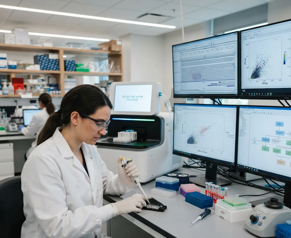
When outfitting a modern genomics laboratory, scientists do not simply replace old technology with new; they utilize all three generations for their respective strengths.
| Specification | 1st Gen (Sanger / CE) | 2nd Gen (NGS / SBS) | 3rd Gen (Long-Read / Nanopore) |
| Typical Read Length | 800 – 1,000 base pairs | 150 – 300 base pairs | 10,000 – 100,000+ base pairs |
| Throughput per Run | Very Low (Kilobases) | Massive (Gigabases to Terabases) | Moderate to High (Gigabases) |
| Accuracy Rating | 99.99% (The Gold Standard) | ~99.9% (Highly accurate consensus) | ~90% – 99% (Improving rapidly) |
| Detection Method | Laser optical fluorescence | Laser/CMOS optical imaging | Electrical current or Real-time optics |
| Primary Lab Utility | Single-gene validation, plasmid verification | NIPT, tumor panels, whole genomes | De novo assembly, structural variations |
Frequently Asked Questions (FAQ)
Q1: Why hasn’t Next-Generation Sequencing (NGS) completely replaced Sanger sequencing?
While NGS is incredibly powerful for screening the whole genome, it is inefficient and expensive if a doctor only wants to check one specific location on one specific gene. Setting up an NGS run requires complex library preparation and expensive flow cells. If a lab only needs to confirm whether a patient carries a single known mutation (like the BRCA1 breast cancer mutation), a Sanger sequencing run is much faster, cheaper, and slightly more accurate for that localized target.
Q2: What is the difference between Whole Genome Sequencing (WGS) and Whole Exome Sequencing (WES)?
Whole Genome Sequencing (WGS) reads the entire 3 billion base pairs of the patient’s DNA, including the vast non-coding regions (introns). It is comprehensive but generates massive data storage costs. Whole Exome Sequencing (WES) uses chemical probes to pull out and sequence only the “exons”—the 1% to 2% of the genome that actually provides instructions for building proteins. WES is highly cost-effective for diagnosing rare genetic diseases because the vast majority of known disease-causing mutations occur within protein-coding regions.
Q3: How is a DNA sample prepared before it enters an NGS machine?
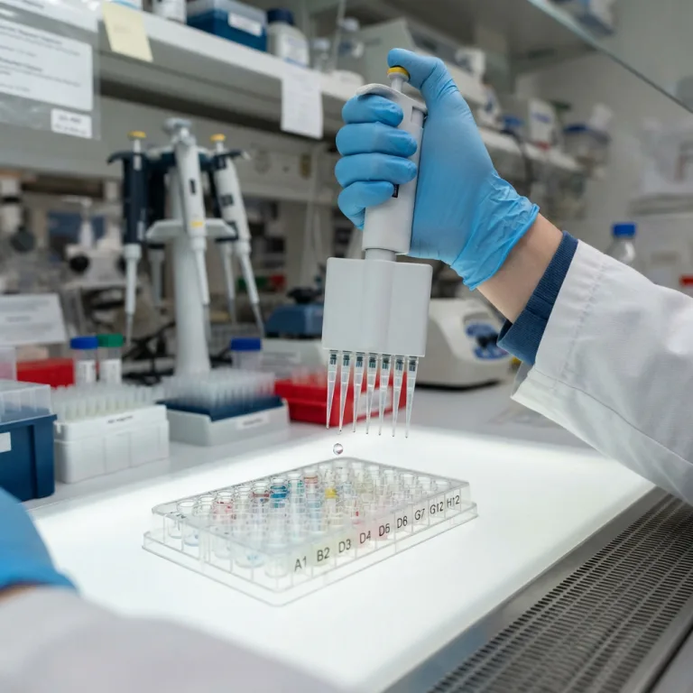
DNA extracted from a patient cannot just be poured into a sequencer. It must undergo Library Preparation. This involves using enzymes or sound waves to chop the long DNA into short fragments, repairing the jagged ends, and ligating (gluing) synthetic “adapters” to the ends of the fragments. These adapters act as handles that allow the DNA to stick to the sequencer’s flow cell and provide a starting point for the sequencing enzymes.
Q4: What does “Depth of Coverage” mean in NGS reports?
Because NGS involves chopping DNA into random fragments and sequencing them, the machine might sequence a specific gene 50 times by overlapping different fragments. A 50x Depth of Coverage means that, on average, every letter in the patient’s genome was read 50 independent times. High coverage (e.g., 100x to 500x) is critical in oncology because a tumor sample is often mixed with healthy cells; finding a rare cancer mutation requires reading the sequence hundreds of times to prove the mutation is real and not an optical error.
Q5: Can DNA sequencers read RNA directly?
Historically, sequencing RNA required reverse-transcribing the unstable RNA into complementary DNA (cDNA) first, and then sequencing the cDNA. However, modern Third-Generation nanopore sequencers have the unique physical capability to pull native RNA strands directly through the electrical pores, allowing scientists to sequence RNA directly and observe biological modifications that are usually lost during cDNA conversion.
Summary
The evolution of DNA sequencing from First-Generation Sanger Capillary Electrophoresis to Massively Parallel Next-Generation Sequencing (NGS) and Third-Generation Long-Read technologies represents the most profound technological leap in modern biology. By replacing slow, single-tube reactions with high-density optical flow cells and nanoscale electrical sensors, modern diagnostic laboratories can decode the human genome in hours, transforming oncology, prenatal care, and infectious disease tracking.