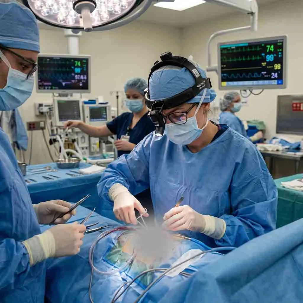
In modern healthcare, rapid diagnostic capability directly impacts clinical outcomes. For critically ill patients in the Intensive Care Unit (ICU), emergency department (ED), or operating room (OR), transporting a hemodynamically unstable individual to a centralized radiology department presents significant clinical risks.
The introduction of the portable CT scanner (also referred to as portable CT) has reshaped point-of-care diagnostics by delivering high-resolution cross-sectional imaging directly to the bedside.
1. What Is a Portable CT Scanner and How Does It Work?
A portable CT scanner is a self-contained, compact computed tomography system equipped with motorized wheels, internal battery reserves, and radiation shielding designed to navigate standard hospital corridors, elevators, and room doorways.
Unlike traditional fixed CT installations—which require dedicated lead-lined rooms, high-voltage 3-phase power, and heavy floor reinforcement—point-of-care CT systems operate on standard wall outlets or battery power and feature self-shielded gantries to minimize scatter radiation.
Key Technological Features
-
Compact Gantry Design: Optimized bore sizes designed specifically for head and neck imaging or full-body critical care scanning.
-
Internal Battery & Power Systems: Integrated drive motors and battery packs allow effortless transport between ICU rooms, ER trauma bays, and surgical suites.
-
Low-Dose Radiation Shielding: Built-in lead curtains and targeted collimation maintain safety for adjacent beds and clinical staff.
-
DICOM & PACS Connectivity: Seamless wireless connection with hospital radiology network systems for instant image retrieval and radiologist review.
2. Point-of-Care Imaging: Comparing Diagnostic Modalities
When medical equipment procurement teams evaluate diagnostic imaging solutions, understanding the operational boundaries between primary modalities is essential.
| Feature / Modality | Portable CT Scanner | Mobile X-Ray Machine / Portable X-Ray | Point-of-Care Ultrasound (POCUS) | Magnetic Resonance Imaging (MRI) |
|---|---|---|---|---|
| Primary Output | 3D Cross-sectional anatomical slices | 2D Projection radiograph | Real-time dynamic acoustic imaging | High-contrast soft tissue 3D slices |
| Radiation Risk | Low-to-moderate ionizing radiation | Low ionizing radiation | Non-ionizing acoustic waves | Non-ionizing magnetic field |
| Main Clinical Focus | Acute hemorrhage, stroke, trauma, bone detailed fractures | Chest screening, line placement, basic bone fracture | Abdominal, vascular, OB/GYN, cardiac assessment | Brain tumors, spinal cord, ligament/tendon detail |
| Bedside Mobility | High (within ICU/OR/ER) | Extremely High | Ultra-compact / Handheld | Highly restricted (Fixed/Specialized) |
CT Scan vs Ultrasound
When weighing ct scan vs ultrasound, key differences lie in speed, tissue visualization, and operator dependency:
-
Ultrasound is non-ionizing, highly portable, and ideal for dynamic vascular flow, organ structure, and soft tissue checks.
-
CT Scanning provides comprehensive 3D density mapping, quickly detecting acute intracranial hemorrhage, bone fractures, and deep internal bleeding where ultrasound waves are blocked by bone or air.
Ultrasound vs MRI
In the decision framework of ultrasound vs mri:
-
MRI offers superior soft tissue resolution (such as spinal cord micro-lesions or deep brain pathology), but requires longer scan times and strict magnetic safety protocols.
-
Ultrasound allows real-time, instantaneous bed-side assessment at a fraction of the capital investment, without radiation or magnetic interference.
3. Complementary Diagnostic Equipment in Point-of-Care Workflow
A modern emergency or critical care suite relies on an integrated ecosystem of diagnostic tools working alongside portable CT scanners.
A. Mobile X-Ray Equipment
Alongside CT technology, mobile x-ray machine units and portable x-ray machines remain the frontline workhorses for daily chest radiographs, verifying endotracheal tube positions, and assessing orthopedic alignment right in the patient room.
B. Ultrasound Systems & Transducers
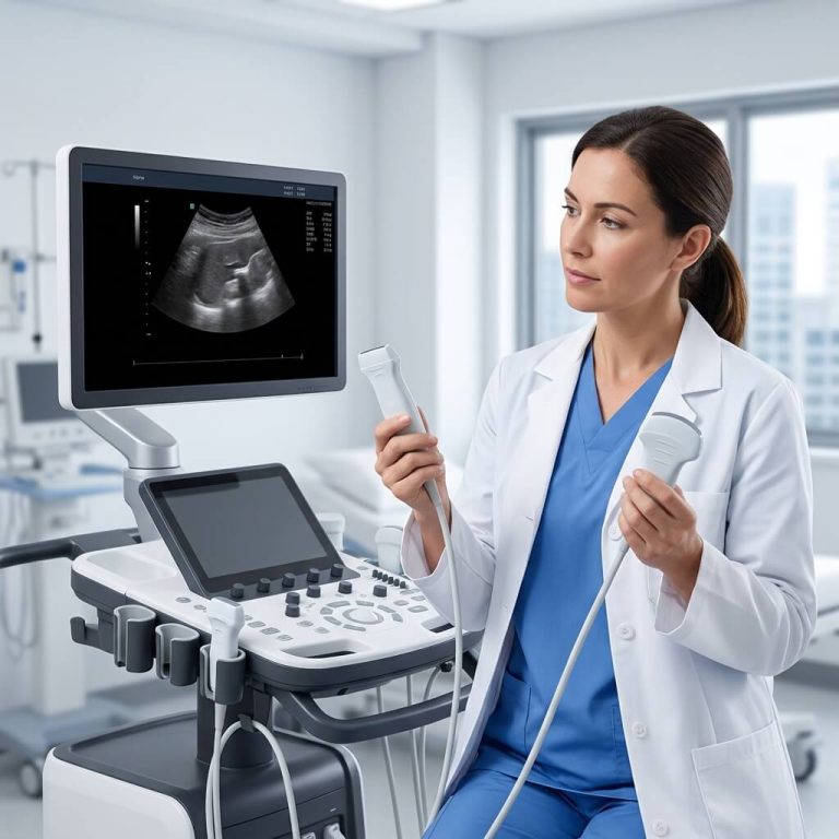
Ultrasound technology ranges from cart-based diagnostic platforms to compact hand held ultrasound machine and mobile ultrasound devices.
Key Ultrasound Probes & Components:
-
Ultrasound Probes / Transducers: Linear probes (vascular/superficial), Convex probes (abdominal), and Phased Array probes (cardiac).
-
Ultra Sound Wand Accessories: Ergonomic transducers with broad-frequency ranges for targeted clinical accuracy.
-
Advanced Handheld Units: Leading handheld innovations like the GE Vscan Air or general Vscan series demonstrate how ultra-portable units empower emergency clinicians to conduct bedside triage.
Patient Preparation & Clinical Clinical Questions:
-
What diseases can be detected by ultrasound? Abdominal aortic aneurysms, gallstones, fatty liver disease, deep vein thrombosis (DVT), pericardial effusion, focal organ masses, and kidney stones.
-
Pregnancy Ultrasound Confirmation: Used for early intrauterine pregnancy validation, gestational age estimation, and observing a week 13 sonogram for early fetal developmental screening.
-
Liver Ultrasound Prep & Troubleshooting: Proper liver ultrasound prep requires fasting for 6–8 hours to reduce bowel gas interference. Understanding signs of a bad abdominal ultrasound (e.g., severe acoustic shadowing from air or incomplete organ visualization due to patient body habitus) helps clinicians decide when to escalate to a portable CT scan.
-
Turnaround Times: Hospital administrators often ask how long do ultrasound results take? Bedside ultrasound provides immediate real-time answers, while formal radiology reports are typically finalized within 1 to 4 hours depending on hospital workflow.
C. Vital Signs & Patient Monitoring
During patient transport and intra-operative imaging, monitoring core physiological parameters is vital. Integrating ECG EKG machines and vital signs equipment ensures real-time oversight of oxygen saturation, heart rhythm, and arterial blood pressure while CT acquisition takes place.
4. Financial & Market Evaluation: Pricing and Cost Factors
Purchasing advanced radiology hardware requires evaluating total cost of ownership (TCO).
Ultrasound & Imaging Capital Expenditure
-
Ultrasound Machines for Sale: Market prices range from $5,000 for primary handheld transducers to $80,000+ for high-end cart-based multi-specialty consoles.
-
Ultrasonography Machine Cost Factors: Overall investment varies based on probe compatibility (e.g., single-crystal vs. piezoceramic arrays), Doppler software packages, AI-assisted diagnostic tools, and warranty terms.
Portable CT Scanner Cost Drivers
The acquisition cost of a portable ct scanner is influenced by key operational factors:
-
Detector Array & Slice Count: Systems configured for 8-slice, 16-slice, or 32-slice capture affect scan speed and image resolution.
-
Clinical Application Focus: Dedicated head/neck neuro-CT systems vs. large-bore full-body portable units.
-
Shielding & Room Requirements: Portable CTs eliminate expensive structural lead room renovations, drastically reducing total facility setup costs.
-
Maintenance & Service Contracts: Ongoing tube replacement costs, detector calibrations, and annual service agreements.
5. Strategic Buying Guide for Hospitals and Medical Equipment Distributors
When evaluating global suppliers—such as Mindray, United Imaging, GE Healthcare, SonoScape, or specialized manufacturers—buyers should follow a standardized procurement framework.
Technical & Clinical Evaluation Checklist
-
[Gantry & Bore Size: Does the inner diameter accommodate patient head frames, stabilization gear, or critical care tubing?
-
Radiation Safety Compliance: Are lead drapes, scatter shields, and low-dose dose-reduction algorithms (e.g., iterative reconstruction) included?
-
Power & Battery Efficiency: How many scans can the internal battery run on a single charge?
-
Software Integration: Full support for DICOM export, PACS compatibility, and worklist integration.
-
[ ] After-Sales & Technical Support: Availability of local service engineers, spare parts inventory, and remote software diagnostic capability.
6. Why Partner with UMY Medical Equipment Co., Ltd.?
Navigating complex medical equipment sourcing requires an experienced international supply partner. Guangzhou Umy Medical Equipment Co., Ltd. delivers comprehensive, reliable healthcare technology solutions tailored for global distributors, private clinics, and hospital projects.
Core Strengths of UMY Medical:
-
Over 20 Years of Industry Expertise: Extensive experience in medical imaging, operating room equipment, and diagnostic tools.
-
One-Stop Procurement Solution: Complete supply portfolio covering X-ray systems, ultrasound scanners, surgical equipment, and patient monitors.
-
OEM/ODM & Customization Capability: Tailored configurations, branding, and packaging to meet specific regional regulatory and market demands.
-
Global Logistics & Export Support: Experienced handling of international shipping documentation, compliance certificates, and technical support.
Conclusion
The adoption of portable CT scanner technology represents a major advance in point-of-care critical care. By bringing fast, precise cross-sectional imaging directly to the patient’s bedside, healthcare facilities can minimize transport risks, accelerate treatment decisions, and optimize ICU workflow.
Whether you are upgrading an emergency department or outfitting a modern surgical center, selecting the right combination of portable CT, mobile X-ray, and diagnostic ultrasound equipment is key to long-term clinical success.