The Microscopic World That Cannot Be Glued
In a molecular biology laboratory, extracting a patient’s DNA and separating it by size on an agarose gel is only the first step in diagnosing complex genetic disorders. To actually identify a specific disease-causing mutation hidden among the 3 billion base pairs of the human genome, scientists must “probe” the DNA using complimentary genetic sequences.
However, you cannot probe DNA while it is suspended in a fragile, jelly-like agarose gel. The gel would melt or tear during the rigorous washing steps required for hybridization.
To solve this, scientists transfer the DNA from the gel onto a sturdy, paper-like nylon membrane. But this creates a new critical vulnerability: the DNA is only resting on the surface of the membrane, held by weak electrostatic forces. If you attempt to wash this membrane with hybridization buffers at 65 degrees Celsius, the patient’s DNA will simply wash down the drain, destroying a week’s worth of clinical work. You cannot use glue, tape, or thermal melting to secure molecules at the nanometer scale.
The only way to permanently secure these microscopic strands of life without destroying their ability to be analyzed is through photochemistry. By utilizing specific wavelengths of ultraviolet light, scientists manipulate the atomic structure of the DNA itself, turning light into an unbreakable chemical anchor.
1. The Biology of UV-C Light and Pyrimidine Dimers
To understand how nucleic acid fixation works, we must look at how ultraviolet light interacts with the organic chemistry of the DNA double helix.
DNA is composed of four nitrogenous bases: Adenine (A), Guanine (G), Cytosine (C), and Thymine (T). Cytosine and Thymine belong to a chemical family called pyrimidines, which possess a single-ring carbon-nitrogen structure.
The 254 nm Absorption Peak
Not all ultraviolet light affects DNA equally. The UV spectrum is divided into UV-A (long wave, 315-400 nm), UV-B (medium wave, 280-315 nm), and UV-C (short wave, 100-280 nm).
Nucleic acids possess a natural maximum light absorption peak at approximately 260 nm. Therefore, exposing DNA to 254 nm UV-C light delivers the maximum amount of photonic energy directly into the molecular backbone.
The Photochemical Reaction
When a 254 nm photon strikes a DNA strand, the energy is absorbed by the pyrimidine rings. If two Thymine bases are sitting adjacent to each other on the same DNA strand, the absorbed UV energy causes the carbon double bonds within their rings to break and instantly reform with each other.
This creates a rigid, covalent ring structure connecting the two adjacent bases, known as a Thymine Dimer (or pyrimidine dimer). In the context of a nylon membrane (which is coated with positively charged amine groups), the UV irradiation also strips highly reactive radicals that form unbreakable covalent bonds between the thymine bases and the membrane surface.
Once this 30-to-60 second light exposure is complete, the DNA is physically welded to the nylon matrix. It can withstand boiling water, harsh detergents like SDS (Sodium Dodecyl Sulfate), and extreme alkaline conditions without detaching.
2. Southern Blotting: Hunting for Genetic Mutations
The ability to fix DNA to a membrane using UV light forms the absolute foundation of the Southern Blot, named after the British biologist Edwin Southern who invented the technique in 1975.
While modern Next-Generation Sequencing (NGS) and PCR have replaced Southern blotting for routine fast diagnostics, the Southern blot remains the undisputed “gold standard” for diagnosing specific large-scale genetic structural variations that modern sequencers struggle to read.
How the Southern Blot Works
-
Digestion: The patient’s DNA is cut into thousands of fragments using restriction enzymes.
-
Separation: The fragments are separated by length via gel electrophoresis.
-
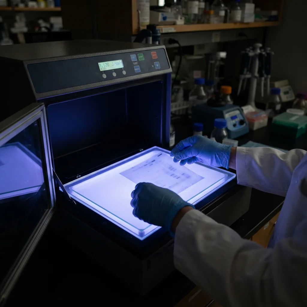
Transfer & Fixation: The DNA is transferred to a nylon membrane and permanently anchored using 254 nm UV irradiation.
-
Hybridization: The membrane is bathed in a solution containing a specific “probe”—a short, synthetic piece of DNA designed to perfectly match the target disease gene. This probe is tagged with a radioactive isotope or a fluorescent dye.
-
Detection: The membrane is washed rigorously. Because the patient’s overall DNA is UV-fixed to the membrane, it stays put. The probe binds only to the mutated sequence. When exposed to X-ray film or an imaging sensor, a dark band appears, confirming the presence and exact size of the genetic target.
Clinical Application: Fragile X Syndrome
A classic example of Southern blotting’s enduring clinical value is the diagnosis of Fragile X Syndrome, the most common inherited cause of intellectual disability and autism.
Fragile X is caused by a massive expansion of a specific repeating sequence (CGG) in the FMR1 gene. A healthy person has 5 to 40 repeats. A patient with the full mutation may have over 200 repeats. Standard PCR struggles to amplify through such a massive, highly repetitive, GC-rich region, often leading to false-negative results. The Southern blot easily accommodates these massive fragments. By UV-fixing the DNA and probing the FMR1 region, clinical geneticists can visually measure the exact length of the trinucleotide expansion, providing a definitive diagnosis for the family.
3. Northern Blotting: Decoding Gene Expression and Oncology
While the Southern blot targets DNA to find out what genes a patient has, the Northern Blot targets RNA to find out what those genes are actually doing.
In human biology, every cell in the body contains the exact same DNA. A liver cell and a brain neuron share the same genetic blueprint. The difference lies in gene expression—which genes are actively being transcribed into messenger RNA (mRNA) to build proteins.
Fixing RNA to Nylon Membranes
RNA is significantly more fragile than DNA. It is single-stranded and highly susceptible to rapid degradation by RNase enzymes present on human skin and in the environment.
During a Northern blot, RNA is separated by size and transferred to a membrane. Just like DNA, the RNA must be immobilized. Because RNA contains Uracil (U) instead of Thymine (T), the 254 nm UV light triggers the formation of Uracil Dimers, covalently crosslinking the RNA strands to the nylon support.
Clinical Application: Oncology and Tumor Markers
Northern blotting allows oncologists and researchers to observe the abnormal behavior of cancer cells. For example, if a researcher is studying breast cancer, they can extract RNA from both healthy breast tissue and a malignant tumor, run them side-by-side on a Northern blot, UV-fix the RNA, and probe for an oncogene (such as HER2).
If the resulting imaging shows a massive, dark band in the tumor lane and a faint band in the healthy lane, it definitively proves that the tumor is over-expressing the HER2 gene. This biological insight dictates the clinical treatment pathway, indicating that the patient will likely respond well to targeted monoclonal antibody therapies.
4. The Dual Purpose of UV-C: PCR Decontamination
The photochemical power of 254 nm ultraviolet light provides a second, equally critical function in the molecular diagnostic laboratory: DNA Destruction for Contamination Control.
Polymerase Chain Reaction (PCR) is highly sensitive; it can detect a single molecule of a pathogen’s DNA and amplify it into billions of copies within an hour. However, this extreme sensitivity is a double-edged sword. If even a microscopic aerosol droplet containing previously amplified DNA (an amplicon) floats across the laboratory and lands in a fresh, healthy patient’s sample tube, the PCR machine will amplify that contaminant. The result is a false-positive diagnosis, which could lead to unnecessary medical treatments or quarantine.
How Pyrimidine Dimers Prevent Amplification
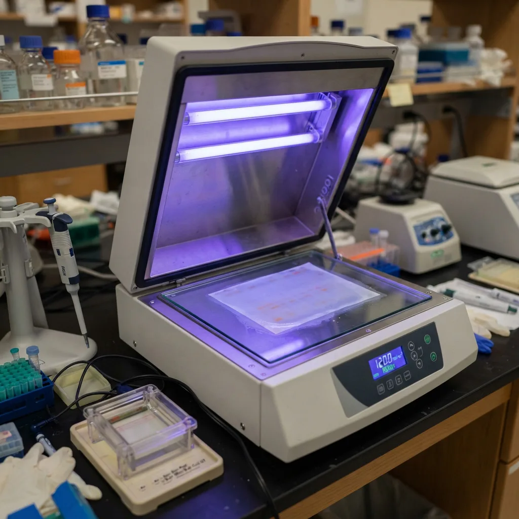
To prevent this, clinical laboratories utilize intense 254 nm UV irradiation inside their PCR preparation cabinets and on their equipment.
The biological mechanism is identical to membrane fixation, but the outcome is different because the DNA is suspended in liquid or resting on a stainless steel benchtop rather than a nylon membrane. When stray DNA is bombarded with 254 nm UV light, massive amounts of thymine dimers form along the DNA backbone. These dimers create physical “kinks” or roadblocks in the molecular structure.
When a stray, UV-irradiated DNA molecule accidentally enters a PCR tube, the Taq polymerase enzyme attempts to copy it. However, when the enzyme encounters the rigid thymine dimer roadblock, it physically stalls and falls off the DNA strand. Because the DNA cannot be copied, the contaminant is rendered completely “dead” to the PCR reaction. A 10-to-15 minute exposure to 254 nm UV light ensures that a diagnostic laboratory remains a sterile, zero-contamination environment.
Frequently Asked Questions (FAQ)
Q1: Why is 254 nm specifically used for nucleic acid fixation instead of 365 nm?
Nucleic acids (DNA and RNA) have a distinct absorption spectrum that peaks strongly around 260 nm. The 254 nm wavelength (UV-C) is generated efficiently by low-pressure mercury vapor lamps and matches this absorption peak almost perfectly. Longer wavelengths, such as 365 nm (UV-A), pass through the DNA without being absorbed strongly enough to break the carbon bonds required to form thymine dimers, making them useless for fixation.
Q2: Can you over-expose DNA during the UV fixation process?
Yes. Photochemical fixation requires a precise dose of energy. If the UV exposure is too short, the DNA fails to form covalent bonds with the membrane and washes away during hybridization, resulting in a blank test. If the exposure is too long (over-crosslinking), excessive thymine dimers and backbone breakages occur. This damages the target sequences so severely that the fluorescent or radioactive probes can no longer recognize and bind to the DNA, also resulting in a blank test.
Q3: What is the difference between a Southern blot and a Western blot?
The distinction lies in the biological molecule being analyzed.
-
A Southern blot analyzes DNA (to find gene mutations or structural variations).
-
A Northern blot analyzes RNA (to measure gene expression).
-
A Western blot analyzes Proteins (to detect specific protein levels or antibody presence, such as in HIV diagnostics). Western blots rely on electrical transfer and hydrophobic interactions rather than UV photochemistry for membrane fixation.
Q4: Does UV-C light destroy RNase enzymes on laboratory equipment?
While 254 nm UV-C light is highly effective at destroying stray DNA and RNA molecules (by forming pyrimidine dimers) and killing live bacteria and viruses, it is generally ineffective at destroying RNase enzymes. RNases are incredibly robust proteins that do not rely on nucleic acid backbones. To eliminate RNase contamination, laboratories must use chemical decontaminants (like diethyl pyrocarbonate – DEPC) or severe heat baking rather than UV irradiation.
Q5: Is UV nucleic acid fixation dangerous to laboratory personnel?
Shortwave 254 nm UV-C light is highly energetic and dangerous to human tissue. Direct exposure can cause severe photokeratitis (welder’s flash) in the eyes and erythema (sunburn-like damage) to the skin within minutes, as it induces the exact same thymine dimers in human skin cells, potentially leading to mutagenesis. Therefore, the crosslinking process must always be performed inside enclosed, shielded chambers featuring safety interlocks that instantly cut power to the bulbs if the door is opened.
Summary
The ability to diagnose complex genetic disorders like Fragile X Syndrome, or to track the specific gene expression of malignant tumors, relies fundamentally on the physics of light interacting with biology. By exposing DNA and RNA to 254 nm UV-C light, clinical scientists force adjacent pyrimidine bases to form covalent thymine and uracil dimers, permanently fixing the genetic material to nylon membranes. This elegant photochemical reaction ensures that the microscopic blueprints of life remain stable during the harsh chemical washes of Southern and Northern blotting, enabling accurate and life-saving molecular diagnostics.