Introduction
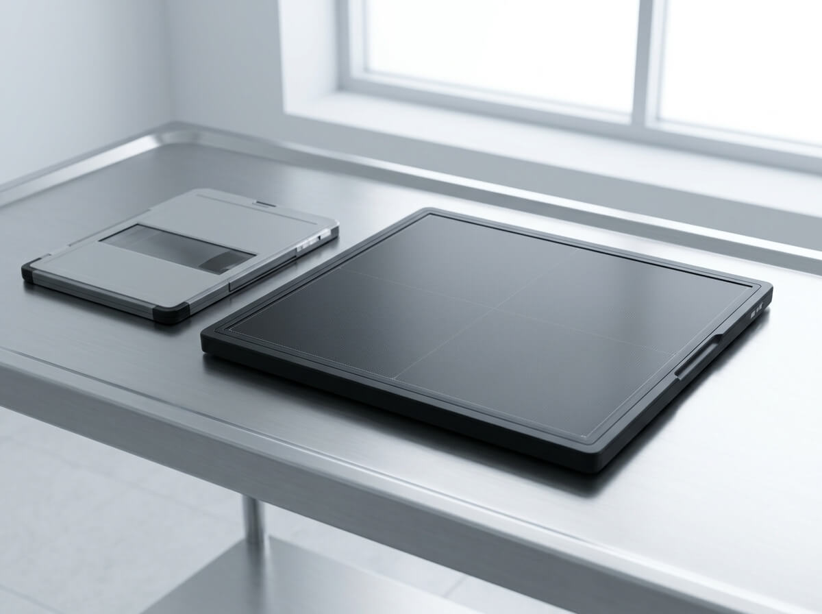
A flat panel detector, commonly called an FPD, is one of the key components used in modern digital X-ray imaging. It captures X-ray information and converts it into a digital image that can be viewed, processed, stored, and transferred through a computer-based imaging system.
Unlike traditional X-ray film, a digital X-ray detector allows the image to be handled electronically. Compared with computed radiography (CR), a flat panel detector is designed for direct digital radiography workflows without the separate image-reading step associated with a CR imaging plate. The International Atomic Energy Agency identifies CR and flat panel detector-based direct radiography as two common forms of digital image receptors.
Today, flat panel detectors are widely used in digital radiography, mobile X-ray systems, hospital radiology departments, clinics, emergency imaging, orthopedic imaging, veterinary X-ray, and X-ray retrofit systems.
For hospitals, distributors, medical equipment dealers, and X-ray system integrators, understanding the basic technology and specifications of an FPD is important before choosing a detector.
This guide explains what a flat panel detector is, how it works, the main FPD types, common detector sizes, wired and wireless options, important specifications, applications, and the difference between FPD and CR.
What Is a Flat Panel Detector?
A flat panel detector (FPD) is a digital X-ray image receptor used to capture X-rays and create a digital radiographic image.
In simple terms:
X-ray tube → Patient → Flat panel detector → Digital image → Workstation
During an X-ray examination, X-rays pass through the body. Different tissues absorb different amounts of X-ray radiation. The remaining X-ray pattern reaches the flat panel detector.
The detector converts this X-ray pattern into electronic information. The system then processes the information and displays a digital X-ray image on a monitor.
This basic workflow is why an FPD is also commonly described as a:
- digital X-ray detector
- digital radiography detector
- DR detector
- DR flat panel detector
- X-ray flat panel detector
- digital radiography panel
- X-ray detector panel
- digital imaging detector
- flat panel X-ray detector
- wireless DR detector
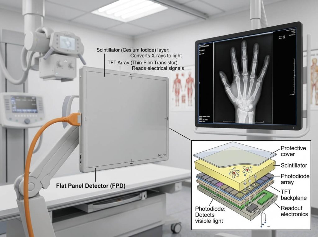
The exact detector construction varies between products. Modern medical flat panel detectors can use different scintillators and sensor technologies, including combinations involving cesium iodide (CsI), gadolinium oxysulfide (GOS), amorphous silicon (a-Si), and other detector architectures. FDA documentation for digital flat panel X-ray detectors describes systems using TFT arrays, amorphous silicon technology, and scintillators such as CsI and Gadox.
How Does a Flat Panel Detector Work?
Understanding how an FPD works does not require advanced engineering knowledge.
The process can be explained in several steps.
1. The X-Ray Tube Produces X-Rays
An X-ray generator supplies power to the X-ray tube. The tube produces X-rays that travel toward the patient.
2. X-Rays Pass Through the Patient
Different parts of the body absorb different amounts of X-ray energy.
For example, bones generally absorb more X-rays than soft tissue. This produces differences in the X-ray pattern reaching the detector.
3. The Detector Captures the X-Ray Pattern
The flat panel detector receives the X-rays.
Depending on the detector design, X-ray energy may first be converted into visible light through a scintillator, or X-rays may be converted more directly into electrical charge.
In many indirect-conversion systems, a scintillator such as CsI or GOS converts X-rays into light. An underlying sensor array then converts that light into electrical signals.
4. The Sensor Array Reads the Signal
The detector contains a large matrix of pixels. Each pixel collects information from a small area of the image.
Amorphous silicon TFT technology is widely used in flat panel detector designs. FDA documentation describes each pixel as incorporating a light-sensitive photodiode and a thin-film transistor switching element in the sensor array.
5. The Digital Image Is Processed
Electronic information from the detector is transferred to an image acquisition system.
The software can perform image correction and processing before presenting the image to the operator. Some detector systems include dedicated acquisition and preprocessing software for this purpose.
6. The Image Can Be Viewed and Stored
The resulting digital X-ray image can be displayed on a workstation and integrated into a wider hospital imaging workflow.
Depending on the system, images can be stored, transferred, reviewed, printed, or connected with healthcare IT systems.
This workflow makes digital radiography different from traditional film-based radiography and from CR workflows that require an imaging plate and reader.
What Is a DR Detector?
DR detector means Digital Radiography detector.
In many medical equipment discussions, the terms flat panel detector, DR detector, digital detector, and digital X-ray detector are used in closely related ways.
A DR detector captures X-ray information electronically and supports digital radiography.
For this reason, a buyer searching for:
DR detector
may be looking for the same general product category as someone searching for:
flat panel detector
However, the exact product specifications can vary significantly between manufacturers and models.
A DR detector may be:
- wired
- wireless
- portable
- fixed
- cassette-sized
- designed for retrofit applications
- designed for a mobile X-ray system
- designed for a radiography room
- designed for specialized applications
Therefore, the term DR detector describes the digital radiography function, while flat panel detector describes the physical detector technology or form factor commonly used for that function.
What Is the Difference Between FPD and CR?
One of the most common questions from people upgrading an X-ray system is:
What is the difference between FPD and CR?
Both can be used for digital radiography, but their image acquisition workflows are different.
Flat Panel Detector
With a flat panel detector, X-ray information is captured electronically by the detector and transferred into the digital imaging system.
The image can therefore enter a digital workflow without scanning a separate imaging plate.
Computed Radiography
CR uses a cassette containing a photostimulable imaging plate. After exposure, the cassette is processed by a CR reader, which reads the stored image information and converts it into a digital image. The IAEA describes this cassette-and-reader workflow for CR and contrasts it with flat panel detector-based direct radiography.
FPD vs CR at a Glance
| Feature | Flat Panel Detector | CR |
|---|---|---|
| Image receptor | Flat panel detector | Imaging plate |
| Separate reader | Generally no | Yes |
| Digital workflow | Direct digital acquisition | Cassette must be processed |
| Workflow speed | Designed for rapid digital workflow | Additional reading step |
| Wireless option | Available on many models | Usually cassette-based |
| Retrofit potential | Available for suitable systems | Common retrofit approach |
| Image handling | Electronic detector workflow | Imaging plate workflow |
For facilities looking to move from CR to DR, detector compatibility, mechanical integration, software, generator communication, image acquisition, and overall system configuration should be evaluated before purchasing.
What Are the Main Types of Flat Panel Detectors?
There is no single type of flat panel detector suitable for every application.
FPDs can be classified in several different ways.
Wired Flat Panel Detector
A wired flat panel detector connects to the imaging system using a cable.
Potential advantages include a stable physical connection and continuous power depending on the system configuration.
Wired detectors can be suitable for:
- fixed X-ray rooms
- radiography tables
- wall stands
- hospitals
- clinics
- stationary digital X-ray systems
A wired detector can also be useful where the imaging workflow does not require frequent detector movement.
Wireless Flat Panel Detector
A wireless flat panel detector sends image information to the acquisition workstation without requiring a permanent data cable between the detector and workstation.
Wireless DR detectors are particularly useful when the detector needs to be moved between different imaging positions.
Common applications include:
- mobile X-ray
- bedside radiography
- emergency imaging
- ICU imaging
- orthopedic imaging
- portable radiography
- veterinary radiography
Some commercial detector systems are available in wireless formats with different sizes and scintillator options. For example, current manufacturer specifications include wireless 14 × 17 inch and 17 × 17 inch detectors using either GOS or CsI configurations.
A wireless detector normally requires more than wireless communication alone. Buyers should also consider:
- battery
- battery charging
- wireless transmission
- connection stability
- detector weight
- detector durability
- software compatibility
- image transfer workflow
- backup wired connection, when available
Portable Flat Panel Detector
A portable flat panel detector is designed to be moved between different imaging positions or locations.
Portability is especially important for:
- mobile X-ray machines
- bedside X-ray
- emergency departments
- operating rooms
- veterinary clinics
- field imaging
- multi-room clinical use
The practical value of a portable DR detector depends on factors such as weight, dimensions, battery design, housing, wireless communication, durability, and workflow.
A lightweight detector can be easier to position, but buyers should evaluate the complete specification rather than focusing only on weight.
What Are 14 × 17 and 17 × 17 Flat Panel Detectors?
Detector size is one of the first specifications that medical equipment buyers usually check.
Two common sizes are:
14 × 17 inch
and
17 × 17 inch
These approximate sizes correspond to commonly used radiography image-receptor formats.
A 14 × 17 inch detector is often used for general radiography and mobile applications where cassette-style dimensions and portability are important.
A 17 × 17 inch detector provides a larger square imaging format and can be suitable for applications where a larger field of view is useful.
Manufacturer specifications demonstrate that both 14 × 17 inch and 17 × 17 inch wireless DR detectors are commercially available for mobile and general radiography systems.
Smaller detector formats also exist for specific imaging requirements.
For example, approximately 24 × 30 cm formats are available in some commercial detector lines.
The correct detector size depends on:
- X-ray system design
- Bucky tray dimensions
- wall stand
- tabletop
- patient positioning
- imaging anatomy
- mobile workflow
- required field of view
- detector weight
- available space
A larger detector is not automatically better for every application.
What Is CsI in a Flat Panel Detector?
CsI stands for Cesium Iodide.
CsI is used as a scintillator in some indirect-conversion X-ray detectors.
Its role is to convert incoming X-ray energy into visible light, which can then be detected by the underlying sensor array.
CsI is commonly discussed when comparing different flat panel detector technologies.
Some commercial medical DR detectors are offered with CsI scintillators, while other models use GOS. Current manufacturer specifications show both CsI and GOS options across similar detector formats.
A buyer should therefore compare the complete detector specification rather than assuming that the scintillator material alone determines the overall image performance.
Important factors also include:
- pixel pitch
- detector efficiency
- image noise
- spatial resolution
- active area
- sensor technology
- image processing
- X-ray dose
- application
- system integration
What Is GOS in a Flat Panel Detector?
GOS means Gadolinium Oxysulfide.
GOS can also be used as a scintillator in indirect-conversion flat panel detectors.
Like CsI, GOS converts X-ray energy into light before the signal is captured by the detector sensor.
Commercial DR detector specifications demonstrate that GOS and CsI detector versions can be offered in comparable detector sizes.
When comparing CsI vs GOS flat panel detectors, it is better to consider the complete design rather than choosing solely from the material name.
For procurement decisions, compare:
- image quality
- pixel pitch
- DQE
- detector weight
- durability
- power requirements
- battery
- dimensions
- software
- compatibility
- price
- service support
These factors determine whether a detector is suitable for a particular X-ray system.
What Is a Pixel Pitch in an X-Ray Detector?
Pixel pitch is the distance from the center of one detector pixel to the center of the neighboring pixel.
It is commonly expressed in micrometers, or μm.
For example, a detector may have a pixel pitch of:
- 100 μm
- 120 μm
- 140 μm
- 143 μm
- 150 μm
- 160 μm
- 180 μm
Pixel pitch is an important specification because smaller pixels can support finer sampling.
However, smaller pixel pitch does not automatically mean that a detector is the right choice for every application.
Image quality depends on multiple factors, including detector design, X-ray dose, detector efficiency, image processing, exposure conditions, and the clinical task.
Commercial detector specifications illustrate this range. For example, one current mobile DR detector series lists a pixel pitch of 0.15 mm, or 150 μm.
Another FDA-documented flat panel detector uses a 100 μm pixel pitch.
Therefore, when comparing two X-ray detectors, pixel pitch should be considered together with other specifications instead of being treated as the only measure of image quality.
What Is the Active Area of a Flat Panel Detector?
The active area is the portion of the detector that actually collects the X-ray image.
It can differ slightly from the total physical size of the detector.
For example, a detector may have an external dimension that is slightly larger than its active imaging area.
This is important when checking:
- image coverage
- detector replacement
- Bucky compatibility
- table compatibility
- wall stand compatibility
- X-ray system integration
For retrofit projects, buyers should confirm both the physical dimensions and the active imaging area.
A detector fitting physically into a tray does not automatically mean that every aspect of the system is compatible.
Flat Panel Detector for Mobile X-Ray
Mobile X-ray is one of the major applications for portable and wireless FPD technology.
A mobile X-ray detector allows the radiographer to position the image receptor near the patient without moving the patient to a fixed radiography room.
This can be valuable in situations involving:
- hospitalized patients
- ICU patients
- emergency cases
- bedside imaging
- operating room imaging
- patients with limited mobility
- veterinary patients
Wireless DR detectors can help simplify positioning because the detector does not always need to remain physically connected to the acquisition workstation.
Some mobile DR systems are specifically designed to convert existing analog mobile X-ray units into digital radiography systems using wireless flat panel detectors.
For buyers considering a mobile DR detector, useful specifications include:
detector size + detector weight + battery life + wireless communication + durability + image preview + software compatibility
Flat Panel Detector for Hospital X-Ray
In a hospital, an FPD may be used in a fixed radiography room, mobile X-ray system, wall stand, radiography table, or other digital imaging configuration.
Hospital buyers generally need to consider more than image quality.
Important procurement factors include:
- daily workload
- detector durability
- patient throughput
- cleaning requirements
- detector weight
- battery management
- connection stability
- system compatibility
- service support
- replacement availability
- image workflow
- hospital IT integration
For a high-use radiology department, operational reliability and workflow should be evaluated together with technical specifications.
Flat Panel Detector for Veterinary X-Ray
Veterinary clinics are another important application for digital X-ray detectors.
A veterinary flat panel detector may be used for:
- small animals
- dogs
- cats
- horses
- orthopedic imaging
- chest imaging
- abdominal imaging
- limb imaging
Veterinary applications may require flexible positioning because animals cannot always be positioned like human patients.
A portable wireless DR detector can therefore be useful for veterinary imaging.
When selecting a veterinary X-ray detector, buyers should consider:
- detector size
- weight
- durability
- waterproof or protection rating
- wireless function
- battery
- software
- image quality
- cleaning requirements
- compatibility with the existing veterinary X-ray system
Flat Panel Detector for Orthopedic Imaging
Orthopedic imaging often requires detailed visualization of bones and joints.
A digital X-ray detector may be used for:
- extremity imaging
- hand X-ray
- wrist X-ray
- elbow X-ray
- shoulder X-ray
- knee X-ray
- ankle X-ray
- foot X-ray
- spine imaging
For orthopedic applications, detector size and pixel pitch can be important considerations.
Smaller detectors may be convenient for extremity imaging, while larger detectors can be useful for full-size radiographic examinations.
The optimal choice depends on the anatomy being examined and the configuration of the X-ray system.
Flat Panel Detector in Emergency Imaging
Emergency imaging often requires fast workflow and flexible positioning.
A portable DR detector can be used with a mobile X-ray system to capture images at the patient’s location.
Common emergency imaging scenarios include:
- trauma imaging
- chest X-ray
- bedside imaging
- emergency department radiography
- ICU radiography
A wireless detector can reduce the need to manage a detector cable during positioning.
However, wireless capability is only one part of the overall solution. A hospital should also evaluate battery capacity, transmission reliability, detector protection, software integration, and cleaning procedures.
Can a Flat Panel Detector Retrofit an Existing X-Ray Machine?
Yes, depending on the design of the existing X-ray system and the detector.
A DR retrofit upgrades an existing analog or CR-based workflow by introducing a digital detector and image acquisition system.
A retrofit project may involve:
- flat panel detector
- image acquisition workstation
- detector interface
- generator communication
- exposure trigger
- software
- mounting accessories
- cables
- wireless communication
- DICOM integration
- PACS connectivity
The exact requirements depend on the original X-ray machine.
Some commercial systems are specifically marketed for transforming existing mobile analog X-ray units into digital radiography systems.
For this reason, a buyer should provide the existing X-ray system model and mechanical configuration to the supplier before ordering a replacement or retrofit detector.
What Specifications Should You Check When Buying an FPD?
If you are comparing flat panel detectors, do not look at only one specification.
A practical evaluation should include the following.
Detector Size
Common formats include:
- 14 × 17 inch
- 17 × 17 inch
- 24 × 30 cm
- other application-specific sizes
Pixel Pitch
Pixel pitch affects image sampling and should be considered together with the intended imaging application.
Scintillator
Common terms include:
- CsI
- GOS
Wired or Wireless
Consider whether the detector will mainly be used in:
- fixed rooms
- mobile X-ray
- bedside imaging
- multiple rooms
- veterinary imaging
Detector Weight
Weight becomes more important when the detector must be frequently moved or positioned under patients.
Battery
For wireless detectors, check:
- operating time
- battery type
- charging time
- number of batteries
- replacement battery availability
Durability
For mobile applications, consider:
- shock resistance
- waterproofing
- ingress protection
- load resistance
- housing design
- drop resistance
Compatibility
Verify compatibility with:
- X-ray generator
- Bucky
- wall stand
- radiography table
- mobile X-ray unit
- detector tray
- image acquisition software
Software
Check support for:
- image acquisition
- image processing
- calibration
- image storage
- DICOM
- PACS
- workstation integration
These details can have a major effect on the total usability of a digital radiography detector.
Does a Flat Panel Detector Improve X-Ray Workflow?
A major reason healthcare facilities move toward digital radiography is workflow.
With a digital flat panel detector, the image can enter an electronic acquisition workflow without the separate film development or CR image-reading process.
The practical benefits may include:
faster image availability
electronic image storage
easier image transfer
digital image processing
simplified workflow
integration with hospital imaging systems
However, workflow performance depends on the complete X-ray system rather than the detector alone.
Generator, detector, workstation, software, network, PACS, operator workflow, and system configuration all contribute to the final experience.
Flat Panel Detector vs Digital X-Ray Detector
These terms are often used interchangeably, but there is a small difference in emphasis.
Flat panel detector generally refers to the detector hardware.
Digital X-ray detector describes the detector’s role in creating digital X-ray images.
DR detector emphasizes its use in digital radiography.
So you may see product pages using several related terms:
Flat Panel Detector
Digital X-Ray Detector
DR Detector
Digital Radiography Detector
Wireless DR Detector
X-Ray Flat Panel Detector
All of these terms are useful when researching digital X-ray detector solutions.
How Long Does a Flat Panel Detector Last?
There is no single lifespan that applies to every flat panel detector.
Service life depends on:
- detector design
- workload
- exposure frequency
- handling
- shock and impact
- cleaning
- operating environment
- battery condition
- maintenance
- calibration
- manufacturer specifications
A detector used several times a day in a low-volume clinic may experience a very different operating environment from a detector used continuously in a busy hospital.
For procurement, ask the manufacturer about the expected operating conditions, warranty, service policy, calibration requirements, spare parts, and replacement support.
How Much Does a Flat Panel Detector Cost?
The price of a flat panel detector can vary considerably.
Factors that influence the cost include:
- detector size
- wired or wireless design
- scintillator technology
- pixel pitch
- image performance
- detector weight
- battery system
- durability
- software
- accessories
- certification
- system integration
- quantity
- warranty
- after-sales support
For this reason, a price comparison should not be based only on the detector name.
Two products with the same nominal detector size may have very different specifications and total ownership costs.
For hospitals and distributors, it is usually more useful to compare the complete solution, including detector, software, accessories, warranty, technical support, and compatibility.
What Is the Best Flat Panel Detector?
There is no single flat panel detector that is the right choice for every hospital, clinic, mobile X-ray system, or veterinary application.
The right detector depends on the intended use.
For example:
A mobile X-ray system may prioritize low weight, wireless operation, battery performance, and durability.
A fixed X-ray room may place greater emphasis on size, image quality, system integration, and workflow.
A veterinary clinic may need flexibility, durability, easy cleaning, and portability.
A hospital radiology department may focus on workload, reliability, compatibility, image workflow, service, and long-term support.
Instead of asking only:
What is the best DR detector?
a more useful question is:
Which flat panel detector is suitable for my X-ray system and intended application?
Flat Panel Detector FAQ
What does FPD stand for in X-ray?
FPD stands for Flat Panel Detector. It is a digital X-ray image receptor used to capture radiographic images electronically.
Is a flat panel detector the same as a DR detector?
In many medical imaging contexts, these terms are used to describe closely related products. A flat panel detector is commonly used as the image receptor in a digital radiography system.
What is a digital X-ray detector?
A digital X-ray detector captures X-ray information electronically and produces a digital image for processing and display.
How does an X-ray flat panel detector work?
The detector receives X-rays after they pass through the patient and converts the X-ray information into electronic signals that are processed into a digital image.
What is the difference between DR and CR?
DR uses an electronic digital detector, while CR uses a cassette containing an imaging plate that must be processed by a CR reader.
What is a wireless DR detector?
A wireless DR detector is a digital X-ray detector that can transfer image data wirelessly to an acquisition workstation, depending on the system design.
What is a 14 × 17 flat panel detector?
It is a commonly used detector format measuring approximately 14 × 17 inches and is widely associated with general radiography and mobile X-ray applications.
What is a 17 × 17 flat panel detector?
It is a larger square detector format of approximately 17 × 17 inches, suitable for applications requiring a larger square imaging field.
Is CsI better than GOS?
CsI and GOS are different scintillator technologies. Neither should be considered automatically better for every application. Buyers should compare the complete detector specifications and intended use.
What does pixel pitch mean?
Pixel pitch is the distance between neighboring pixel centers in a detector array. It is commonly specified in micrometers.
Does smaller pixel pitch always mean better image quality?
No. Pixel pitch is an important specification, but overall image performance also depends on detector efficiency, noise, X-ray dose, image processing, and other system factors.
Can an FPD be used with an existing X-ray machine?
Potentially, yes. Compatibility must be evaluated based on the existing X-ray system, detector tray, generator, communication method, software, and other integration requirements.
Can an FPD replace a CR system?
A suitable flat panel detector can be part of a transition from CR to DR, but the complete system configuration and compatibility should be checked before replacement.
Can a flat panel detector be used for veterinary X-ray?
Yes. Digital flat panel detectors are used in veterinary radiography, including applications for small and large animals.
Are wireless flat panel detectors rechargeable?
Many wireless detectors use rechargeable battery systems, but the exact battery design varies by model.
What should I consider when buying a flat panel detector?
Consider detector size, pixel pitch, scintillator, wired or wireless design, weight, battery, durability, software, compatibility, service support, warranty, and total cost.
Who supplies flat panel detectors?
Flat panel detectors are manufactured and supplied by medical imaging manufacturers, detector technology companies, X-ray equipment manufacturers, and specialized medical equipment suppliers.
Conclusion
A flat panel detector is a central component of modern digital X-ray imaging. It enables an X-ray system to capture radiographic information electronically and convert it into a digital image.
The term FPD is closely associated with DR detector, digital X-ray detector, digital radiography detector, wireless DR detector, and X-ray flat panel detector.
When choosing an FPD, however, the product name is only the starting point.
Important factors include:
detector size
pixel pitch
CsI or GOS scintillator
wired or wireless communication
detector weight
battery performance
durability
image quality
software
system compatibility
DICOM/PACS integration
warranty
technical support
For hospitals, clinics, veterinary facilities, distributors, and X-ray equipment integrators, the right flat panel detector should be selected according to the existing X-ray system and the intended imaging workflow.
As digital radiography continues to replace traditional imaging workflows, understanding FPD technology can help buyers compare products more effectively and select a detector that fits their application.
Looking for a flat panel detector for an existing X-ray system, mobile DR system, hospital, clinic, or veterinary application? Compare the detector size, technology, compatibility, image specifications, and support services before making a purchasing decision.