Choosing a flat panel detector (FPD) is not simply a matter of selecting a detector size.
A medical X-ray detector is a combination of several technologies and specifications. The detector material, conversion method, pixel pitch, active area, image matrix, detector efficiency, signal-to-noise performance, weight, battery, and communication system can all affect how the product performs in a real digital radiography workflow.
This is why buyers searching for a flat panel detector, DR detector, or digital X-ray detector often encounter technical terms such as:
- CsI
- GOS
- a-Si
- a-Se
- direct conversion
- indirect conversion
- pixel pitch
- pixel matrix
- spatial resolution
- DQE
- MTF
- NPS
- active area
- detector sensitivity
- dynamic range
- image bit depth
These specifications can look complicated, but they can be explained in relatively simple terms.
This guide compares the main flat panel detector technologies and specifications so hospitals, clinics, distributors, medical equipment buyers, engineers, and X-ray system integrators can understand what each specification means and how it may affect detector selection.
What Are the Main Flat Panel Detector Technologies?
Flat panel detectors can be broadly discussed according to how X-rays are converted into an electrical signal.
Two important categories are:
Indirect-conversion flat panel detectors
and
Direct-conversion flat panel detectors
The difference is what happens between the incoming X-ray photons and the electronic signal used to create the image.
What Is an Indirect Flat Panel Detector?
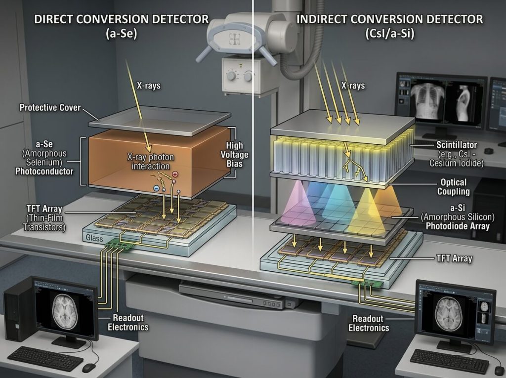
An indirect flat panel detector converts X-rays into light first.
The basic process is:
X-rays → Scintillator → Light → Photodiode → Electrical Signal → Digital Image
A scintillator is a material that converts X-ray energy into visible light.
Common scintillator materials include:
- Cesium Iodide (CsI)
- Gadolinium Oxysulfide (GOS)
The light generated by the scintillator is then detected by a sensor array.
Many commercial digital radiography detectors use an indirect-conversion architecture involving a scintillator and an amorphous silicon sensor array. FDA documentation for digital flat panel detectors describes systems using a scintillator layer together with an amorphous silicon TFT sensor array. (accessdata.fda.gov)
Indirect conversion is therefore an important concept when comparing medical X-ray flat panel detectors.
What Is a Direct Flat Panel Detector?
A direct flat panel detector converts incoming X-rays into electrical charge without first converting the radiation into visible light.
The simplified process is:
X-rays → Photoconductor → Electrical Signal → Digital Image
A material such as amorphous selenium (a-Se) may be used as the photoconductor in direct-conversion detector designs.
Direct-conversion technology is therefore often associated with:
- amorphous selenium
- a-Se detector
- direct X-ray conversion
- direct digital detector
The choice between direct and indirect technologies depends on the detector architecture and intended application.
It should not be assumed that one conversion method is automatically superior for every clinical or industrial requirement.
Direct vs Indirect Flat Panel Detector
This is one of the most common technical comparison topics.
| Feature | Indirect Conversion | Direct Conversion |
|---|---|---|
| First conversion step | X-ray to light | X-ray to electrical charge |
| Common materials | CsI, GOS | a-Se |
| Typical sensor concepts | Photodiode/TFT array | Photoconductor/TFT structure |
| Intermediate light | Yes | No |
| Common use | Digital radiography and other X-ray imaging | Specialized direct-conversion detector applications |
| Main comparison factors | Scintillator, light spread, efficiency, pixel pitch | Photoconductor properties, charge collection, pixel design |
The important point is that conversion architecture is only one part of detector performance.
A complete technical comparison should also examine:
DQE + MTF + noise + pixel pitch + active area + dynamic range + application + detector design
What Is CsI in a Flat Panel Detector?
CsI, or cesium iodide, is a commonly discussed scintillator for indirect-conversion X-ray detectors.
Its job is to convert X-ray energy into light.
One important characteristic of CsI is its structured crystal design in many detector implementations. The structure can help control lateral light spread compared with scintillator structures that have more isotropic light propagation.
For buyers, the practical question is not simply:
Is this detector CsI?
A more useful question is:
What are the complete performance specifications of the CsI detector?
For example, compare:
- pixel pitch
- active area
- DQE
- MTF
- detector sensitivity
- image noise
- weight
- dimensions
- durability
- operating conditions
- software
- system compatibility
Commercial detector products illustrate the use of CsI in medical DR systems, including portable and wireless detector configurations. (fujifilm.com)
What Is GOS in a Flat Panel Detector?
GOS is short for gadolinium oxysulfide.
It is another scintillator material used in indirect-conversion flat panel detectors.
A GOS scintillator converts X-rays into light, which is then detected by the sensor layer.
GOS detectors are available in different detector sizes and technical configurations. Commercial specifications show GOS options in both general and mobile digital radiography products. (fujifilm.com)
When comparing GOS and CsI detectors, buyers should consider the entire detector architecture.
Important questions include:
- What is the pixel pitch?
- What is the DQE?
- What is the MTF?
- What is the detector weight?
- What is the active area?
- What exposure range is supported?
- Is the detector wireless?
- What is the battery life?
- What X-ray systems are compatible?
CsI vs GOS: Which One Should You Choose?
The question CsI vs GOS is common among X-ray equipment buyers.
However, there is no universal answer that one scintillator material is the right option for every application.
The practical choice depends on the complete product design.
When comparing a CsI flat panel detector and a GOS flat panel detector, evaluate:
Image Performance
Look at:
- DQE
- MTF
- noise
- spatial resolution
- low-contrast performance
Physical Design
Look at:
- detector thickness
- detector weight
- housing
- dimensions
- active area
Workflow
Look at:
- wired or wireless communication
- battery
- preview time
- image transfer
- workstation software
Application
Consider whether the detector will be used for:
- general radiography
- mobile X-ray
- bedside imaging
- emergency imaging
- orthopedic imaging
- veterinary X-ray
- retrofit DR
Budget
Finally, compare the detector price and total ownership cost.
In other words:
CsI vs GOS should be a complete system comparison, not a material-only comparison.
What Is Amorphous Silicon in an FPD?
Amorphous silicon, often abbreviated as a-Si, is a semiconductor material commonly associated with indirect-conversion flat panel detector sensor arrays.
In this type of detector, the scintillator converts X-rays into light. The light is then detected by photodiodes in the amorphous silicon sensor array.
Thin-film transistor technology allows the detector to read the signals generated across the pixel array.
FDA documentation for several digital flat panel detector systems identifies amorphous silicon TFT arrays as part of the detector architecture. (accessdata.fda.gov)
This is why a buyer may encounter the term:
a-Si flat panel detector
when researching digital radiography technology.
What Is Amorphous Selenium?
Amorphous selenium, or a-Se, is associated with direct-conversion X-ray detector architectures.
Instead of using a scintillator to convert X-rays to light first, the photoconductor generates electrical charge from X-ray exposure.
This allows the detector to use a direct X-ray-to-charge conversion process.
When comparing:
a-Si vs a-Se
the key difference is usually the conversion architecture.
a-Si
Commonly associated with:
Indirect conversion
X-rays → Scintillator → Light → a-Si sensor
a-Se
Commonly associated with:
Direct conversion
X-rays → a-Se photoconductor → Electrical charge
However, the material name itself does not provide enough information to predict the complete performance of a detector.
Other factors still need to be considered.
a-Si vs a-Se Flat Panel Detector
| Specification Concept | a-Si | a-Se |
|---|---|---|
| Typical architecture | Indirect conversion | Direct conversion |
| Scintillator | Usually required | Not used for direct X-ray conversion |
| Typical role | Digital radiography sensor array | Direct-conversion photoconductor |
| Common search term | a-Si detector | a-Se detector |
| Main technical issue | Light conversion and sensor response | Direct charge generation and collection |
For a procurement decision, it is better to compare actual manufacturer data instead of selecting only from the semiconductor material.
What Is Pixel Pitch?
Pixel pitch is one of the most frequently listed specifications on an X-ray detector datasheet.
It represents the distance between neighboring pixel centers.
Pixel pitch is normally expressed in:
micrometers (μm)
or:
millimeters (mm)
For example:
0.10 mm = 100 μm
0.14 mm = 140 μm
0.15 mm = 150 μm
Commercial digital X-ray detectors are available with different pixel pitches, including 100 μm and 150 μm examples. (accessdata.fda.gov)
Does Smaller Pixel Pitch Mean Better Resolution?
Generally, a smaller pixel pitch allows the detector to sample image information at a finer spatial scale.
But there is an important distinction:
pixel pitch is not exactly the same thing as system spatial resolution.
For example, a detector with 100 μm pixels does not automatically guarantee 100 μm system resolution in every imaging condition.
Actual image resolution can be affected by:
- detector design
- scintillator properties
- optical spread
- pixel response
- image processing
- focal spot size
- geometric magnification
- motion
- exposure conditions
- display
- reconstruction or processing algorithms
Therefore:
Smaller pixel pitch is one specification, not the entire image-quality specification.
This is particularly important for buyers comparing two DR detectors only because one product has a smaller pixel size.
Common Flat Panel Detector Pixel Pitch
Depending on the detector and application, buyers may encounter values such as:
- 100 μm
- 120 μm
- 125 μm
- 140 μm
- 143 μm
- 150 μm
- 160 μm
- 180 μm
- 200 μm
There is no single pixel pitch suitable for every radiography application.
General radiography, orthopedic imaging, mobile X-ray, veterinary imaging, and specialized applications may have different design priorities.
What Is Pixel Matrix?
The pixel matrix describes how many pixels are arranged across the detector.
For example, a specification may show something similar to:
3072 × 3072 pixels
or:
2480 × 3072 pixels
The matrix is related to:
- detector dimensions
- pixel pitch
- image resolution
- data size
- image processing
The basic relationship can be understood as:
Active width ÷ pixel pitch ≈ number of pixels across the detector
Therefore, detector size and pixel pitch work together to determine the pixel matrix.
A large detector with a small pixel pitch can produce a large image matrix.
This can also increase image data size and processing requirements.
What Is Spatial Resolution?
Spatial resolution describes how well an imaging system can distinguish small or closely spaced structures.
In digital radiography, spatial resolution is affected by many components:
- focal spot
- detector pixel pitch
- scintillator
- detector design
- signal processing
- image reconstruction
- geometry
- motion
It is therefore important not to confuse:
pixel pitch
with:
measured spatial resolution
A detector data sheet may provide an MTF value or line-pair measurement as additional information.
What Is MTF in a Flat Panel Detector?
MTF stands for Modulation Transfer Function.
It describes how well an imaging system transfers object contrast at different spatial frequencies.
In simpler language:
MTF helps describe how well fine image details are preserved.
A detector with stronger signal transfer at a particular spatial frequency may preserve more information about structures at that scale.
MTF is therefore commonly used in technical evaluation of digital X-ray detector image quality.
When comparing two detectors, an MTF curve can be more informative than looking at pixel pitch alone.
However, MTF should also be interpreted with DQE and noise-related measurements.
What Is DQE?
DQE means Detective Quantum Efficiency.
It is an important measure used in evaluating the efficiency of an X-ray imaging detector.
In simplified terms, DQE describes how effectively the detector uses the available X-ray signal while considering image noise.
Higher DQE can indicate that the detector transfers useful X-ray information more efficiently under the tested conditions.
DQE is commonly discussed together with:
- MTF
- NPS
- spatial resolution
- image noise
Research on digital X-ray detectors commonly evaluates detector performance using measures such as DQE, MTF and noise-related metrics. (pubmed.ncbi.nlm.nih.gov)
For procurement, however, DQE should not be viewed in isolation.
What Is NPS?
NPS stands for Noise Power Spectrum.
It characterizes image noise as a function of spatial frequency.
In technical detector evaluation, NPS can help explain how noise is distributed across different spatial scales.
This can be particularly useful for engineers and imaging specialists comparing detector performance.
For a normal equipment buyer, the most important point is simply:
NPS is a technical image-noise measurement used together with DQE and MTF.
DQE vs MTF vs Pixel Pitch
These three terms are often confused.
Pixel Pitch
Describes the physical spacing of detector pixels.
MTF
Describes how effectively image detail or modulation is transferred at different spatial frequencies.
DQE
Describes how efficiently the imaging system uses the incoming X-ray signal while accounting for noise.
Therefore:
Pixel pitch ≠ MTF ≠ DQE
They provide different pieces of information.
A professional detector comparison should ideally review all relevant specifications rather than selecting a detector based on one number.
What Is Dynamic Range?
Dynamic range refers to the range of signal levels that a detector can capture and represent.
A wide usable dynamic range can be useful because different areas of the radiographic image may receive substantially different levels of X-ray exposure.
The actual usable range depends on detector design, electronics, software processing, exposure conditions, and other system parameters.
When reviewing detector specifications, it can be useful to ask how the manufacturer defines dynamic range and under what test conditions the value was obtained.
What Is Detector Bit Depth?
Digital X-ray images are represented using digital values.
Detector and imaging system specifications may mention:
- 12-bit
- 14-bit
- 16-bit
Bit depth indicates how many digital intensity levels can be represented.
For example:
12-bit = 4,096 levels
14-bit = 16,384 levels
16-bit = 65,536 levels
However, a larger bit depth does not automatically mean a visibly better clinical image.
Actual image quality depends on detector signal performance, noise, processing, display, and the imaging task.
What Is the Active Area of an FPD?
The active area is the portion of the detector used to acquire the image.
It is not always identical to the detector’s total external dimensions.
For example, a detector may have a physical size slightly larger than its active imaging area because of its housing and electronics.
Active area matters when checking:
- anatomical coverage
- image field
- Bucky compatibility
- table integration
- wall stand installation
- mobile X-ray use
When purchasing a replacement FPD, always check both:
physical dimensions
and
active imaging area
Flat Panel Detector Dimensions
Common FPD dimensions include formats such as:
- 14 × 17 inch
- 17 × 17 inch
- 24 × 30 cm
- 35 × 43 cm
- other application-specific sizes
The detector format should match the intended examination and mechanical configuration.
A 14 × 17 inch detector can be useful where a cassette-style format and portability are important.
A 17 × 17 inch detector provides a square imaging format and may be selected for applications requiring broader coverage.
The physical dimensions should also be checked against the:
- Bucky tray
- wall stand
- X-ray table
- mobile X-ray tray
- detector holder
Detector Weight
Weight is often underestimated during detector selection.
For a fixed radiography room, detector weight may not be a major issue.
For mobile X-ray or veterinary imaging, the detector may be handled many times throughout the day.
A portable wireless detector should therefore be evaluated for:
- total weight
- battery weight
- housing
- grip
- thickness
- handling
- positioning
Commercial mobile detector specifications show that weight differs between detector formats and models, demonstrating why size alone does not determine portability. (fujifilm.com)
Flat Panel Detector Battery
Wireless DR detectors normally require a battery system.
Battery-related specifications can include:
- battery capacity
- operating time
- charging time
- battery type
- battery quantity
- removable battery
- charging station
- battery replacement
For hospitals with high daily usage, battery workflow can have a direct effect on detector availability.
A buyer should ask:
How many examinations can the detector perform on one charge?
The practical answer depends on exposure conditions, wireless transmission, standby time, battery age, and operating environment.
Wireless Communication
Wireless flat panel detectors transmit image data to a workstation or imaging system.
Depending on the product, specifications may mention:
- wireless communication standard
- frequency band
- transmission speed
- connection range
- wired backup
- network security
- connection mode
However, wireless performance is not simply a matter of transmission speed.
A hospital should also evaluate the stability of the wireless network and how the detector integrates with the existing imaging workflow.
Image Acquisition Time and Preview Time
Some detector specifications list:
image acquisition time
or:
preview time
These terms describe how quickly an image can become available after exposure.
A shorter image preview time can contribute to a faster radiography workflow, especially in mobile or emergency applications.
However, the total workflow also includes:
- patient positioning
- exposure
- image transfer
- image processing
- operator review
- retake decisions
- documentation
Therefore, detector speed should be viewed within the complete imaging workflow.
Detector Durability
A medical detector may be exposed to frequent handling, cleaning, movement, and accidental impacts.
Durability specifications may include:
- shock resistance
- vibration resistance
- drop resistance
- static load
- waterproofing
- dust protection
- IP rating
For mobile X-ray and veterinary use, durability can be especially important.
A detector used in a fixed room may have different physical requirements from a detector carried between wards.
What Does IP Rating Mean?
An IP rating indicates the level of protection provided by an enclosure against ingress from solids and liquids under defined testing conditions.
For example, buyers may see specifications such as:
IP54
IP65
IP67
The exact protection level depends on the product.
An IP rating can be useful when choosing a detector for environments where cleaning, moisture, dust, or fluid exposure may occur.
However, buyers should always check the manufacturer’s detailed operating and cleaning instructions instead of assuming that any IP-rated detector can be exposed to all liquids or cleaning chemicals.
Flat Panel Detector Resolution
When buyers search for:
flat panel detector resolution
they may actually be looking for several different specifications.
Resolution can refer to:
- pixel pitch
- image matrix
- spatial resolution
- MTF
- line-pair resolution
These are related but not identical.
For example, two detectors may have similar pixel pitches but different image performance due to differences in scintillator, detector design, electronics, processing, and test conditions.
Therefore, when comparing detector resolution, review the manufacturer’s complete test data where available.
How to Compare Two Flat Panel Detectors
Suppose you are comparing two wireless 14 × 17 inch DR detectors.
Detector A:
- 14 × 17 inch
- CsI
- 140 μm pixel pitch
- wireless
- 3.0 kg
Detector B:
- 14 × 17 inch
- GOS
- 150 μm pixel pitch
- wireless
- 3.2 kg
It would be tempting to decide based only on:
140 μm vs 150 μm
But that is not enough.
A more complete comparison should include:
| Specification | Detector A | Detector B |
|---|---|---|
| Detector size | 14 × 17 | 14 × 17 |
| Scintillator | CsI | GOS |
| Pixel pitch | Compare | Compare |
| DQE | Compare | Compare |
| MTF | Compare | Compare |
| Active area | Compare | Compare |
| Weight | Compare | Compare |
| Battery | Compare | Compare |
| Wireless | Yes/No | Yes/No |
| IP rating | Compare | Compare |
| Software | Compare | Compare |
| Compatibility | Verify | Verify |
| Warranty | Compare | Compare |
| Price | Compare | Compare |
The right decision should be based on the actual application and complete specifications.
Which Flat Panel Detector Specifications Matter Most?
Not every buyer needs to study every engineering parameter.
For a hospital procurement team, the following specifications are usually a practical starting point:
Essential Specifications
Detector size
Pixel pitch
Scintillator
Wired or wireless
Detector weight
Battery
Active area
Compatibility
Software
Warranty
Technical Specifications
For more detailed evaluation, also review:
DQE
MTF
NPS
spatial resolution
dynamic range
matrix
bit depth
sensitivity
image acquisition time
The importance of each specification depends on the clinical application and system design.
Which Specifications Matter for Mobile X-Ray?
For mobile X-ray, prioritize:
- Detector weight
- Wireless connectivity
- Battery performance
- Durability
- Detector size
- Image preview speed
- Software compatibility
- Cleaning and protection
- System integration
A detector with excellent technical specifications but inconvenient weight or weak workflow integration may not be the most practical option for a mobile system.
Which Specifications Matter for a Fixed X-Ray Room?
A fixed radiography room may place more emphasis on:
- detector size
- active area
- pixel pitch
- image quality
- DQE
- MTF
- workflow
- Bucky compatibility
- wall stand compatibility
- software
- PACS/DICOM integration
- workload
- serviceability
The detector does not need to be evaluated in isolation.
The complete radiography system should be considered.
Which Specifications Matter for Veterinary X-Ray?
Veterinary imaging often requires flexible positioning.
Important specifications may include:
- detector size
- detector weight
- wireless operation
- durability
- IP protection
- cleaning requirements
- battery
- software
- image quality
- portability
For large-animal applications, detector size and physical durability may become particularly important.
What Should Medical Equipment Buyers Ask the Supplier?
Before purchasing a flat panel detector, prepare a list of technical questions.
Compatibility
Is the detector compatible with my X-ray machine?
Can it work with my current generator?
Does it fit my Bucky or wall stand?
Does the system require an additional interface?
Technology
Is the detector CsI or GOS?
Is it direct or indirect conversion?
What is the pixel pitch?
What is the active area?
Workflow
Is the detector wired or wireless?
How long does image transmission take?
How many examinations can the battery support?
Is a spare battery available?
Image Quality
What are the DQE and MTF specifications?
What is the spatial resolution?
What is the matrix?
Physical Design
What is the detector weight?
What is the IP rating?
What is the load capacity?
What is the operating temperature range?
Service
What is the warranty?
Are replacement batteries available?
Is technical support available?
Can spare parts be supplied?
These questions can make supplier comparisons much more meaningful.
Flat Panel Detector Specification Checklist
Before sending a purchase order, review:
Detector type
Direct or indirect conversion
CsI or GOS
a-Si or a-Se
Detector size
Active area
Pixel pitch
Pixel matrix
Spatial resolution
DQE
MTF
NPS
Dynamic range
Bit depth
Detector weight
Battery
Wireless communication
IP rating
Load capacity
Operating conditions
Software
DICOM
PACS integration
X-ray system compatibility
Warranty
Technical support
Frequently Asked Questions
What are the main types of flat panel detectors?
Flat panel detectors can be categorized by conversion technology, including indirect-conversion detectors and direct-conversion detectors. They can also be classified by detector format, communication method, scintillator, and application.
What is the difference between direct and indirect flat panel detectors?
Indirect detectors convert X-rays into light before converting the light into an electrical signal. Direct detectors convert X-rays into electrical charge without the intermediate light conversion step.
Is CsI a flat panel detector?
CsI is not the entire detector. It is a scintillator material used in some indirect-conversion flat panel detectors.
Is GOS a detector material?
GOS is a scintillator material used in some indirect X-ray detector designs.
What is the difference between CsI and GOS?
Both can serve as scintillators in indirect-conversion detectors. Their characteristics and overall detector performance depend on the complete detector design.
Is CsI better than GOS?
Neither should automatically be considered better for every application. Compare actual detector specifications and intended use.
What is a-Si in an X-ray detector?
a-Si means amorphous silicon. It is commonly used in sensor arrays in indirect-conversion flat panel detector designs.
What is a-Se in an X-ray detector?
a-Se means amorphous selenium. It is associated with direct-conversion X-ray detector technology.
What is pixel pitch in a DR detector?
Pixel pitch is the distance between the centers of adjacent detector pixels, normally expressed in micrometers.
Is 100 μm better than 150 μm?
A 100 μm pixel pitch provides finer sampling than 150 μm, but smaller pixel pitch does not by itself determine total clinical image quality.
What is DQE in digital radiography?
DQE, or Detective Quantum Efficiency, is a technical measure of how efficiently a detector uses incident X-ray information while considering noise.
What is MTF in digital X-ray?
MTF, or Modulation Transfer Function, describes image-signal transfer as a function of spatial frequency and is used to evaluate image-detail performance.
What does active area mean on a detector datasheet?
Active area is the portion of the detector that is used to acquire the image.
What is the difference between detector size and active area?
Detector size describes the overall physical format, while active area describes the effective imaging region.
What is a pixel matrix?
The pixel matrix describes the number and arrangement of pixels across the active imaging area.
Does a higher bit depth mean better X-ray images?
Not necessarily. Bit depth represents the number of digital intensity levels, while overall image quality also depends on detector signal, noise, processing, exposure, and display.
What is the most important flat panel detector specification?
There is no single specification that is most important for every application. Detector size, pixel pitch, detector technology, image performance, weight, compatibility, battery, durability, and workflow should be considered together.
What is the best pixel pitch for a DR detector?
The appropriate pixel pitch depends on the imaging application and detector design. A smaller pixel pitch is not automatically the right choice for every X-ray system.
What should I compare when buying a wireless DR detector?
Compare detector size, pixel pitch, scintillator, active area, DQE, MTF, weight, battery life, wireless communication, durability, software, compatibility, warranty, and price.
Can any flat panel detector work with any X-ray machine?
No. Detector compatibility depends on mechanical dimensions, detector interface, generator communication, image acquisition software, Bucky or tray design, and other system requirements.
How do I choose between a 14 × 17 and 17 × 17 detector?
Consider the anatomy, required field of view, X-ray system configuration, detector weight, available tray space, and intended workflow.
Is a wireless detector better than a wired detector?
Not in every situation. Wireless detectors offer greater mobility, while wired detectors can be practical for fixed imaging rooms. The appropriate choice depends on workflow.
Conclusion
Flat panel detector specifications can look complicated, but the main concepts are easier to understand when they are separated into several categories.
Detector technology includes:
- direct conversion
- indirect conversion
- CsI
- GOS
- a-Si
- a-Se
Image specifications include:
- pixel pitch
- pixel matrix
- spatial resolution
- DQE
- MTF
- NPS
- dynamic range
Physical specifications include:
- detector size
- active area
- weight
- thickness
- durability
- IP rating
Workflow specifications include:
- wired or wireless communication
- battery
- image transmission
- preview time
- software
- DICOM
- PACS compatibility
No single specification should be used to judge an entire detector.
For a hospital, clinic, mobile X-ray provider, veterinary facility, distributor, or medical equipment integrator, the best approach is to match detector technology and specifications to the actual application.
When comparing two flat panel detectors, look beyond the product name and compare the complete specification sheet.
A practical evaluation can start with:
size → technology → pixel pitch → image performance → weight → battery → durability → compatibility → software → service → total cost
That approach makes it easier to identify a detector that fits the X-ray system and the daily imaging workflow.
For buyers planning a new digital radiography system or upgrading an existing X-ray machine, the next important step is understanding how to select the right detector based on application, compatibility, size, technology, workflow, and budget.