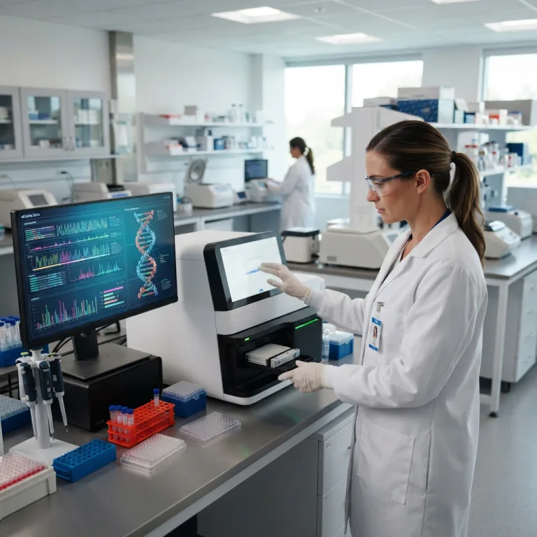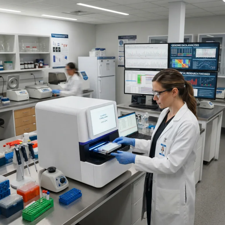
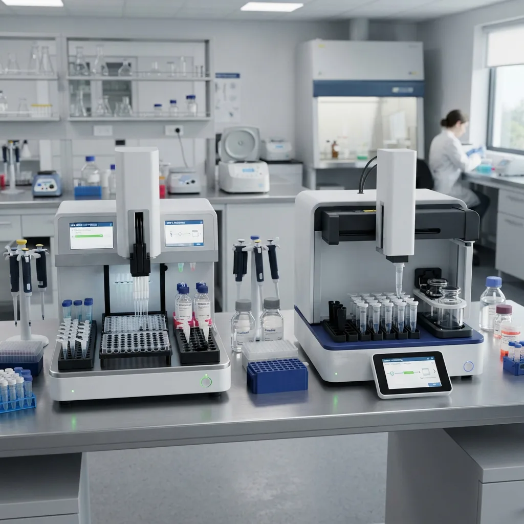
The “Garbage In, Garbage Out” Law of Molecular Diagnostics
In the rigorous discipline of molecular biology, there is an inescapable rule: Garbage In, Garbage Out.
A hospital laboratory can invest millions of dollars into acquiring the most advanced Next-Generation Sequencing (NGS) platform on the planet. They can hire the most brilliant bioinformaticians to analyze the data. However, if the DNA extracted from a patient’s whole blood tube or a formalin-fixed tumor biopsy is chemically fragmented, highly degraded, or contaminated with residual cellular proteins, the resulting sequencing data will be complete garbage. The sequencer will simply read the damaged fragments and output a false-negative clinical report.
Extracting pure nucleic acids from complex biological matrices is a violent chemical war waged at the microscopic level. Before a scientist can read the genetic code, they must forcefully break open the patient’s cells, destroy the destructive enzymes that naturally hunt DNA, and wash away chemical inhibitors that paralyze diagnostic equipment.
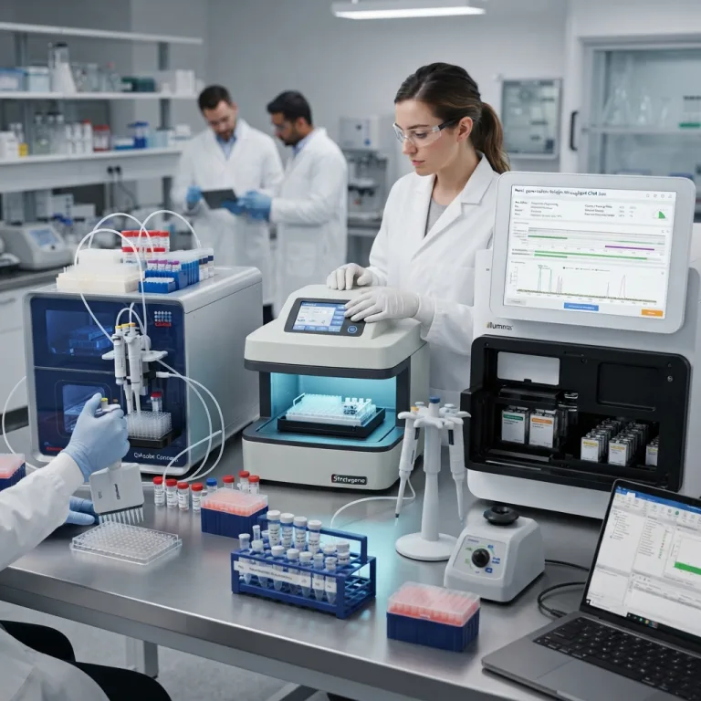
This guide explores the intense biological challenges of nucleic acid purification, the biochemical chemistry of cellular lysis, and why clinical laboratories have universally shifted from manual centrifugation to automated magnetic bead extraction.
1. The Biological Fortress: Lysis and Nuclease Destruction
To extract a human genome, one must first breach a series of formidable biological fortresses.
Human DNA does not float freely in blood or tissue. It is tightly spooled around basic proteins called histones to form nucleosomes, compacted into dense chromatin, sealed inside a double-layered nuclear envelope, and surrounded by a cellular phospholipid bilayer.
Chemical Lysis: Breaching the Cell
The first step of any extraction protocol is Lysis (breaking open the cell).
-
Detergents: Technicians introduce powerful detergents (such as Sodium Dodecyl Sulfate, SDS, or Triton X-100). These surfactants aggressively dissolve the lipid bilayers of the cell membrane and the nuclear envelope, spilling the internal contents into the liquid buffer.
-
Proteinase K: DNA is entangled in structural proteins. The extraction buffer contains Proteinase K, a highly robust serine protease enzyme that shreds histone proteins into short peptides, allowing the tightly coiled DNA helix to unfurl and float freely in the solution.
The Immediate Threat: Endogenous Nucleases
The moment a cell is lysed, the DNA is in mortal danger. Human cells contain endogenous enzymes called DNases (Deoxyribonucleases) and RNases (Ribonucleases). In a living cell, these enzymes act as a defense mechanism, hunting and chopping up foreign viral DNA. When the cell membrane is artificially ruptured during laboratory extraction, these nucleases are released into the same liquid soup as the patient’s genomic DNA. If left unchecked, they will chop the three-billion-base-pair genome into useless tiny fragments within minutes.
To neutralize this threat, extraction buffers are loaded with Chaotropic Salts (such as Guanidinium Thiocyanate). These harsh chemicals instantly strip the hydration shell (water molecules) away from the nuclease enzymes, causing their 3D protein structures to denature and unfold permanently. Denatured nucleases are biologically dead, securing the survival of the patient’s DNA.
2. The Invisible Enemy: PCR Inhibitors in Clinical Samples
Extracting DNA is only half the battle; the DNA must also be perfectly pure. Biological samples are loaded with powerful chemical compounds that, if accidentally carried over into the final purified DNA tube, will completely paralyze Taq polymerase enzymes during downstream PCR testing.
These compounds are known as PCR Inhibitors, and failing to wash them away is the leading cause of false-negative clinical diagnoses.
                  COMMON CLINICAL PCR INHIBITORS BY SAMPLE TYPE
+-------------------------------------------------------------------------+
| WHOLE BLOOD SAMPLES                                                     |
| - Heme / Hemoglobin: The iron-carrying pigment in red blood cells.      |
|   Even a slight reddish tint in the final DNA elution indicates severe  |
|   heme contamination, which instantly blocks polymerase activity.       |
| - Immunoglobulin G (IgG): Plasma antibodies that bind and block enzymes.|
|                                                                         |
| TISSUE & FORENSIC SAMPLES                                               |
| - Melanin: Found in high concentrations in skin and hair samples.       |
| - Collagen: Found in cartilage, bone, and connective tissues.           |
|                                                                         |
| GASTROINTESTINAL & ENVIRONMENTAL SAMPLES                                |
| - Bile Salts & Complex Polysaccharides: Present in stool (feces) samples|
| - Humic Acid: Present in soil, deeply inhibiting environmental PCR.     |
|                                                                         |
| PATHOLOGY BIOPSIES (FFPE)                                               |
| - Formalin: Formaldehyde used to preserve tissues causes severe DNA     |
|   crosslinking, requiring intense heat and chemical reversal to extract.|
+-------------------------------------------------------------------------+
An extraction technology is ultimately judged by its ability to efficiently bind the DNA while aggressively washing away 100% of these specific biological inhibitors.
3. The Legacy Bottleneck: Silica Spin Column Technology
For the past thirty years, the standard method for purifying DNA has been Solid-Phase Extraction (SPE) using silica-gel membranes packed into small plastic centrifuge tubes, known as Spin Columns.
The Principle of Silica Binding
In the presence of high concentrations of chaotropic salts (which create a low-pH, dehydrated environment), nucleic acids undergo a conformational change. The negative charges on the DNA phosphate backbone bind tightly to the positively charged silica membrane via a combination of hydrogen bonding and cation bridges. Meanwhile, cellular proteins, lipids, and carbohydrates fail to bind to the silica and remain in the liquid.
The Mechanical Nightmare of Centrifugation
To force the biological sample through the microscopic pores of the silica filter, the technician must place the tubes into a high-speed microcentrifuge.
-
The Workflow: The technician adds the sample to the column, closes the lid, places it in the centrifuge, balances the rotor, spins it at 10,000 to 14,000 rpm for one minute, takes it out, discards the waste liquid, adds a wash buffer, and repeats the spinning process three to four times.
-
The Drawbacks:
-
Zero Automation: A technician’s hands are tied to the centrifuge for an hour just to process 24 samples.
-
Centrifugal Shearing Forces: Spinning liquid through a tightly packed silica filter creates immense fluidic shear stress. This mechanical violence physically snaps ultra-long DNA strands. If a laboratory requires High Molecular Weight (HMW) DNA greater than 50,000 base pairs for Third-Generation Long-Read Sequencing, spin columns will routinely destroy the sample.
-
Clogging Risks: If a blood or stool sample is highly viscous or contains unlysed tissue debris, it will physically clog the silica membrane. The liquid will refuse to spin through, ruining the sample entirely.
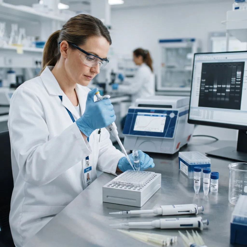
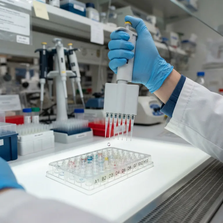
A molecular biologist carefully pipettes a purified DNA sample into a micro-volume spectrophotometer to determine nucleic acid concentration and purity before downstream applications.
-
4. The Automation Standard: Magnetic Bead Technology
To eliminate the mechanical damage of centrifuges and enable high-throughput robotic automation, the molecular diagnostics industry engineered a revolutionary solid-phase extraction medium: Paramagnetic Nanoparticles (Magnetic Beads).
These microscopic spheres (typically 1 to 5 micrometers in diameter) consist of a solid iron oxide core ($Fe_3O_4$) surrounded by a synthetic polymer shell coated in Silica or Carboxyl functional groups. Because they are paramagnetic, the beads only exhibit magnetism when exposed to an external magnetic field. The moment the magnet is removed, they instantly lose all magnetic attraction and disperse freely in the liquid without clumping together.
                  THE 4-STEP MAGNETIC BEAD EXTRACTION WORKFLOW
+-------------------------------------------------------------------------+
| STEP 1: LYSIS & BINDING                                                 |
| - Patient sample, lysis buffer, and magnetic beads are mixed in a well. |
| - Chaotropic salts force the DNA to bind to the floating magnetic beads.|
| - No filter is involved; the beads have massive 3D surface area.        |
|                                                                         |
| STEP 2: MAGNETIC CAPTURE                                                |
| - An external magnet is applied to the side or bottom of the well.      |
| - The beads (carrying the DNA) instantly snap to the magnet.            |
| - The dirty liquid (containing proteins and inhibitors) is aspirated    |
|   and thrown away.                                                      |
|                                                                         |
| STEP 3: WASHING (Repeated 2-3 Times)                                    |
| - The magnet is removed. Alcohol-based wash buffers are added.          |
| - The beads are gently resuspended (mixed) to wash away trapped salts.  |
| - The magnet is reapplied, and the wash waste is discarded.             |
|                                                                         |
| STEP 4: ELUTION                                                         |
| - A highly purified, low-salt aqueous buffer (or TE buffer) is added.   |
| - In the absence of chaotropic salts, the DNA releases from the beads.  |
| - The magnet captures the bare beads, leaving pure, liquid DNA behind.  |
+-------------------------------------------------------------------------+
Why Magnetic Beads Dominate Modern Diagnostics
-
Zero Shear Stress for Intact DNA: Because there is no centrifuge and no silica filter to squeeze through, the DNA is never subjected to violent mechanical shearing. Magnetic beads float gently in the liquid, preserving massive fragments of High Molecular Weight (HMW) DNA essential for advanced genomics.
-
Immunity to Clogging: Viscous samples, heavy mucous, or fatty tissue homogenates that would instantly clog a spin column pose zero threat to magnetic beads. The beads simply swim through the debris to capture the DNA.
-
Infinite Scalability: Unlike a centrifuge rotor that only holds 24 tubes, a robotic liquid handler can drop a 96-well plate of magnetic beads onto a magnetic block, processing 96 patient samples simultaneously in exactly the same amount of time it takes to process one sample.
5. Assessing DNA Yield and Quality Metrics (A260/A280)
Once an automated extractor completes its run, a laboratory technician must verify that the extraction was successful before wasting thousands of dollars loading the DNA onto a Next-Generation Sequencer.
The industry standard for evaluating nucleic acid purity relies on UV-Vis Spectrophotometry, measuring how the liquid absorbs ultraviolet light at three specific wavelengths: 230 nm, 260 nm, and 280 nm.
The A260 / A280 Ratio (Protein Contamination)
-
Nucleic acids maximally absorb UV light at 260 nm.
-
Proteins (specifically the aromatic amino acids tyrosine and tryptophan) maximally absorb UV light at 280 nm. By dividing the absorbance reading at 260 nm by the reading at 280 nm, the spectrophotometer calculates the A260/A280 ratio.
-
Pure DNA: Should yield a ratio between 1.7 and 1.9 (ideal is 1.8).
-
Pure RNA: Should yield a ratio of roughly 2.0.
-
Failed Extraction (Less than 1.6): A low ratio indicates that the automated extractor failed to wash away cellular proteins. This DNA must be re-purified before downstream testing.
The A260 / A230 Ratio (Chemical Contamination)
-
Organic compounds, chaotropic salts (Guanidinium), and phenol maximally absorb UV light at 230 nm.
-
Pure Nucleic Acid: Should yield an A260/A230 ratio greater than 2.0.
-
Failed Extraction (Less than 1.5): Indicates that the magnetic beads were not washed properly, and harsh extraction chemicals carried over into the final elution buffer. These salts will severely inhibit PCR amplification.
Frequently Asked Questions (FAQ)
Q1: Why do some magnetic bead protocols require heating during the elution step?
Heat acts as a thermodynamic catalyst to break the electrostatic bonds between the negatively charged DNA and the silica-coated magnetic beads. While DNA will naturally elute in a low-salt buffer at room temperature, heating the elution buffer to 60 or 70 degrees Celsius significantly increases the kinetic energy, driving the final DNA yield up by 15 percent to 30 percent and ensuring that heavy, long-strand DNA detaches completely from the nanoparticles.
Q2: What happens if I accidentally leave trace amounts of ethanol in the final DNA sample?
During the magnetic bead washing steps, 70 percent to 80 percent ethanol is used to strip away salts while keeping the DNA tightly bound to the beads. Before the final elution buffer is added, the protocol mandates an “Air Dry” step (usually 3 to 5 minutes) to evaporate the residual alcohol. If a technician rushes this step, trace ethanol will carry over into the final DNA tube. Ethanol is a potent PCR inhibitor; even a 2 percent ethanol carryover will severely suppress Taq polymerase, causing PCR assays to fail.
Q3: Can magnetic bead extractors isolate RNA just as easily as DNA?
Yes. The physical properties of magnetic bead extraction apply equally to both DNA and RNA. The only difference lies in the specific chemistry of the binding buffer and the absolute necessity to eliminate RNase enzymes. Because RNA is incredibly fragile, RNA extraction kits use harsher lysis buffers containing Beta-mercaptoethanol to instantly obliterate RNases, and utilize specialized silica or carboxyl beads optimized to bind single-stranded nucleic acids.
Q4: Are magnetic beads reusable?
No. Paramagnetic nanoparticles used in clinical diagnostics and molecular biology are strictly single-use consumables. Attempting to wash and reuse beads from one patient on a second patient guarantees massive cross-contamination and false-positive clinical results. Furthermore, the silica or carboxyl functional groups on the surface of the beads degrade after exposure to harsh lysis buffers.
Q5: Can I extract DNA from old, dried blood spots on filter paper?
Yes. Forensic laboratories and newborn screening programs routinely extract high-quality DNA from dried blood spots (such as FTA cards) that are decades old. The dried punch is incubated in a heated lysis buffer to rehydrate the cellular material and dissolve the paper matrix. Once the DNA is pushed into the liquid phase, standard magnetic bead technology efficiently captures the DNA while washing away the heavy paper cellulose fibers and dried heme proteins that would normally clog a traditional spin column.
Summary
The foundation of all modern genomics, oncology sequencing, and viral tracking relies on the absolute purity of the biological sample. While manual spin columns served the industry for decades, the intense mechanical shearing and manual labor bottlenecks rendered them obsolete for high-throughput clinical diagnostics. By harnessing paramagnetic nanoparticles, automated extraction eliminates centrifuge damage, protects High Molecular Weight DNA, aggressively washes away PCR inhibitors, and allows laboratories to process 96 patients simultaneously with perfect reproducibility.