The Heart of Modern Microsurgery
The evolution of modern surgery is defined by the shift toward minimally invasive techniques. Whether a neurosurgeon is clipping a tiny cerebral aneurysm, an ophthalmologist is performing cataract surgery, or an endodontist is navigating a calcified root canal, human vision alone is no longer sufficient.
The operating microscope (surgical microscope) is the central piece of capital equipment that makes these intricate procedures possible. By providing brilliant stereoscopic (3D) magnification, deep depth of field, and intense coaxial illumination, surgical microscopes allow doctors to operate on blood vessels and nerves thinner than a human hair.
For hospital procurement officers, clinical directors, and medical equipment distributors, purchasing an operating microscope is a major capital investment. The market is filled with varying specifications, ranging from basic dental microscopes to highly complex neurosurgical platforms. This comprehensive buyer’s guide breaks down the core hardware configurations, optical parameters, and illumination technologies to help you make a financially sound and clinically optimal purchasing decision.
2. Anatomy of a Surgical Microscope: The Core Components
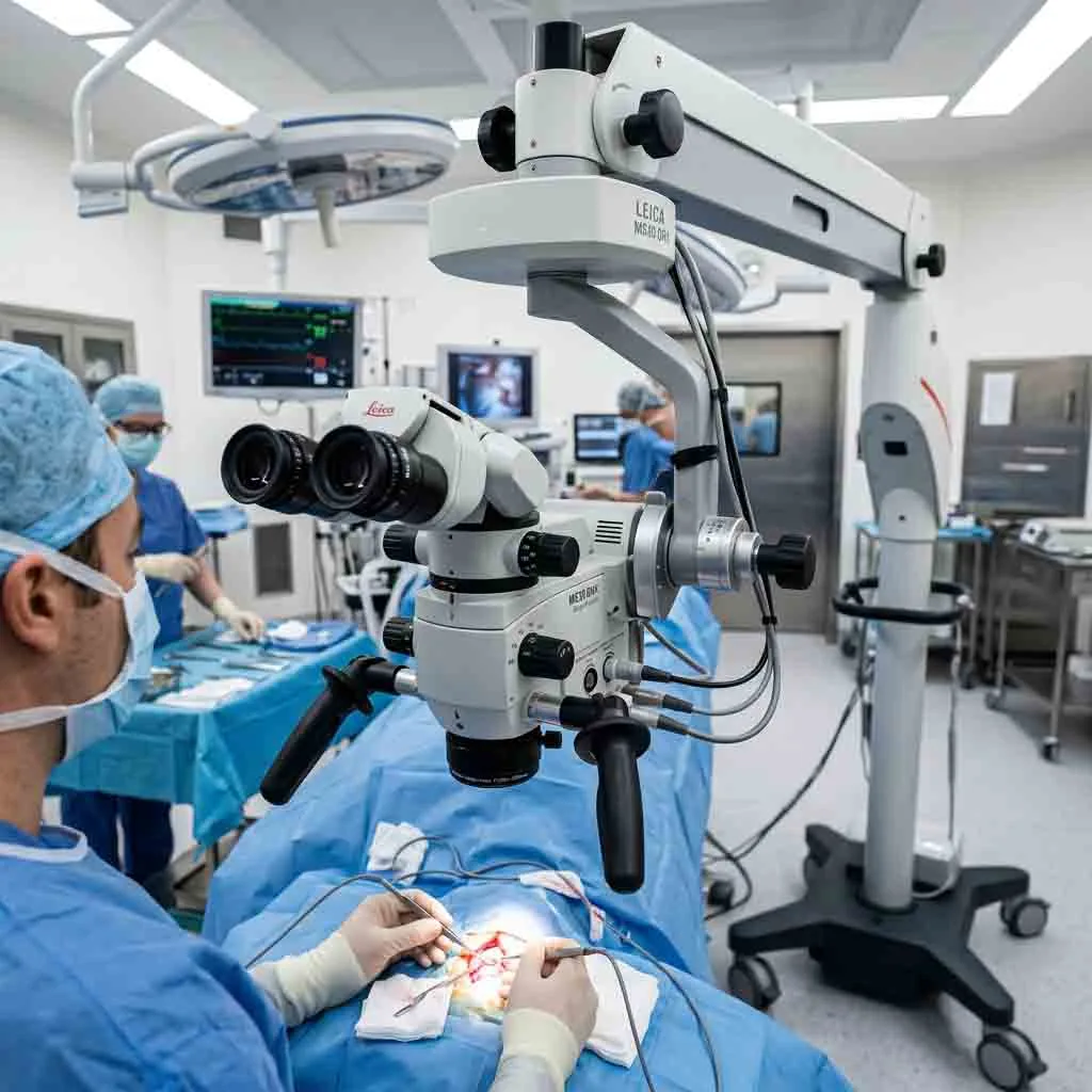
Before diving into complex specifications, it is essential to understand the basic anatomy of a surgical microscope system. A standard operating microscope consists of three primary subsystems:
-
The Optical Head (Microscope Body): The core component housing the objective lens, the magnification changer, the binocular tube, and the eyepieces.
-
The Illumination System: The light source (LED, Xenon, or Halogen), typically housed in the microscope base or the optical head, which delivers light directly to the surgical field via a fiber-optic cable and coaxial prisms.
-
The Suspension Arm and Mounting Stand: The mechanical framework that holds the optical head. It utilizes counterbalanced spring or pneumatic arms to allow the surgeon to move the heavy optical head with a single finger.
3. Demystifying Optical Specifications
The primary reason a hospital invests in a high-end surgical microscope is optical clarity. When comparing spec sheets, procurement teams must look beyond just “magnification power” and evaluate the quality of the glass and the design of the optical pathways.
A. Apochromatic Optics (The Gold Standard)
Standard glass lenses bend different colors of light at slightly different angles. In a basic microscope, this creates “chromatic aberration”—a distracting colored halo or blur around the edges of tissue.
-
The Solution: Premium surgical microscopes utilize Apochromatic lenses. These complex, multi-element lenses are precisely engineered to focus all wavelengths of the color spectrum onto the exact same focal plane.
-
The Clinical Benefit: Apochromatic optics deliver true-to-life color rendering, high-contrast borders, and razor-sharp resolution. This is critical when a surgeon needs to differentiate between a healthy nerve and a similar-colored tumor margin.
B. Working Distance and Objective Lenses
The working distance is the physical space between the bottom of the microscope’s objective lens and the patient’s surgical site.
-
Fixed Objective Lenses: Basic microscopes come with a fixed focal length (e.g., 200 mm, 250 mm, or 300 mm). If the surgeon needs to look deeper into a cavity, they must physically move the entire microscope head up or down.
-
Variable Working Distance (Vario Objective): Advanced microscopes feature an adjustable objective lens (often motorized). The surgeon can adjust the focal distance smoothly (e.g., from 200 mm to 400 mm) using a foot pedal or hand grip without moving the microscope head. This is absolutely essential in spine and neurosurgery, where cavity depth changes constantly.
C. Magnification Changer: Step vs. Continuous Zoom
Surgeons need to switch between a wide overview of the surgical field and extreme magnification for fine suturing.
-
3-Step or 5-Step Manual Magnification: A mechanical dial switches between fixed magnification levels (e.g., 0.4x, 0.6x, 1.0x, 1.6x, 2.5x). This is highly reliable, cost-effective, and standard for dental operating microscopes and basic ENT procedures.
-
Continuous Motorized Zoom: The magnification increases or decreases seamlessly without any blackout or clicking between steps. This is controlled via foot pedals or handgrips, allowing the surgeon to keep their eyes on the tissue. Continuous zoom is mandatory for high-end ophthalmic, reconstructive, and neurosurgical microscopes.
D. Depth of Field and Field of View
-
Depth of Field (DOF): Refers to how much of the surgical cavity is in sharp focus vertically at one time. A large depth of field means the surgeon does not have to constantly readjust the focus when operating on uneven tissue layers.
-
Field of View (FOV): The diameter of the circular image the surgeon sees. A wide field of view provides better anatomical context, improving surgical safety.
4. Illumination Technologies: LED vs. Xenon vs. Halogen
Even the best optics are useless without proper lighting. The surgical cavity is often deep, narrow, and dark. Operating microscopes use coaxial illumination, meaning the light path runs perfectly parallel to the surgeon’s visual path, eliminating shadows inside deep cavities.
When selecting an operating microscope, buyers must choose between three distinct light sources:
| Feature / Light Source | Halogen Illumination | Xenon Illumination | Medical LED Illumination |
| Light Color (Temperature) | Warm yellow/white (3000K – 4000K) | Bright daylight white (5500K – 6000K) | Pure daylight white (5000K – 6000K) |
| Tissue Contrast | Average | Excellent (Standard for Neurosurgery) | Excellent |
| Heat Generation | High (Requires heavy cooling fans) | Very High (Requires IR filters) | Low (Cold light technology) |
| Bulb Lifespan | ~50 to 100 hours | ~500 hours | 50,000+ hours |
| Maintenance Cost | Moderate (Frequent bulb changes) | Very High (Expensive bulbs) | Virtually Zero (Lifetime bulb) |
| Buyer Recommendation | Phasing out. Avoid for new purchases. | Good for older premium models. | The Modern Industry Standard. |
Why Medical LED is the Future of Microsurgery
For new equipment procurement, Medical LED (Light Emitting Diode) is the overwhelming choice. While Xenon bulbs provide brilliant light, they are incredibly expensive to replace and burn out quickly. LEDs offer 80,000+ Lux of intense, shadow-free illumination that matches the color temperature of natural daylight, reducing surgeon eye strain. Furthermore, because LEDs generate very little heat, there is less risk of thermal tissue damage during prolonged procedures.
5. Ergonomics: Protecting the Surgeon
Microsurgery is physically demanding. Surgeons often sit in fixed positions for 4 to 8 hours. Poor microscope ergonomics lead to severe neck, back, and shoulder pain. When buying an operating microscope, ergonomic configurations are non-negotiable.
Inclinable Binocular Tubes
Older microscopes featured fixed straight or fixed 45-degree binocular tubes, forcing the surgeon to bend their neck to meet the eyepieces. Modern surgical microscopes feature 0-180 degree inclinable binocular tubes. This allows the surgeon to sit upright with perfect posture, adjusting the eyepieces to their eyes rather than bending their body to the machine.
Interpupillary and Diopter Adjustments
Every surgeon has a different facial structure and visual acuity. High-quality eyepieces offer wide-range interpupillary distance adjustments and diopter settings (usually -6 to +6), allowing surgeons who normally wear glasses to operate comfortably without them.
6. Mounting Options: Floor Stand vs. Ceiling vs. Wall
The physical layout of the operating room (OR) dictates the type of mounting stand required. The suspension arm must provide perfect counterbalance, ensuring the optical head feels weightless and stays exactly where the surgeon leaves it (no drifting).
A. Mobile Floor Stand
-
Description: A heavy, weighted base on caster wheels.
-
Pros: Highly versatile. Can be rolled between different operating rooms or pushed into a corner when not in use. Ideal for hospitals with shared equipment.
-
Cons: Takes up valuable floor space. Power cables can become a tripping hazard if not managed properly.
B. Ceiling Mount
-
Description: The microscope arm is bolted directly to a reinforced ceiling plate.
-
Pros: Leaves the OR floor completely clear. No cables on the ground. Integrates beautifully into modern digital operating theaters.
-
Cons: Requires structural architectural planning and ceiling reinforcement. Once installed, it cannot be moved to another room.
C. Wall Mount
-
Description: Bolted to a reinforced wall bracket.
-
Pros: Excellent for small, dedicated spaces where floor space is limited.
-
Cons: Limited reach compared to ceiling mounts.
-
Ideal For: Dental clinics, outpatient ENT exam rooms, and ophthalmic screening rooms.
7. Direct Sourcing: The UMY Medical Equipment Co Ltd Advantage
Procuring high-end operating microscopes has historically meant paying massive premiums for legacy Western brands. However, savvy hospital procurement networks and medical equipment distributors are increasingly sourcing directly from certified, high-quality manufacturers to drastically reduce their Total Cost of Ownership (TCO).
UMY Medical Equipment Co Ltd is a premier global manufacturer of advanced medical optical equipment, specializing in surgical microscopes tailored for demanding clinical environments.
Why Global Procurement Teams Choose UMY Medical:
-
Premium Optical Engineering: UMY Medical surgical microscopes feature true apochromatic lens systems and advanced anti-reflective coatings. This guarantees the high-fidelity color rendering and wide field of view required for complex neurovascular and ophthalmic procedures.
-
ISO 13485 & CE Certification: All operating microscopes are manufactured in state-of-the-art facilities certified under ISO 13485 Quality Management Systems and hold CE marking. This guarantees frictionless importation and full compliance with international hospital safety standards.
-
Factory-Direct Cost Efficiency: By operating as a direct-factory source, UMY Medical eliminates third-party distributor markups. This allows hospitals to acquire premium continuous-zoom, LED-illuminated surgical microscopes at highly competitive wholesale prices.
-
Custom OEM & Stand Configurations: UMY Medical offers extensive customization. Whether a clinic requires a compact wall-mounted unit for dental endodontics or a heavy-duty floor stand with integrated beam splitters and 4K camera adapters for an operating theater, UMY provides tailored turnkey solutions.
Frequently Asked Questions (FAQ)
Q1: What is a beam splitter on a surgical microscope?
A beam splitter is an optical accessory inserted between the binocular tube and the microscope body. It “splits” a percentage of the light (e.g., 20% or 50%) and directs it to a side port. This allows the attachment of a 4K digital camera, a video recording system, or a co-observation tube for an assistant surgeon.
Q2: How long does a surgical microscope last?
A well-maintained operating microscope is a long-term asset. With proper routine maintenance, regular optical cleaning, and professional mechanical balancing, a high-quality surgical microscope can provide reliable clinical service for 10 to 15 years.
Q3: Why is continuous zoom more expensive than 5-step magnification?
Continuous zoom requires highly complex internal motorized lens arrays that slide smoothly along a track to change magnification without losing focus (parfocality). 5-step systems use a simpler mechanical rotating drum with fixed glass lenses, which is cheaper to manufacture but requires the surgeon to briefly lose sight of the tissue when clicking between magnification levels.
Q4: Can I upgrade a floor stand microscope to a ceiling mount later?
In most cases, the optical head and suspension arm can be detached and retrofitted to a ceiling mount bracket. However, this requires purchasing the specific ceiling column hardware from the manufacturer and ensuring your operating room ceiling can bear the structural weight load.
Q5: How can my hospital request a customized quote from UMY Medical Equipment Co Ltd?
Hospital procurement officers, clinical directors, and B2B medical distributors can contact UMY Medical Equipment Co Ltd directly via their official international sales portal. You can request customized product configurations, technical spec sheets, and direct factory wholesale pricing.
Summary
Selecting the right operating microscope is a critical decision that directly impacts surgical outcomes, physician comfort, and hospital capital budgets. By prioritizing apochromatic optics, medical LED illumination, and ergonomic mounting, buyers can future-proof their operating theaters. Partnering directly with a verified manufacturer like UMY Medical Equipment Co Ltd ensures that your facility receives certified, world-class optical technology without the inflated costs of legacy supply chains.