
For dental clinic owners, orthodontists, oral surgeons, and hospital procurement officers, investing in a high-performance cephalometric x-ray machine is essential for precision orthodontic diagnosis and maxillofacial treatment planning. Cephalometric imaging provides detailed, standardized lateral and anteroposterior (AP) views of the craniofacial skeleton, allowing clinicians to measure jaw relationships, track dental growth, and evaluate airway structures.
This purchasing guide breaks down essential technical specifications, imaging modalities, pricing structures, and key procurement considerations to help healthcare buyers select the optimal imaging platform.
Technical Specifications & System Comparisons
Selecting the right cephalometric x-ray machine requires balancing diagnostic scope, patient volume, and clinic space:
| System Category | Key Imaging Capabilities | Sensor & Detector Technology | Average Price Range (USD) | Primary Clinical Settings |
| 2D Panoramic + Ceph Combo | 2D Panoramic, Lateral Ceph, AP/PA Ceph | High-resolution Digital CMOS Sensor | $22,000 – $45,000 | Orthodontic Clinics, General Dental Practices |
| 3D CBCT + Ceph All-in-One | 3D Cone Beam CT, 2D Pano, One-Shot Ceph | Dual Flat Panel Detector (FPD) / CMOS | $55,000 – $110,000+ | Oral Surgery Centers, Dental Hospitals, Complex Orthodontics |
| Stand-Alone Ceph Extension | Dedicated Lateral / AP Cephalometric Scanning | Scanning Slot / One-Shot Detector | $15,000 – $30,000 | Modular Upgrades for Existing Pano/CBCT Units |
Key Technical Factors to Evaluate Before Purchasing
When reviewing quotes from global dental equipment vendors, procurement teams must assess four critical engineering parameters:
1. One-Shot vs. Scanning Cephalometric Technology
-
One-Shot Ceph: Captures the full cephalometric exposure in less than 1 second, virtually eliminating motion blur artifacts caused by patient movement—ideal for pediatric orthodontic patients.
-
Scanning Ceph: Uses a line sensor that moves across the head over 8–12 seconds. While offering a lower initial purchase price, patient motion can occasionally compromise image quality.
2. Sensor Ergonomics and Dual-Sensor Setup
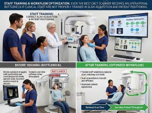
Systems equipped with dual dedicated sensors allow seamless toggling between panoramic and cephalometric modes without requiring manual sensor transfers, preventing accidental drops and protecting sensitive CMOS detectors.
3. Software & Cephalometric Tracing Integration
Integrated cephalometric analysis software enables automated landmark detection, rapid tracing, and overlay features for soft tissue profile analysis, cutting diagnostic workflow time from 20 minutes down to seconds.
Why Partner with UMY Medical for Cephalometric & Dental Imaging?
As a leading global developer and OEM supplier of digital radiography and advanced diagnostic systems, UMY Medical (www.umymedical.com) supplies reliable, high-resolution imaging equipment tailored to modern clinical demands.
Why orthodontic practices and medical equipment distributors partner with UMY Medical:
-
Versatile Diagnostic Portfolio: UMY Medical offers an extensive line of imaging platforms—from high-definition digital radiography (DR) suites to multi-functional panoramic and cephalometric imaging solutions.
-
Exceptional Image Quality & Value: Built with high-sensitivity digital sensors and ultra-fine focal spot X-ray generators, UMY Medical devices deliver crisp craniofacial imaging at low radiation doses with competitive factory-direct pricing.
-
Full B2B Support & Customization: UMY Medical backs global healthcare partners with full OEM/ODM options, international regulatory compliance assistance, long-term warranty coverage, and responsive technical service.
Frequently Asked Questions (FAQ)
Q1: What is the main difference between panoramic and cephalometric X-rays?
A panoramic X-ray captures a curved, full-mouth 2D view of the teeth, jawbones, and sinus cavities. A cephalometric X-ray captures a precise 1:1 scale lateral or anteroposterior view of the entire head, showing the relationship between jawbones, facial profile, and cranial anatomy required for orthodontics.
Q2: How does a 3D CBCT system enhance cephalometric analysis?
While traditional 2D cephalometric images superimpose anatomical structures, 3D CBCT imaging generates volumetric slices. This allows clinicians to assess severe skeletal asymmetry, impacted teeth, and airway volume with 3D cephalometric landmark placement.
Q3: What space requirements are needed for a cephalometric X-ray unit?
Because cephalometric machines require an extended arm and head positioner (cephalostat), they require a larger room footprint (typically at least 2.0 m x 1.5 m) compared to standard stand-alone panoramic units, along with proper lead shielding.
Request a Custom B2B Equipment Quote Today
Looking to upgrade your orthodontic practice or expand your dental distribution catalog?
Contact the UMY Medical sales engineering team today to request technical data sheets, dimensional layout guides, and factory-direct B2B quotation packages.