Why Technical Specifications Matter in Mammography
In digital mammography, the margin between an early breast cancer detection and an overlooked lesion often comes down to millimeter-level spatial resolution and subtle tissue contrast. Breast tissue consists of fibroglandular structures and fat with very similar X-ray attenuation properties. Furthermore, microcalcifications—one of the earliest signs of breast malignancy—are frequently as small as 0.1 mm to 0.2 mm.
For biomedical engineers, radiology administrators, and medical equipment procurement managers, evaluating a mammography machine requires looking beyond broad feature sets. You must examine core hardware specifications: detector architecture, Detective Quantum Efficiency (DQE), pixel pitch, X-ray tube target/filter combinations, and breast compression mechanics.
This technical guide breaks down every critical spec parameter to help clinical buyers make data-driven, future-proof equipment purchasing decisions.
2. Flat-Panel Detector Technology: Direct (a-Se) vs. Indirect (CsI) Conversion
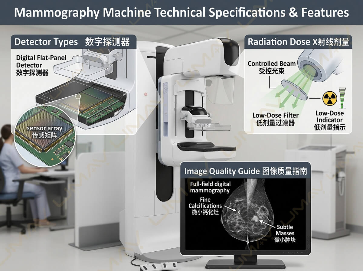
The digital flat-panel detector (FPD) is the central element of any modern Full-Field Digital Mammography (FFDM) or 3D Digital Breast Tomosynthesis (DBT) system. Detectors fall into two main technical categories: Direct Conversion and Indirect Conversion.
  DIRECT CONVERSION (Amorphous Selenium)
  X-Ray Photons ───────────────────────────► Electrical Signal (No Light Scattering)

  INDIRECT CONVERSION (Cesium Iodide)
  X-Ray Photons ──► Scintillator (Light) ──► Photodiode Array ──► Electrical Signal
Direct Conversion Detectors (Amorphous Selenium / a-Se)
Direct conversion detectors utilize a layer of Amorphous Selenium (a-Se) deposited onto a Thin-Film Transistor (TFT) array.
-
Working Principle: Incoming X-ray photons interact directly with the selenium layer, creating electron-hole pairs that are drawn vertically by a high-voltage bias field directly to the readout electrode.
-
Advantage: Because there is no intermediate conversion of X-rays into visible light, there is zero light diffusion (scattering). This results in exceptionally high spatial resolution, ultra-sharp edge definition, and superior detection of fine microcalcifications.
-
Clinical Best Use: Specialized breast diagnostic centers, 3D tomosynthesis, and academic research institutions where maximal spatial sharpness is mandatory.
Indirect Conversion Detectors (Cesium Iodide / CsI)
Indirect conversion detectors use a Cesium Iodide (CsI) scintillator paired with an Amorphous Silicon (a-Si) or CMOS photodiode matrix.
-
Working Principle: X-rays strike the needle-structured CsI scintillator, converting X-ray energy into visible light photons. The photodiode array then converts light into electrical signals.
-
Advantage: CsI needle structures guide light downward to minimize lateral blur. Indirect detectors exhibit high X-ray absorption efficiency, robust operational durability, and excellent Detective Quantum Efficiency (DQE) at lower exposure levels.
-
Clinical Best Use: High-throughput screening clinics, mobile mammography units, and regional general hospitals seeking a durable, cost-effective imaging platform.
| Specification Parameter | Direct Conversion (a-Se) | Indirect Conversion (CsI) |
| Primary Material | Amorphous Selenium (a-Se) | Cesium Iodide (CsI) + a-Si / CMOS |
| Intermediate Light Stage | None (Direct to Charge) | Yes (X-ray → Light → Charge) |
| Spatial Sharpness / Edge Clarity | Industry Leading | High |
| DQE at Low Radiation Doses | High | Superior |
| Sensitivity to Temperature Drops | Moderate (Requires Climate Control) | Low (Highly Durable) |
| Ideal Primary Application | Microcalcification Analysis & 3D DBT | High-Volume Screening & Mobile Clinics |
3. Image Quality Metrics: Spatial Resolution, Pixel Pitch, and DQE
When reviewing manufacturer datasheets, three closely linked imaging metrics determine a machine’s diagnostic capabilities: Pixel Pitch, Spatial Resolution (lp/mm), and Detective Quantum Efficiency (DQE).
┌────────────────────────────────────────────────────────────────────────┐
│                        CORE IMAGE QUALITY TRINITY                      │
├──────────────────────────┬───────────────────────┬─────────────────────┤
│       PIXEL PITCH        │  SPATIAL RESOLUTION   │      DQE RATING     │
│   50 µm to 85 µm sizes   │  10 lp/mm to 20 lp/mm │ Dose efficiency &   │
│  Determines detail limit │  Measures fine edges  │signal-to-noise ratio│
└──────────────────────────┴───────────────────────┴─────────────────────┘
1. Pixel Pitch (Microns / µm)
Pixel pitch refers to the distance between the centers of two adjacent pixels on the detector surface.
-
Typical mammography pixel sizes range from 50 µm to 85 µm.
-
Smaller pixel pitch (e.g., 50 µm) provides finer sampling grid density, enabling visualization of smaller structures. However, smaller pixels require careful noise management from the readout electronics.
2. Spatial Resolution (Line Pairs per Millimeter – lp/mm)
Spatial resolution quantifies how close two tiny line objects can be placed before they blur into a single object.
-
Standard digital mammography systems deliver 10 lp/mm to 20 lp/mm of spatial resolution.
-
Higher lp/mm values allow radiologists to evaluate microcalcification boundary shapes and spiculated mass margins with greater confidence.
3. Detective Quantum Efficiency (DQE)
DQE measures how effectively a detector converts incoming X-ray photons into a useful, noise-free diagnostic image. It is expressed as a percentage across spatial frequencies.
-
High DQE (>65% at 0 lp/mm): Means the detector produces clear, high-contrast images while exposing the patient to significantly lower radiation doses.
-
Low DQE: Requires higher X-ray exposure to achieve acceptable image contrast, increasing overall radiation dose.
4. Radiation Dose Management & X-Ray Tube Specifications
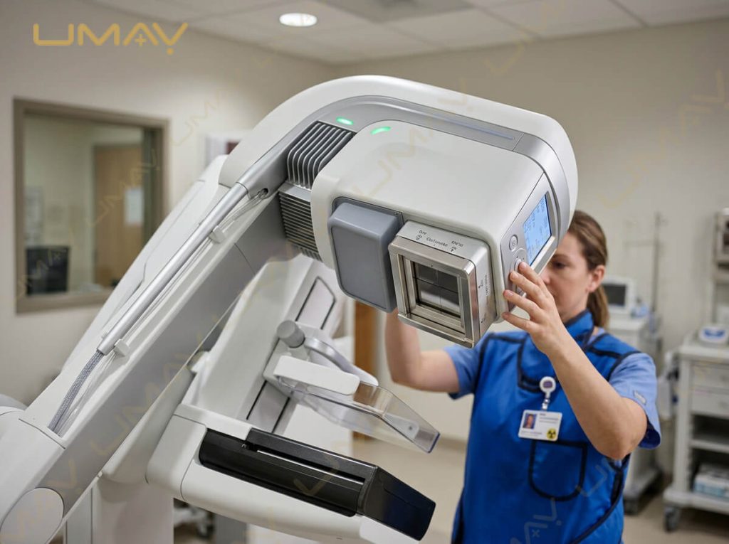
Radiation safety in breast imaging is strictly regulated (e.g., FDA MQSA, EUREREF standards). Modern systems rely on intelligent tube assemblies and automated filtration to minimize the Average Glandular Dose (AGD).
Target and Filter Material Combinations
X-ray tubes generate different energy spectra depending on the target material (anode) and filter selection:
-
Molybdenum / Molybdenum (Mo/Mo): Ideal for thin to average uncompressed breasts (under 4 cm). Provides soft X-ray spectrum for maximum tissue contrast.
-
Rhodium / Rhodium (Rh/Rh): Harder X-ray beam; penetrates denser or thicker breast tissue (4 cm to 6 cm) effectively.
-
Tungsten / Rhodium or Silver (W/Rh or W/Ag): Modern gold standard for high-end digital mammography and 3D tomosynthesis. Tungsten targets produce higher photon flux while significantly reducing radiation exposure in dense tissue.
               ┌─────────────────────────────────────────────────┐
               │    X-Ray Tube Filtration vs. Breast Thickness   │
               └────────────────────────┬────────────────────────┘
                                        │
           ┌────────────────────────────┴────────────────────────────┐
           ▼                                                         ▼
┌──────────────────────────────────────┐  ┌──────────────────────────────────────┐
│  Thin / Average Breast (< 4 cm)      │  │  Dense / Thick Breast (> 5 cm)       │
├──────────────────────────────────────┤  ├──────────────────────────────────────┤
│ • Molybdenum (Mo) Target             │  │ • Tungsten (W) / Rhodium (Rh) Target │
│ • Soft X-ray energy spectrum         │  │ • Hardened X-ray beam profile        │
│ • Maximum low-contrast detail        │  │ • Reduced radiation dose (AGD)       │
└──────────────────────────────────────┘  └──────────────────────────────────────┘
Automatic Exposure Control (AEC) Algorithms
Modern mammography systems use smart digital AEC systems that perform a microsecond pre-shot prior to the main exposure:
-
The pre-shot measures local tissue density and breast thickness under compression.
-
The AEC system instantly selects optimal kVp, mAs, and filter combination.
-
This prevents over-exposure of fatty tissue or under-penetration of dense fibroglandular regions.
5. Ergonomic Compression Systems & Patient Comfort Features
Breast compression is necessary to flatten tissue, reduce scatter radiation, decrease motion blur, and lower required dose levels. However, compression anxiety is a major reason women delay routine mammograms.
Smart Compression Paddles
-
Flexible / Tilting Compression Paddles: Automatically pivot to match the natural anatomical contour of the patient’s breast, distributing pressure evenly from base to chest wall.
-
Speed-Controlled Motorized Compression: Fast initial paddle positioning smoothly transitions to micro-controlled, pressure-sensitive motorized compression as contact is made.
-
Automatic Decompression: The paddle immediately releases pressure the instant X-ray exposure concludes, minimizing duration of discomfort.
-
Custom Paddle Sizes: Includes standard 24×30 cm paddles, small 18×24 cm paddles, spot compression paddles, and dedicated magnification paddles.
6. Gantry Mechanics, C-Arm Movement, and Magnification
A versatile C-arm design speeds up technologist positioning workflows and simplifies imaging for wheelchair-bound or mobility-impaired patients.
┌────────────────────────────────────────────────────────────────────────┐
│                   GANTRY & C-ARM MECHANICAL SPECS                      │
├──────────────────────────┬───────────────────────┬─────────────────────┤
│    ISOCENTRIC ROTATION   │     MOTORIZED HEIGHT  │ SID (SOURCE-TO-IMAGE│
│    -135° to +180° range  │     Smooth vertical   │ DISTANCE)           │
│    Fast view changes     │     travel speed      │ Standard 65 – 70 cm │
└──────────────────────────┴───────────────────────┴─────────────────────┘
Key Mechanical Parameters:
-
Isocentric C-Arm Rotation: Allows the gantry to rotate smoothly around a fixed central point without altering the height of the object table between Cranio-Caudal (CC) and Medial-Lateral Oblique (MLO) projections.
-
Source-to-Image Distance (SID): Typically 65 cm to 70 cm. A larger SID provides comfortable space for technologist positioning and accommodates stereotactic biopsy accessories.
-
Magnification Capability: Magnification stands (typically 1.5x and 1.8x to 2.0x) elevate the breast above the detector surface to enlarge suspicious focal areas for detailed diagnostic analysis.
-
Anti-Scatter Grid Technology: High-transmission cellular grids strip scatter radiation during 2D imaging. For 3D tomosynthesis, specialized systems temporarily retract the grid to preserve low-dose exposure levels.
7. Technical RFP Purchasing Checklist for Buyers
Use this technical checklist when comparing quotes and specs from mammography manufacturers or third-party refurbishers:
-
[x] Detector Conversion Type: Clarify if detector is Direct a-Se or Indirect CsI based on your diagnostic vs screening priorities.
-
[x] Pixel Pitch Rating: Verify detector pixel size is 85 µm or smaller (50–70 µm preferred for 3D tomosynthesis).
-
[x] DQE Performance Curve: Confirm peak DQE is >60% at low spatial frequency.
-
[x] Anode Target & Filter Setup: Ensure the system offers a Tungsten (W) anode with Rhodium/Silver filters for low-dose dense breast imaging.
-
[x] C-Arm Motion Range: Verify motorized C-arm handles at least -135° to +180° isocentric rotation.
-
[x] Compression Safety Controls: Confirm presence of emergency manual pressure release and automated comfort-sensing paddle software.
8. Frequently Asked Questions (FAQ)
Q1: What detector pixel size is best for digital mammography?
A pixel pitch between 50 µm and 85 µm is ideal. Smaller pixel pitch improves spatial detail for spotting microcalcifications, provided the system maintains a high DQE and low electronic noise floor.
Q2: Why is Detective Quantum Efficiency (DQE) important when evaluating mammography equipment?
DQE indicates how efficiently the detector converts radiation into a sharp image. A system with high DQE produces clear diagnostic images at lower radiation doses, protecting patient health while maintaining clinical clarity.
Q3: What target/filter combination offers the lowest radiation dose in dense breasts?
A Tungsten (W) target paired with Rhodium (Rh) or Silver (Ag) filters delivers a hardened X-ray beam that penetrates dense fibroglandular tissue effectively with lower Average Glandular Dose (AGD) compared to older Molybdenum targets.
Q4: How do flexible compression paddles improve image quality?
Flexible tilting paddles conform to the natural taper of the breast, preventing over-compression at the chest wall while ensuring proper flattening near the nipple. This eliminates tissue thickness gradients and reduces motion artifacts.