Introduction to Modern Digital Mammography Systems
Breast cancer remains one of the most common cancers diagnosed among women worldwide. Early detection through precision imaging is the gold standard for improving survival rates. At the center of modern early detection protocols is the mammography machine—a specialized low-dose X-ray system designed specifically to evaluate breast tissue density, microcalcifications, and soft tissue masses.
Over the past two decades, breast imaging technology has transitioned from traditional screen-film mammography to Full-Field Digital Mammography (FFDM) and advanced 3D Digital Breast Tomosynthesis (DBT).
For hospital administrators, radiology department directors, and clinical procurement teams, investing in the right digital mammography system requires balancing diagnostic accuracy, patient comfort, workflow speed, and overall capital investment. This guide breaks down the key technical differences, clinical applications, and decision frameworks needed to choose the optimal mammography machine for your medical facility.
2. Main Types of Mammography Machines Explained
When evaluating digital breast imaging equipment, systems generally fall into three core categories: 2D Full-Field Digital Mammography (FFDM), 3D Digital Breast Tomosynthesis (DBT), and Mobile/Portable Mammography Units.
                           ┌─────────────────────────────────────────┐
                           │      Mammography Equipment Types        │
                           └────────────────────┬────────────────────┘
                                                │
         ┌──────────────────────────────────────┼──────────────────────────────────────┐
         ▼                                      ▼                                      ▼
┌─────────────────────────┐            ┌─────────────────────────┐            ┌─────────────────────────┐
│     2D FFDM Systems     │            │   3D Tomosynthesis (DBT)│            │   Mobile/Portable Units │
├─────────────────────────┤            ├─────────────────────────┤            ├─────────────────────────┤
│ • Standard 2D Projection│            │ • Multi-angle 3D Slices │            │ • Vehicle-mounted / Bus │
│ • Cost-effective        │            │ • Reduces tissue overlap│            │ • High screening access │
│ • High patient speed    │            │ • High diagnostic detail│            │ • Compact footprints    │
└─────────────────────────┘            └─────────────────────────┘            └─────────────────────────┘
1. 2D Full-Field Digital Mammography (FFDM)
2D FFDM systems utilize direct or indirect digital detectors to produce two-dimensional projection images of the breast (typically Cranio-Caudal [CC] and Medial-Lateral Oblique [MLO] views).
-
Primary Uses: Routine screening programs, general clinical evaluations, and healthcare facilities with moderate budgets.
-
Key Strengths: Lower initial purchase price, fast acquisition speed, lower raw data storage demands, and straightforward operator workflow.
-
Primary Limitations: Overlapping dense breast tissue can sometimes obscure small lesions or create false-positive readings.
2. 3D Digital Breast Tomosynthesis (DBT)
3D tomosynthesis represents a major technological jump. During a tomosynthesis scan, the X-ray tube moves in an arc over the compressed breast, capturing multiple low-dose images at varying angles. Advanced software algorithms then reconstruct these projections into thin, high-resolution 1 mm cross-sectional slices.
-
Primary Uses: Diagnostic workups, dense breast screening, high-volume academic hospitals, and specialized breast health centers.
-
Key Strengths: Significantly reduces tissue overlap, increases invasive cancer detection rates, and lowers patient recall rates (false positives).
-
Primary Limitations: Higher initial system cost, larger digital storage requirements (PACS capacity), and slightly longer operator reading times.

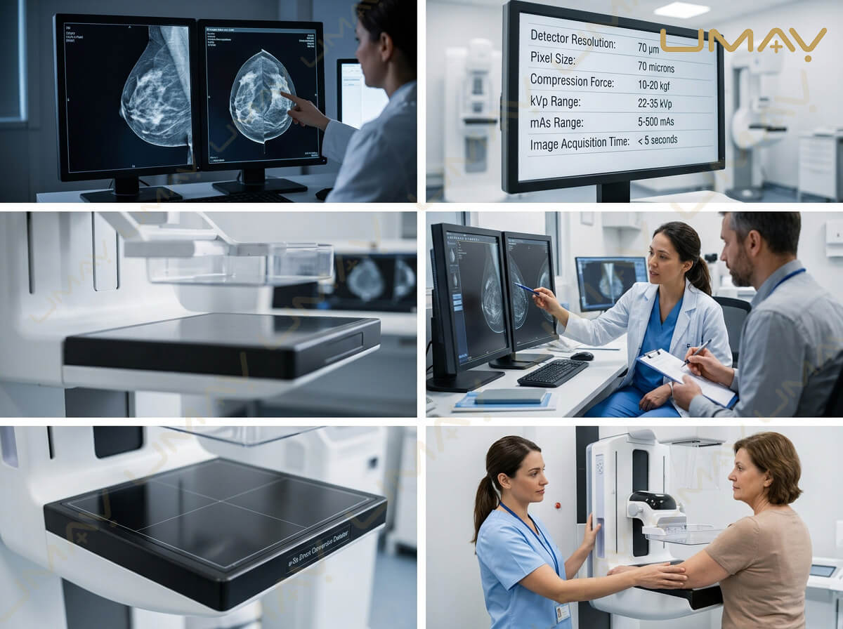
Professional radiographer assisting a female patient with proper positioning on a modern digital mammography machine in a hospital clinic.
3. Mobile Mammography Units
Mobile mammography machines are installed inside specialized trucks, buses, or modular transport trailers to deliver community-based screening programs in remote or underserved areas.
-
Primary Uses: Public health initiatives, corporate wellness screenings, and rural healthcare outreach.
-
Key Strengths: Expands clinical reach, brings diagnostic care directly to patients, and maximizes equipment utilization.
-
Primary Limitations: Requires ruggedized mechanical components, specialized vibration-damping mounts, and stable vehicle climate controls.
3. 2D FFDM vs. 3D Tomosynthesis: Side-by-Side Comparison
| Operational Metric | 2D Digital Mammography (FFDM) | 3D Digital Breast Tomosynthesis (DBT) |
| Image Dimensionality | 2D planar projection | 3D reconstructed slices (1 mm thickness) |
| Tissue Overlap Reduction | Minimal | High (eliminates overlapping structure noise) |
| Dense Breast Performance | Moderate | Superior |
| Average Recall Rate | Standard | Reduced by 15% to 30% |
| Cancer Detection Rate | Baseline standard | Up to 20% – 40% increase in invasive cancers |
| Data File Size per Exam | ~30 MB – 50 MB | ~300 MB – 1 GB+ |
| Relative Equipment Cost | $60,000 – $150,000 (Entry to Mid) | $180,000 – $380,000+ (High-End) |
4. Essential Technical Features to Evaluate Before Purchasing
To select a system that delivers reliable long-term service and outstanding diagnostic clarity, procurement teams should audit four primary technical parameters:
1. Detector Technology (Amorphous Selenium vs. CsI)
-
Direct Conversion (Amorphous Selenium – a-Se): Converts X-ray photons directly into electrical charges without converting them to light first. This yields exceptional spatial resolution and sharp edge definition, which is vital for identifying fine microcalcifications.
-
Indirect Conversion (Cesium Iodide – CsI): Converts X-rays into light, which is then recorded by a photodiode array. CsI detectors are robust, offers high Detective Quantum Efficiency (DQE), and performs well under high-throughput conditions.
2. Spatial Resolution and Pixel Pitch
High spatial resolution is essential in mammography because microcalcifications can be as small as 0.1 mm to 0.2 mm. Look for system detector pixel pitch between 50 µm and 85 µm. Finer pixel pitch delivers higher detail resolution, enabling earlier detection of suspicious tissue clusters.
3. Automated Exposure Control (AEC) & Smart Compression
-
Optimized AEC: Automatically adjusts tube voltage (kVp), exposure current (mAs), and filter materials based on individual breast thickness and estimated tissue density. This ensures consistent contrast while minimizing total radiation dose.
-
Ergonomic Compression Paddles: Flexible, tilting paddles distribute compression pressure evenly across the breast tissue, minimizing patient discomfort while maintaining necessary thickness uniformity.
4. Stereotactic Biopsy Capability
If your clinic performs non-surgical diagnostic tissue sampling, verify whether the mammography unit supports an add-on stereotactic biopsy attachment (either upright or lateral access). This feature allows interventional radiologists to pinpoint lesions in 3D space for precise needle placement.
5. Application Scenarios: Screening vs. Diagnostic Workflows
Selecting the right machine architecture depends directly on your clinical workflow:
                          ┌────────────────────────────────────────┐
                          │    Select Clinical Focus Strategy      │
                          └───────────────────┬────────────────────┘
                                              │
                     ┌────────────────────────┴────────────────────────┐
                     ▼                                                 ▼
      ┌─────────────────────────────┐                   ┌─────────────────────────────┐
      │  Routine Mass Screening     │                   │  Diagnostic & Biopsy Center │
      ├─────────────────────────────┤                   ├─────────────────────────────┤
      │ • Focus on rapid throughput │                   │ • Focus on 3D tomosynthesis │
      │ • 2D FFDM or basic DBT      │                   │ • Stereotactic biopsy ready │
      │ • Low dose & auto-paddle    │                   │ • High workstation power    │
      │ • Fast patient turnarounds  │                   │ • Advanced CAD / AI tools   │
      └─────────────────────────────┘                   └─────────────────────────────┘
-
High-Volume Mass Screening Centers: Prioritize rapid exposure speeds, fast gantry movement, automatic compression release, and robust 2D/synthetic 2D imaging capabilities to maximize daily patient volume.
-
Specialized Breast Health Clinics: Require 3D tomosynthesis, high-performance diagnostic reading workstations with dedicated mammography monitors (5MP or 12MP medical displays), AI-assisted computer-aided detection (CAD), and biopsy add-ons.
6. Step-by-Step Decision Framework for Procurement Officers
When preparing a request for proposal (RFP) for a new or refurbished mammography unit, follow this five-step evaluation framework:
-
[x] Step 1: Patient Demographic & Volume Audit: Estimate daily patient numbers and the proportion of patients presenting with dense breast tissue.
-
[x] Step 2: IT & PACS Capacity Assessment: Ensure your Picture Archiving and Communication System (PACS) server bandwidth and archive storage can handle large 3D tomosynthesis datasets.
-
[x] Step 3: Radiation Compliance Check: Review local nuclear safety and health authority guidelines regarding mammography radiation dosage Limits (e.g., ACR/FDA MQSA standards).
-
[x] Step 4: Total Budget Planning: Factor in maintenance service contracts, X-ray tube replacement guarantees, software license renewals, and technologist application training.
-
[x] Step 5: Supplier Support Audit: Choose equipment suppliers that guarantee fast technical response times, local spare parts stock, and certified field service engineers.
7. Frequently Asked Questions (FAQ)
Q1: What is the main difference between 2D and 3D mammography?
A 2D mammogram takes two flat images of each breast, whereas a 3D mammogram (tomosynthesis) takes multiple low-dose x-ray slices from different angles to construct a 3D image. This helps doctors see through dense breast tissue more clearly.
Q2: Is 3D tomosynthesis worth the extra investment for a diagnostic clinic?
Yes. 3D tomosynthesis increases invasive breast cancer detection rates by up to 40% and reduces patient recall rates significantly. For diagnostic centers and specialty clinics, it delivers higher diagnostic accuracy and stronger competitive advantage.
Q3: What monitor resolution is required for reading digital mammograms?
Medical display standards mandate certified high-contrast diagnostic monitors with a minimum resolution of 5 Megapixels (5MP) for 2D images, or specialized 12 Megapixel (12MP) single-surface displays for viewing 3D tomosynthesis slice stacks.
Q4: How long does a digital mammography machine typically last?
With proper routine maintenance, quality control calibrations, and periodic tube maintenance, a quality digital mammography machine offers an operational lifespan of 7 to 10 years.