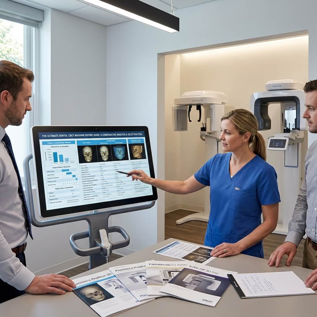
Investing in a 3D Dental Cone Beam Computed Tomography (CBCT) scanner is one of the most significant equipment decisions a modern dental practice, clinic network, or procurement team will ever make. Transitioning from standard 2D panoramic x-rays to high-definition 3D dental imaging fundamentally changes your diagnostic accuracy, treatment acceptance, and surgical precision.
However, navigating the dental imaging market can feel overwhelming. With prices ranging from $30,000 to over $120,000, varying Fields of View (FOV), complex software licensing, and dozens of dental x-ray machine suppliers, choosing the right unit requires a clear, commercial perspective.
This definitive buying guide breaks down everything you need to know about purchasing a dental CBCT machine—without the dense academic jargon.
What is a Dental CBCT Scanner and Why Upgrade Now?
A Dental CBCT machine (Cone Beam Computed Tomography) uses a cone-shaped X-ray beam combined with a flat-panel detector to capture hundreds of 2D images in a single 360-degree rotation. The system’s acquisition software then reconstructs these images into a high-resolution 3D volumetric digital model of the patient’s teeth, jawbone, nerve pathways, and soft tissues.
2D vs. 3D Dental X-Ray
2D Panoramic / Periapical | 3D CBCT Volumetric Scan
Flat, distorted projection | 1:1 true scale 3D anatomy
Superimposed anatomical structures| Cross-sectional views
High margin for diagnostic error | Exact bone density & width
Limited to basic diagnostics | Guided implant & endo planning
Understanding Field of View (FOV): Which Size Do You Need?
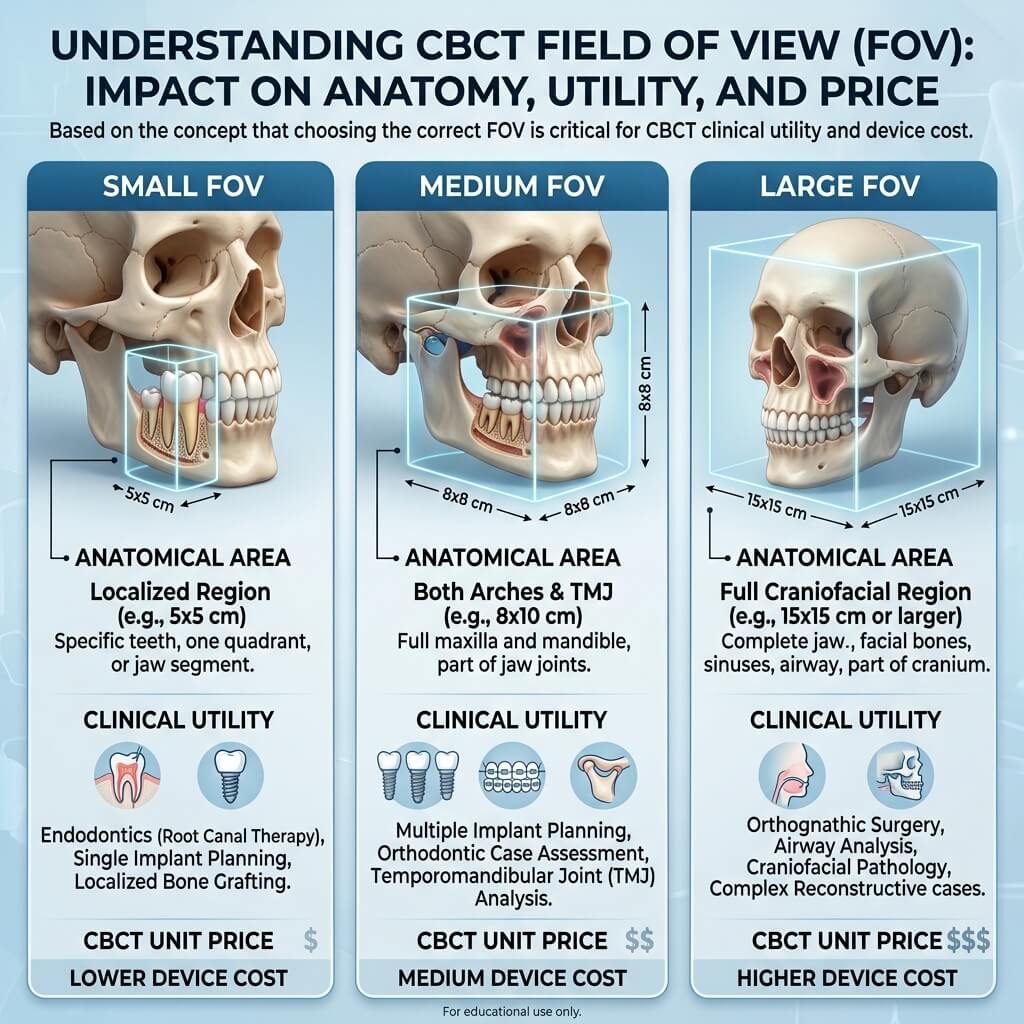
Field of View (FOV) refers to the anatomical area scanned by the 3D X-ray beam. Choosing the correct FOV is the single most important factor determining both the price of the CBCT unit and its clinical utility.
[ Small FOV ] [ Medium FOV ] [ Large FOV ]
5×5 cm – 8×5 cm 8×8 cm – 12×10 cm 15×15 cm – 21×19 cm
(Single site/Endo) (Dual Arch/Implants) (Full Face/Ortho/TMJ)
1. Small FOV (5 * 5 cm to 8 * 5 cm)
-
Best for: Endodontics, localized single-implant planning, impacted wisdom teeth.
-
Pros: Lowest radiation dose, highest spatial resolution, lower purchase price.
-
Cons: Cannot capture a full single arch or dual-arch relationship in one scan.
2. Medium FOV (8 * 8 cm to 12 * 10 cm)
-
Best for: General dentists, routine dental implant placement, dual-arch diagnostic scans.
-
Pros: Captures both upper and lower jaws including the sinus floor; optimal for 90% of general practice needs.
-
Cons: Cannot capture full facial structures or TMJ joints simultaneously.
3. Large FOV (15 * 15 cm to 21 * 19 cm)
-
Best for: Oral and maxillofacial surgeons, orthodontists, airway and sleep apnea analysis, TMJ evaluation.
-
Pros: Captures the entire craniofacial structure, both arches, sinuses, airway, and temporomandibular joints in a single rotation.
-
Cons: Higher initial machine cost, larger image file sizes, and broader legal responsibility to review the entire scanned volume.
Key Hardware & Software Specifications Checklist
When evaluating dental CBCT models and technical specification sheets, pay close attention to these six core hardware and software parameters:
Hardware Specifications
-
Voxel Size (Resolution): Measured in micrometers. Smaller voxels deliver higher image sharpness. Look for an Endo Mode with 75um – 100um voxel size for detailed root canal diagnostics. Standard scans typically run between 150um and 300um.
-
Focal Spot Size: Look for a focal spot of 0.5 mm or lower. A smaller focal spot drastically reduces image blur and produces crisp bone margin details.
-
Sensor Technology: Demand a modern CMOS Flat Panel Detector (FPD) over older Image Intensifier or CCD technologies. CMOS sensors offer longer lifespans, better contrast resolution, and lower radiation absorption.
-
Scan Time vs. Exposure Time: A total scan time might be 10–14 seconds, but actual X-ray exposure time should ideally be under 5 seconds using pulsed radiation technology. This reduces radiation exposure and minimizes motion artifacts caused by patient movement.
Software Capabilities
-
DICOM 3.0 Compatibility: Ensure the acquisition software exports standard, unencrypted DICOM files. This allows seamless integration with 3rd party implant planning software (Exoplan, CoDiagnostiX, BlueSkyPlan), CAD/CAM systems, and 3D printers for surgical guide fabrication.
-
Metal Artifact Reduction (MAR): High-quality MAR algorithms filter out streaks and shadows caused by metallic dental crowns, amalgam fillings, and orthodontic brackets.
-
Airway & TMJ Analysis Tools: Automated segmentation tools for volume measurement of airway passages add value for sleep dentistry practices.
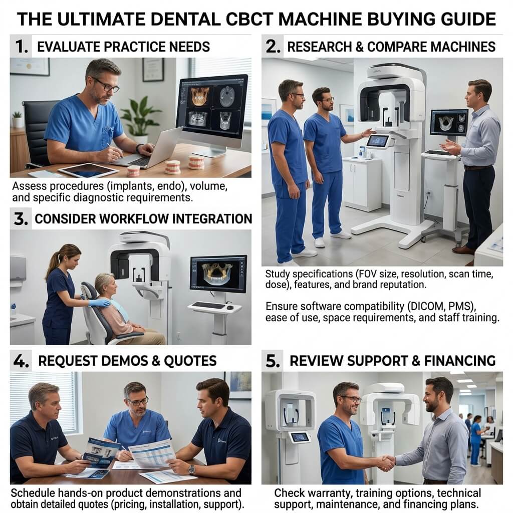
Step-by-Step CBCT Procurement Checklist
Follow this streamlined checklist when sourcing your machine from international manufacturers or local suppliers:
-
Assess Clinical Needs: Determine if your primary focus is general dentistry (8 * 8 FOV), endodontics (5 * 5 FOV), or orthodontics/surgery (15 * 15 FOV).
-
Measure X-Ray Room Dimensions: Verify room footprint (1.5m * 1.5m for standing models) and wall construction for radiation safety compliance.
-
Confirm Power Requirements: Ensure availability of stable 110V/220V dedicated electrical circuits.
-
Evaluate Software Licensing Model: Ask supplier: “Is the software subscription-free, or are there recurring annual user fees?”
-
Verify DICOM Export Freedom: Guarantee that export of 3D DICOM files is open and unrestricted without per-export charges.
-
Inquire About Spare Parts Availability: Check availability and replacement cost of critical components like the X-ray tube and flat panel detector.
-
Request Sample DICOM Files: Ask vendors for raw, unedited sample scans of various FOVs to inspect contrast, clarity, and artifact reduction before signing a sales contract.
Frequently Asked Questions (FAQ)
What is the average lifespan of a dental CBCT machine?
With routine calibration and preventive maintenance, a high-quality dental CBCT system lasts between 7 to 10 years. X-ray tubes and sensors are designed for tens of thousands of exposure cycles.
Can a 3D CBCT machine take normal 2D panoramic images?
Yes! Modern 3D CBCT systems are 2-in-1 or 3-in-1 combo units. They feature dedicated 2D panoramic sensors or reconstruction algorithms that capture traditional extraoral panoramic and cephalometric images without requiring a separate 2D unit.
How quickly can a clinic recover the cost of a CBCT scanner (ROI)?
Most practices achieve full ROI within 8 to 14 months. Charging for diagnostic 3D scans, converting more implant cases in-house, and fabricating surgical guides creates immediate new monthly revenue streams.
Is lead shielding required in the room for a CBCT machine?
In most jurisdictions, yes. Local radiation health regulations usually require lead-lined drywall, lead glass windows, and a dedicated exposure switch outside the scan room. Always consult a certified health physicist prior to equipment installation.
Ready to Upgrade Your Practice’s 3D Diagnostic Power?
Adding a 3D dental CBCT machine to your clinical workflow elevates patient care, enhances diagnostic precision, and drives practice profitability. By matching your practice’s clinical focus with the right Field of View, evaluating total cost of ownership, and choosing a reliable direct supplier, you ensure maximum return on your capital equipment investment.