Choosing between a 2D Panoramic X-Ray machine and a 3D Cone Beam Computed Tomography (CBCT) scanner is one of the most pivotal equipment decisions for any dental clinic, multi-specialty group, or hospital procurement office.
While traditional 2D panoramic imaging (also known as Pan, OPG, or Panorex) has served as the diagnostic benchmark for decades, 3D CBCT technology has rapidly become the standard of care for modern restorative, surgical, and endodontic treatments.
This comprehensive guide compares 2D Panoramic vs. 3D CBCT imaging across diagnostic accuracy, radiation exposure, machine purchase costs, workflow integration, and return on investment (ROI)—helping practice owners and purchasing directors make a confident, future-proof acquisition.
2D Panoramic X-Ray (Extraoral Radiography)
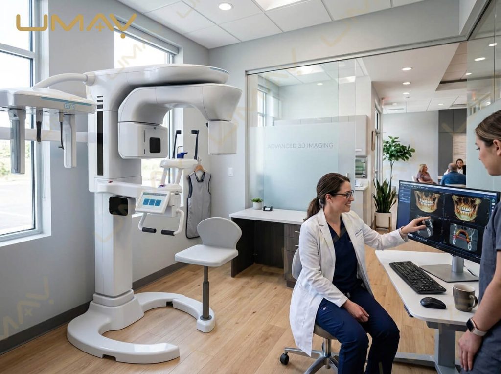
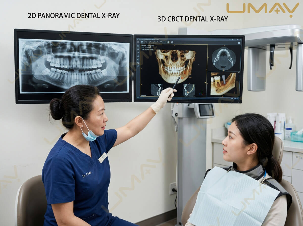
A 2D panoramic unit moves an X-ray tube and sensor around the patient’s head in a fixed semicircular arch. It produces a single flattened image showing the entire maxilla and mandible.
-
The Limitation: Because 3D anatomical structures are flattened onto a 2D plane, images suffer from distortion, magnification errors (typically 15% to 30%), and overlapping anatomical structures (such as cervical spine shadows masking anterior teeth).
3D Dental CBCT Scanner
A 3D CBCT machine uses a cone-shaped X-ray beam and a high-resolution flat panel detector (FPD) to execute a single rotation around the patient (ranging from 180 degrees to 360 degrees). During this rotation, it captures hundreds of individual digital images.
-
The Advantage: Reconstruction software converts these raw projection frames into a 3D volumetric model. Clinicians can slice, rotate, and view the patient’s anatomy in axial, sagittal, and coronal planes at a 1:1 true scale without spatial distortion.
Diagnostic Capabilities: What 2D Misses and What 3D Reveals
Relying solely on 2D panoramic images introduces diagnostic blind spots that can lead to surgical complications, misdiagnoses, or failed treatments.
Dental Implant Planning: Height vs. Width & Density
-
2D Panoramic: Measures vertical bone height from the ridge to the inferior alveolar nerve (IAN) or sinus floor, but provides zero information regarding bone width, ridge curvature, or cortical bone density. Clinicians must guess if sufficient bone thickness exists or perform invasive exploratory flaps.
-
3D CBCT: Displays exact bone width, localized height, bone density gradients, and 3D contours. Allows virtual implant placement, nerve mapping, sinus proximity checks, and instant generation of CAD/CAM surgical guides.
Endodontics: Finding Hidden Canals & Fractures
-
2D Panoramic / Periapical: Canals superimposed over thick cortical bone or roots can easily mask secondary canals (such as the MB2 canal in upper molars), lateral canals, internal resorption, or subtle vertical root fractures.
-
3D CBCT (High-Resolution Endo Mode): Slices tissue down to 75 to 100 microns voxel resolution. Clinicians can isolate individual root canals, evaluate periapical lesions in 3D space, and detect vertical fractures before initiating endodontic therapy.
Impacted Wisdom Teeth & Oral Surgery
-
2D Panoramic: Shows if an impacted third molar is close to the Inferior Alveolar Canal, but cannot reveal whether the nerve runs buccally, lingually, or directly between the root apexes.
-
3D CBCT: Clearly separates the nerve trunk from the root tip in 3D cross-sections, drastically reducing the risk of permanent nerve paresthesia during surgical extractions.
Cost-to-Benefit Analysis: 2D vs. 3D Investment ROI
When evaluating equipment purchases from a procurement perspective, initial capital expenditure must be balanced against revenue potential and diagnostic efficiency.
Price Range Breakdown (2026 Market Standard)
-
Standalone 2D Panoramic Unit: $12,000 to $25,000 USD
-
2D Panoramic with Cephalometric Arm: $20,000 to $35,000 USD
-
Combo 2D/3D CBCT Scanner (Small/Medium FOV): $30,000 to $65,000 USD
-
Premium Large FOV 3D CBCT System (with Ceph): $65,000 to $110,000+ USD
Financial Return on Investment (ROI) Matrix
| Revenue Driver | 2D Panoramic System | 3D CBCT Scanner System |
| Direct Scan Billing | Low fee per scan; limited insurance/out-of-pocket coverage. | High diagnostic billing fee ($150 to $350 USD per scan). |
| In-House Case Retention | Low; complex implant/endo cases often referred out. | High; keep implants, surgical extractions, and endo in-house. |
| Patient Case Acceptance | Moderate; patients struggle to understand 2D X-rays. | High; 3D visual models clearly demonstrate treatment need. |
| Surgical Guide Production | Not possible with 2D data alone. | Unlocks revenue from 3D printed surgical guides and models. |
| Average Payback Period | 24 to 36 months | 8 to 14 months (based on 3 to 5 scans/week) |
Cephalometric Attachments: 2D Ceph vs. 3D Reconstruction
For orthodontic practices and pediatric dental clinics, cephalometric imaging is vital for skull growth analysis and facial profiling.
Traditional 2D Cephalometric Arm
-
Added as a side arm to a 2D panoramic or 3D CBCT base.
-
Takes Lateral (Lat) and Anteroposterior (AP) skull projections.
-
Lower initial machine cost, standard workflow for traditional cephalometric tracing software.
3D Synthetic & True Cephalometric Reconstruction
-
Generated directly from a Large FOV 3D CBCT scan (15×15 cm or larger) without needing a secondary mechanical arm.
-
Eliminates magnification distortion inherent in traditional 2D Ceph projections.
-
Provides full 3D facial asymmetry analysis, soft tissue profiling, and 3D airway measurements in a single rotation.
Summary: Future-Proofing Your Clinical Imaging
While 2D panoramic units remain a cost-effective choice for basic diagnostic screenings, 3D CBCT imaging is the cornerstone of modern, high-growth dental practices. Investing in 3D CBCT technology expands in-house treatment capabilities, eliminates diagnostic errors, increases patient case acceptance, and delivers a rapid return on investment.
When choosing a system, select a unit that offers flexible Field of View options, low-dose exposure protocols, open DICOM file export, and direct manufacturer support for seamless long-term operation.