The Two-Hour Diagnostic Clock: Why Manual Urinalysis Fails at Scale
Leave a freshly collected, unpreserved urine specimen sitting on a clinical laboratory bench at room temperature for more than two hours, and its diagnostic reliability begins to collapse.
Urine is an unstable biological fluid. The moment it leaves the urinary tract, chemical and cellular breakdown begins. Any trace bacteria present in the specimen double in population roughly every 20 to 30 minutes at ambient temperature. Urea-splitting organisms—such as Proteus species—rapidly metabolize urinary urea into free ammonia, driving the specimen’s pH upward into strongly alkaline territory.
This alkaline shift triggers a cascade of microscopic destruction:
-
Erythrocyte Lysis: Red blood cells (RBCs) swell and rupture in alkaline or hypotonic environments, leaving behind invisible “ghost cells” that fail to appear under a manual microscope even while the chemical dipstick tests strongly positive for free hemoglobin.
-
Cast Dissolution: Delicate Tamm-Horsfall mucoprotein matrices—the structural backbone of hyaline, granular, and cellular casts that signal acute kidney injury—dissolve completely in alkaline urine.
-
Chemical Volatility: Unconjugated bilirubin and urobilinogen photo-oxidize when exposed to ambient clinic lighting, ketone bodies evaporate into the headspace of the collection cup, and multiplying bacteria consume urinary glucose, masking poorly controlled diabetes.
In a high-volume hospital laboratory or busy outpatient clinic receiving hundreds of urine cups every morning, manual dipstick timing and traditional 5-minute tube centrifugation create an unavoidable processing bottleneck. Automated urine analyzers solve this time-sensitivity crisis by turning a slow, subjective, labor-intensive manual procedure into a standardized, high-speed diagnostic workflow.
1. The Two Pillars of Modern Automated Urinalysis
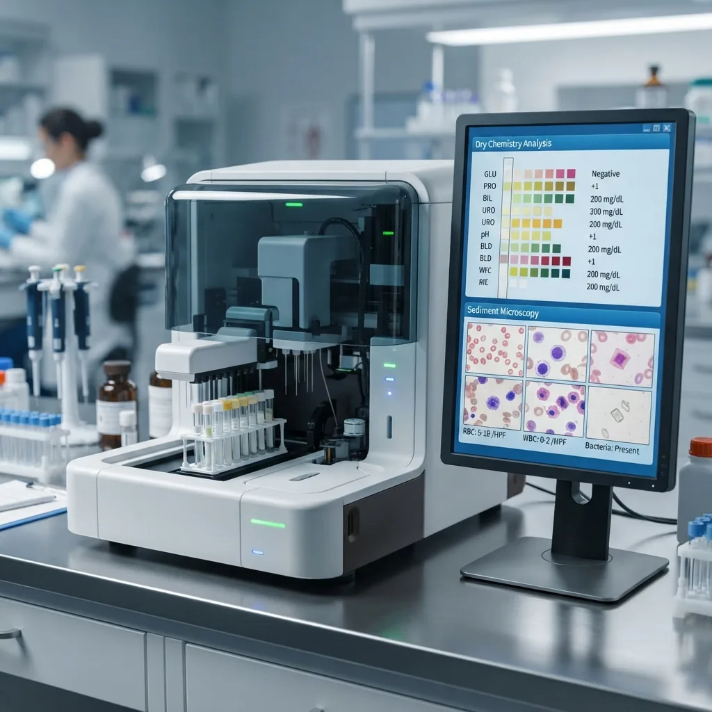
A complete clinical urinalysis evaluates both the dissolved biochemical solutes in the fluid and the suspended microscopic particles. Modern laboratory automation divides this workload into two specialized analytical engines that frequently link together via a robotic track to form an integrated hybrid urinalysis workstation.
Plaintext
                    INTEGRATED AUTOMATED URINALYSIS WORKFLOW
                                       |
         +-----------------------------+-----------------------------+
         |                                                           |
         v                                                           v
[ ENGINE 1: DRY CHEMISTRY MODULE ]            [ ENGINE 2: SEDIMENT MICROSCOPY MODULE ]
- Method: Dual-Wavelength Reflectance         - Method: Planar Flow Digital Microscopy
  Photometry + Optical Refractometry            or Laser Urinary Flow Cytometry
- Target: Dissolved Biochemical Solutes       - Target: Suspended Formed Elements
- Output: 11 to 14 Chemical Parameters        - Output: RBC, WBC, Casts, Crystals,
  (Protein, Glucose, Nitrite, ACR, etc.)        Epithelial Cells, Bacteria, Yeast
         |                                                           |
         +-----------------------------+-----------------------------+
                                       |
                                       v
              [ INTELLIGENT MIDDLEWARE CROSS-CHECK & LIS EXPORT ]
              - Auto-verifies concordant normal results
              - Flags chemical vs. microscopic discrepancies for review
-
Chemical Urinalysis (Dry Chemistry): Utilizes multi-pad reagent test strips (dipsticks) analyzed by optical reflectance photometers to screen for metabolic disorders, hepatic dysfunction, hemolysis, urinary tract infections, and early renal protein leakage.
-
Physical Macroscopic Evaluation: Uses integrated optical refractometers, light-scattering turbidimeters, and RGB spectral sensors to measure exact Specific Gravity (SG), visual clarity (turbidity), and true specimen color prior to chemical testing.
-
Microscopic Sediment Analysis (Formed Elements): Employs automated planar digital microscopy or laser-based flow cytometry to image, classify, and count intact cells, casts, crystals, and microorganisms in uncentrifuged native urine.
2. Inside Dry Chemistry Urine Analyzers: Dual-Wavelength Reflectance Photometry
When a nurse or medical technologist dips a urine test strip by hand and holds it up against the color chart printed on the side of the vial, the interpretation is vulnerable to three human errors: inconsistent reaction timing, variable overhead fluorescent lighting, and individual differences in human color perception.
An automated urine strip analyzer eliminates these variables through dual-wavelength reflectance photometry.
Plaintext
                  OPTICAL REFLECTANCE PHOTOMETRY MECHANISM
+-------------------------------------------------------------------------+
| [ Multi-Wavelength Cold LED Array ] (Emits 470nm, 525nm, 625nm, 850nm)  |
|                  |                                                      |
|                  v (Targeted Light Beam at 45-Degree Angle)             |
| [ Chromogenic Reagent Strip Pad ] (Chemical reaction absorbs specific λ)|
|                  |                                                      |
|                  v (Diffuse Reflected Light)                            |
| [ Spherical Optical Integrator & Photodiode Detector ]                  |
|                  |                                                      |
|                  v                                                      |
| [ Microprocessor Calculation ] -> Compares Test Wavelength against      |
|                                   Reference Infrared Wavelength         |
+-------------------------------------------------------------------------+
How Optical Reflectance Photometry Works
Inside the dry chemistry analyzer, a mechanical transport arm or precision peristaltic micropipette aspirates an exact micro-volume of mixed urine (typically 2 to 10 microliters per pad) and dispenses it onto each dry chemical pad of a multiparameter reagent strip. This point-dispensing method prevents chemical “run-over” (cross-contamination between adjacent acidic and alkaline pads).
Once incubated for an exact, microprocessor-controlled duration (typically 60 seconds), the strip passes beneath an optical reading head:
-
Monochromatic LED Illumination: Cold light-emitting diodes project specific narrow-band wavelengths of light (such as 470 nm blue, 525 nm green, and 625 nm red) onto the wet reagent pad.
-
Selective Light Absorption: As the urinary analyte reacts with the dry chemical reagents in the cellulose pad, a chromogenic (color-producing) dye forms. The darker the color change, the more light of that specific complementary wavelength is absorbed by the pad, and the less light is reflected back toward the optical sensor.
-
Dual-Wavelength Background Compensation: Dark yellow urine, beetroot pigment, or medication metabolites (such as phenazopyridine or rifampin) can stain the reagent pads and distort single-wavelength readings. Automated analyzers counter this by taking two simultaneous readings per pad: one at the reactive test wavelength and a second at an unreactive reference infrared wavelength (typically 810 nm to 850 nm). By subtracting the baseline background absorption, the analyzer calculates true chemical concentration even in darkly pigmented specimens.
3. Complete Guide to the 11–14 Dry Chemistry Urinalysis Parameters
Standard screening urine analyzers evaluate 10 or 11 baseline parameters, while advanced renal-screening configurations expand the panel to 14 parameters by adding Microalbumin, Creatinine, Calcium, and Ascorbic Acid.
4. Automated Urine Sediment Analysis: Digital Microscopy vs. Flow Cytometry
While dry chemistry strips evaluate dissolved biomarkers, they cannot see structural morphology. A dipstick cannot tell a nephrologist whether red blood cells are bleeding smoothly from a bladder stone or squeezing through a ruptured glomerular basement membrane, nor can it identify non-esterase casts or uric acid crystals.
Historically, inspecting urinary sediment required the manual Kova or chamber microscopy protocol: pouring 10 to 12 mL of urine into a conical tube, spinning it in a centrifuge at 400 RCF for 5 minutes, pouring off the supernatant fluid by hand, flicking the tube to resuspend the sediment pellet, pipetting a drop onto a slide, and manually scanning 10 to 20 low-power and high-power optical fields. Every step introduced massive inter-operator variance.

Modern automated urine sediment analyzers bypass manual centrifugation entirely, utilizing native uncentrifuged urine across two primary engineering platforms: Automated Digital Flow Microscopy and Urinary Flow Cytometry.
Plaintext
               THE TWO AUTOMATED SEDIMENT TECHNOLOGIES
                                  |
     +----------------------------+----------------------------+
     |                                                         |
     v                                                         v
[ DIGITAL FLOW MICROSCOPY ]                     [ URINARY FLOW CYTOMETRY ]
- Passes native urine through a planar          -Stains nucleic acids & membranes
  optical flow cell or micro-cuvette             with polymethine fluorescent dyes
- Captures dozens of high-res brightfield       - Hydrodynamically focuses single
  or phase-contrast microscope images             cells past a semiconductor laser
- AI neural network crops and sorts             - Plots scattergrams using FSC, SSC,
  actual visual images of every particle          SFL (Fluorescence) & DSS (Crystals)
- Benefit: On-screen visual morphology          - Benefit: Ultra-fast throughput &
  verification without touching a scope           pinpoint sub-micron bacterial count
Technology A: Automated Digital Flow Microscopy (Planar Cuvette & Stage Imaging)
Digital microscopy analyzers replicate the human microscopic experience using robotics, microfluidics, and computer vision:
-
Laminar Sheath Flow or Micro-Cuvette Settling: In flow-cell systems, native urine is injected into a flat, precision glass flow chamber surrounded by a clear hydrodynamic sheath fluid. This sheath layer forces asymmetric particles (such as flat squamous cells or elongated cylindrical casts) to orient with their broadest surface facing the camera lens and aligns all particles onto a single focal plane. In disposable or reusable micro-cuvette systems, a brief mechanical pulse settles the particles softly onto the bottom optical window of a precision counting chamber.
-
Stroboscopic LED & High-Speed CMOS Capture: As the sample passes beneath a high-numerical-aperture microscope objective lens, a stroboscopic LED flashes hundreds of times per second—freezing particle motion without blur—while a high-definition CMOS digital camera captures 15 to 50 full-field microscope frames per specimen.
-
Convolutional Neural Network (CNN) Classification: Onboard image-processing software segments every individual particle from the background, measures its area, perimeter, circularity, internal optical texture, and transparency, and categorizes it into clinically standardized groups (RBC, WBC, Hyaline Cast, Calcium Oxalate Crystal, Squamous Epithelial Cell, Yeast, etc.).
-
On-Screen Visual Review: Because every counted data point is tied to a real digital photograph, the laboratory technologist can review suspicious pathological elements (such as dysmorphic RBCs, cellular casts, or Trichomonas) directly on a high-resolution monitor from anywhere on the hospital network.
Technology B: Urinary Flow Cytometry (Laser Scatter & Fluorescence)
Adapted from hematology cell counters, urinary flow cytometers analyze tens of thousands of individual particles at extreme speed without capturing traditional camera photographs:
-
Fluorescent Nucleic Acid & Membrane Staining: The aspirated urine sample is automatically mixed with specialized fluorescent polymethine dyes inside a heated reaction chamber. One dye penetrates cell membranes to bind specifically to DNA and RNA (heavily staining bacteria, yeast, and nucleated WBCs), while a second dye binds to cellular membranes and protein matrices (staining anucleate RBCs and hyaline casts).
-
Hydrodynamic Focusing: The stained sample is injected into the center of a rapidly moving sheath fluid stream, narrowing the sample core so that urinary particles pass through a red or blue semiconductor laser beam in strict single file.
-
Four-Dimensional Optical Signal Detection: As each particle intersects the laser beam, optical detectors measure four simultaneous physical signals:
-
Forward Scattered Light (FSC): Proportional to the physical length and surface area of the particle.
-
Side Scattered Light (SSC): Proportional to the internal cellular complexity, cytoplasmic granules, and nuclear lobulation.
-
Side Fluorescent Light (SFL): Measures the exact quantity of stained nucleic acids (DNA/RNA), cleanly separating tiny bacteria from inert amorphous crystal debris.
-
Depolarized Side Scattered Light (DSS): Measures how the particle rotates the polarization plane of the laser light—a physical property known as birefringence, which allows the analyzer to distinguish crystalline minerals (uric acid, calcium oxalate) from biological cells.
-
Head-to-Head Comparison: Manual vs. Digital Microscopy vs. Flow Cytometry
5. Clinical Atlas of Urinary Formed Elements: What Sediment Analyzers Detect
Understanding how an automated urine sediment analyzer adds clinical value requires examining the specific biological particles it identifies and why their presence alters patient management.
Plaintext
                  CLINICAL HIERARCHY OF URINARY PARTICLES
+-------------------------------------------------------------------------+
| CELLS          --> Erythrocytes (Isomorphic vs Dysmorphic), Leukocytes, |
|                    Squamous, Transitional (Urothelial), and RTE Cells   |
| CASTS          --> Hyaline, Finely/Coarsely Granular, RBC, WBC,         |
|                    Epithelial (RTE), Waxy, and Fatty Casts              |
| CRYSTALS       --> Calcium Oxalate, Uric Acid, Triple Phosphate,        |
|                    Cystine, Leucine, Tyrosine, and Drug Crystals        |
| MICROORGANISMS --> Bacilli/Cocci Bacteria, Budding Yeast/Hyphae,        |
|                    Trichomonas vaginalis, Schistosoma haematobium ova   |
+-------------------------------------------------------------------------+
A. Red Blood Cells (Erythrocytes) & Morphological Profiling
Normal urine contains fewer than 3 to 5 RBCs per high-power field (HPF). When microscopic hematuria is detected, automated sediment analyzers evaluate erythrocyte morphology to pinpoint the anatomical source of bleeding:
-
Isomorphic RBCs: Smooth, uniform, biconcave discs. Because these cells entered the urine after the kidney filter, they indicate non-glomerular (urological) bleeding—such as ureteral kidney stones, bladder tumors, prostate enlargement, or acute hemorrhagic cystitis.
-
Dysmorphic RBCs & Acanthocytes (G1 Cells): Twisted, fragmented, ring-shaped red blood cells with Mickey-Mouse-like cytoplasmic blebs. These deformities occur when red blood cells are mechanically squeezed through tiny ruptures in the glomerular basement membrane and exposed to osmotic stress in the renal tubules. Finding greater than 20% acanthocytes or 40% dysmorphic RBCs strongly indicates glomerulonephritis (such as IgA nephropathy or lupus nephritis), directing the patient to a nephrologist rather than a urological cystoscopy.
B. White Blood Cells (Leukocytes)
Primarily composed of polymorphonuclear neutrophils (PMNs), an elevated urinary WBC count (pyuria) indicates inflammation or infection anywhere along the urogenital tract:
-
Glitter Cells: Neutrophils exposed to hypotonic (low specific gravity) urine swell up, causing their cytoplasmic granules to exhibit rapid Brownian motion (“glittering”).
-
WBC Clumps: Aggregated clusters of white blood cells strongly suggest acute upper urinary tract infection (pyelonephritis) and should always be reported separately from dispersed individual leukocytes.
C. Epithelial Cells: Differentiating Contamination from Renal Injury
Automated image classifiers separate urinary epithelial cells into three anatomical tiers based on nuclear-to-cytoplasmic ratio and cell shape:
-
Squamous Epithelial Cells: Large, flat, irregular cells with a tiny central nucleus originating from the distal urethra or vaginal introitus. High numbers indicate a poorly collected “midstream clean-catch” specimen contaminated with skin and mucosal flora.
-
Transitional (Urothelial) Cells: Round, pear-shaped, or caudate (tailed) cells lining the renal pelvis, ureters, and bladder. Elevated sheets of transitional cells without inflammation warrant cytological evaluation for transitional cell carcinoma.
-
Renal Tubular Epithelial (RTE) Cells: Small, round-to-columnar cells with an eccentric nucleus and granular cytoplasm originating directly from the proximal and distal nephron tubules. Finding more than 1 to 2 RTE cells per HPF is a critical clinical warning sign of acute tubular necrosis (ATN) caused by renal ischemia, sepsis, nephrotoxic aminoglycoside antibiotics, radiographic contrast dye, or heavy metal poisoning.
D. Urinary Casts (Cylindruria): Biopsies of the Nephron
Urinary casts are cylindrical molds formed inside the lumen of the distal convoluted tubules and collecting ducts, where Tamm-Horsfall uromodulin protein gels under acidic, concentrated conditions:
-
Hyaline Casts: Transparent, colorless empty protein cylinders with a very low refractive index (easily missed in brightfield manual microscopy, but readily captured by automated phase-contrast optics). Small numbers can appear after vigorous exercise, dehydration, or loop diuretic therapy.
-
Granular Casts (Fine & Coarse): Casts packed with degraded cellular lysosomes and protein aggregates. Dark, coarse “muddy brown” granular casts are pathognomonic for acute tubular necrosis.
-
Cellular Casts:
-
RBC Casts: Diagnostic of proliferative glomerulonephritis or vasculitis.
-
WBC Casts: Confirm that urinary leukocytes originated inside the kidney parenchyma, establishing a definitive diagnosis of acute pyelonephritis or acute interstitial nephritis over simple lower bladder cystitis.
-
RTE Casts: Indicate sloughing of the tubular lining during severe acute kidney injury.
-
-
Waxy & Broad Casts: Wide, brittle cylinders with sharp, cracked margins and high refractive indices formed in dilated, atrophic collecting ducts with severe urinary stasis—classic markers of advanced chronic renal failure.
E. Urinary Crystals: Metabolic and Stone Risk Indicators
Automated sediment systems classify crystals by geometric habit and correlate them with the dry chemistry pH reading:
-
Acidic Urine Crystals (pH 5.0–6.5): Calcium oxalate dihydrate (classic four-sided “envelope” bipyramids), calcium oxalate monohydrate (ovoid or dumbbell shapes often seen in ethylene glycol poisoning), and pleomorphic uric acid rosettes/diamonds (associated with gout, high purine turnover, and tumor lysis syndrome).
-
Alkaline Urine Crystals (pH 7.0–8.5): Triple phosphate (struvite / magnesium ammonium phosphate) presenting as three-dimensional “coffin-lid” prisms (strongly associated with urea-splitting Proteus infections and staghorn calculi), calcium carbonate dumbbells, and ammonium biurate “thorny apples.”
-
Pathological Crystals (Always Abnormal): Hexagonal cystine plates (congenital cystinuria), leucine spheres and tyrosine needles (severe hepatic failure), and drug-induced crystals from high-dose acyclovir, indinavir, or sulfonamides.
6. Clinical Disease Pathways: Turning Analyzer Data into Early Intervention
When dry chemistry and automated sediment microscopy work in tandem, clinical laboratories transform routine screening into targeted disease management across three major medical disciplines.
Plaintext
                  HIGH-IMPACT CLINICAL DECISION PATHWAYS
+-------------------------------------------------------------------------+
| 1. RAPID UTI RULE-OUT (Antimicrobial Stewardship)                       |
|    Negative Nitrite + Negative Leukocyte Esterase + Low Sediment        |
|    Bacteria/WBC Count ---> Rules out UTI in < 5 Mins (Saves 48hr Culture)|
|                                                                         |
| 2. EARLY DIABETIC NEPHROPATHY (Spot Urine ACR)                          |
|    Microalbumin Pad + Creatinine Pad ---> Calculates ACR Ratio          |
|    (ACR 30–300 mg/g detects early glomerular leak years before dipstick)|
|                                                                         |
| 3. HEMATURIA vs. HEMOGLOBINURIA / MYOGLOBINURIA TRIAGE                  |
|    3+ Dry Chemistry Blood + Zero Sediment RBCs ---> Alerts clinician to |
|    Rhabdomyolysis (Muscle crush injury) or Intravascular Hemolysis      |
+-------------------------------------------------------------------------+
Pathway 1: Rapid UTI Screening and Culture Reduction
In hospital microbiology laboratories, urine cultures represent the single highest volume of daily specimens—yet up to 60% to 75% of submitted urine cultures yield negative or contaminated growth after 24 to 48 hours of agar incubation.
-
By combining dry chemistry Nitrite and Leukocyte Esterase readings with quantitative automated sediment WBC and bacterial counts, laboratories establish a rapid negative screening cutoff.
-
If all four parameters fall below validated thresholds, a urinary tract infection can be ruled out with a Negative Predictive Value (NPV) exceeding 98% within 3 minutes of sample arrival. This prevents unnecessary empiric antibiotic prescriptions and frees microbiology staff to focus on true positive infections.
Pathway 2: Spot Urine ACR for Early Kidney Disease
Traditional reagent strip protein pads only turn positive once urinary albumin excretion exceeds 300 mg/L (macroalbuminuria)—a stage where irreversible glomerular scarring has already occurred.
-
Automated urine analyzers equipped with 14-parameter microalbumin and creatinine strips measure trace albumin down to 10–20 mg/L and automatically compute the Albumin-to-Creatinine Ratio (ACR).
-
Because creatinine is excreted at a steady daily rate proportional to muscle mass, dividing microalbumin by creatinine corrects for whether the patient drank three glasses of water before the test or arrived dehydrated. An ACR between 30 and 300 mg/g flags incipient diabetic or hypertensive nephropathy early enough for ACE-inhibitor or SGLT2-inhibitor therapy to halt renal decline.
7. Intelligent Middleware & Cross-Check Verification Rules
The true operational power of an integrated urine analyzer lies in the intelligent cross-check algorithms running inside its workstation software. Rather than treating dry chemistry and sediment microscopy as separate silos, the middleware compares both datasets to catch analytical interferences before results reach the physician’s chart:
-
Rule 1 (Lysed RBCs or Myoglobin Check): If Dry Chemistry Blood is positive (1+ to 3+) but Sediment RBC count is normal, the software flags the specimen. If urine pH is above 7.5 or Specific Gravity is below 1.008, it suggests osmotic RBC lysis; if pH and SG are normal in an emergency department patient, it alerts the doctor to suspect rhabdomyolysis (myoglobinuria) or intravascular hemolysis.
-
Rule 2 (Vitamin C Interference Alert): If the Ascorbic Acid (VC) pad reads positive alongside a high Sediment RBC count (> 20/HPF) but a negative Dry Chemistry Blood pad, the software automatically flags a false-negative chemical blood reaction caused by Vitamin C oxidation inhibition.
-
Rule 3 (Non-Esterase Leukocytes or Trichomonas): If Sediment WBCs are elevated but Dry Chemistry Leukocyte Esterase is negative, the system flags the possibility of mononuclear lymphocytes (seen in renal transplant rejection) or high urinary antibiotic/glucose interference.
Frequently Asked Questions (FAQ)
Q1: Why can dry chemistry blood and microscopic RBC counts disagree on the same urine sample?
The dry chemistry blood pad detects the pseudoperoxidase enzymatic activity of the heme molecule, whereas the sediment analyzer counts physically intact red blood cells. If red blood cells burst (lyse) due to alkaline urine, very dilute urine (low specific gravity), or delayed sample transport, the sediment analyzer will see zero intact cells while the chemical pad turns strongly positive from the released free hemoglobin. Conversely, high doses of dietary Vitamin C (ascorbic acid) can chemically block the reagent pad, causing a negative strip reading despite dozens of intact RBCs appearing on the sediment camera.
Q2: Does automated urine sediment analysis completely eliminate manual microscopy?
Automated sediment analyzers eliminate the need for manual slide preparation in approximately 80% to 90% of routine daily samples. Normal specimens and straightforward urinary tract infections are auto-verified or confirmed directly on the digital monitor in seconds. Manual phase-contrast or polarized microscopy is reserved only for complex pathological flags—such as confirming atypical malignant urothelial cells, rare metabolic crystals, or complex cellular casts in nephrology patients.
Q3: Why is an optical refractometer better than a reagent strip pad for measuring Specific Gravity?
The Specific Gravity (SG) pad on a dry chemistry strip measures ionic concentration via a polyelectrolyte pKa shift. Consequently, it only detects dissolved ionized salts (like sodium and chloride) and completely ignores non-ionic molecules. If a diabetic patient has heavy glycosuria (high glucose) or a nephrotic patient has heavy proteinuria, or if a patient recently received intravenous radiographic contrast dye, an optical refractometer (which measures the physical refractive index of light passing through the liquid) accurately reflects the true total density of the urine, whereas a chemical strip may underestimate it.
Q4: How much urine volume is required for an automated urinalysis workstation?
While manual centrifugation requires 10 to 12 mL of urine to form a standardized sediment pellet, modern automated urine analyzers require only 1.5 to 3.0 mL of native, uncentrifuged urine for a complete dry chemistry and sediment run (and as little as 0.5 to 1.0 mL in pediatric micro-sampling mode). This drastically reduces “Quantity Not Sufficient” (QNS) rejections in neonatal, pediatric, and geriatric wards.
Q5: How do automated analyzers prevent sample-to-sample carryover?
High-precision automated urine analyzers utilize hydrophobic-coated stainless steel sampling probes paired with multi-stage peristaltic wash stations. Between every aspiration, both the interior lumen and exterior surface of the probe are flushed with pressurized wash buffer, ensuring a sample carryover rate of less than 0.05% for both biochemical solutes and cellular particles.
Summary
Automated urine analyzers have transformed urinalysis from a subjective, time-consuming manual chore into a high-speed, precision diagnostic science. By combining dual-wavelength reflectance photometry for 14-parameter dry chemistry with automated digital flow microscopy or laser flow cytometry for formed element analysis, clinical laboratories eliminate sample degradation delays, standardize microscopic reporting, and empower physicians to detect urinary tract infections, metabolic disorders, and early chronic kidney disease with unprecedented speed.