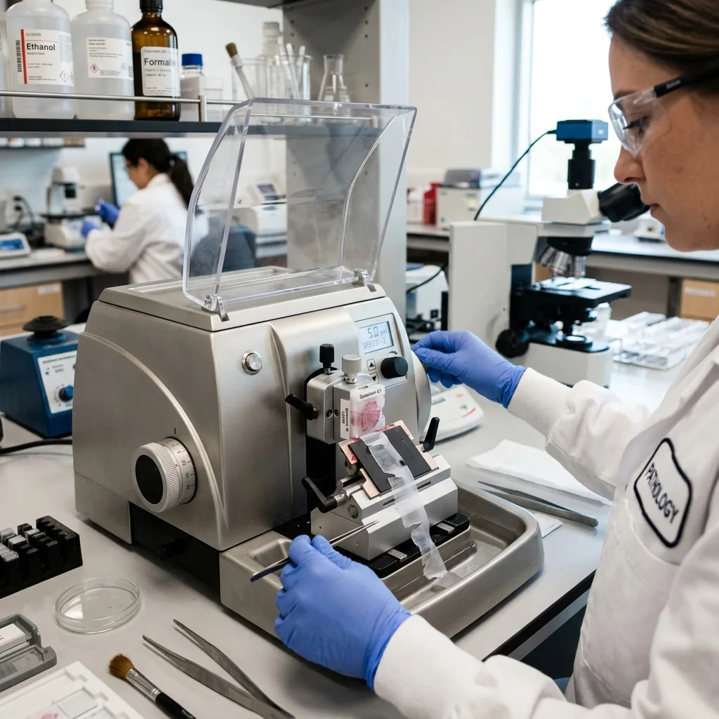
What Is a Microtome?
A microtome is a laboratory instrument designed to produce thin, uniform sections of tissue or other specimens.
The word is closely associated with microtomy, the process of preparing thin sections for microscopic examination.
A typical microtome contains a specimen holder, a cutting mechanism, a blade or knife holder, a section-thickness adjustment system, and controls for moving the specimen toward the cutting edge.
During sectioning, the specimen moves toward or across a very sharp cutting surface. A precisely controlled movement determines how much material is removed with each cutting cycle.
For routine paraffin histology, the specimen is usually a hardened paraffin-embedded tissue block.
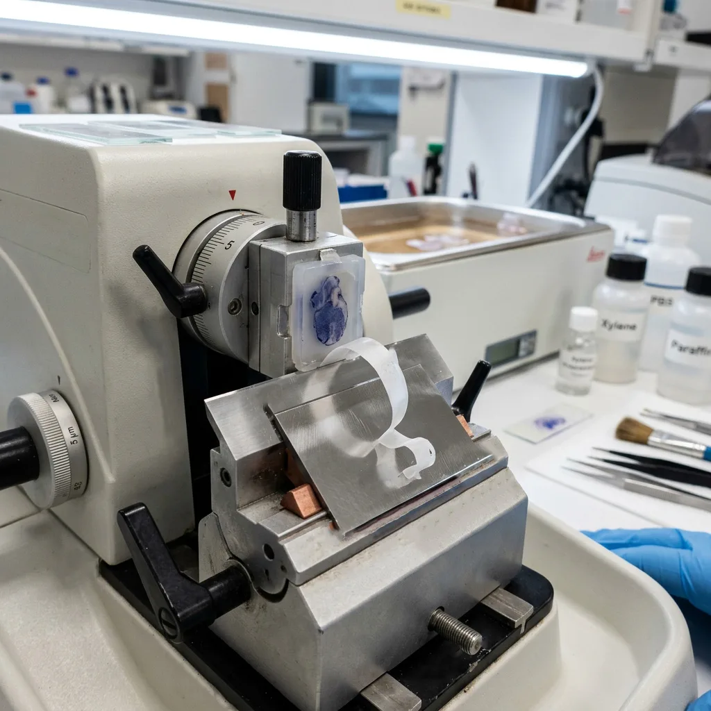
The resulting sections can form a continuous strip called a tissue ribbon. The ribbon can then be floated on water and transferred to microscope slides before staining.
This simple process requires considerable precision. Section thickness, tissue preparation, paraffin quality, blade condition, specimen orientation, cutting technique, and instrument adjustment can all affect the final section.
What Does a Microtome Do?
The main job of a microtome is:
To cut a tissue block into thin, controlled sections for microscopic examination.
This makes the microtome an important link between tissue preparation and microscopy.
A routine histology laboratory may use a microtome to prepare samples for:
- Histopathology
- Histology
- Surgical pathology
- Anatomic pathology
- Research
- Drug development
- Toxicology
- Veterinary pathology
- Teaching laboratories
- Immunohistochemistry
- Special staining
- Digital pathology
The final section must be thin enough to allow useful microscopic observation while remaining intact.
For modern digital pathology, section quality becomes even more important because folds, chatter, compression, uneven thickness, and other artifacts can affect the quality of scanned whole-slide images. A recent review of digital histopathology describes typical paraffin sections as approximately 2–5 µm and notes that sectioning artifacts can affect subsequent digital image analysis.
How Does a Microtome Work?
The operating principle is easier to understand with a simple example.
Imagine a paraffin block attached securely to a specimen holder.
The block is moved toward a stationary blade by a controlled mechanical movement. At each cutting cycle, the specimen advances by a predetermined amount. The blade removes a thin layer from the block.
Repeated cutting creates multiple sections.
When the conditions are suitable, the sections connect edge to edge and form a paraffin ribbon.
A simplified process looks like this:
1. Mount the tissue block
The embedded specimen is securely positioned in the specimen clamp.
2. Install the blade
A suitable microtome blade is positioned in the blade holder.
3. Orient the specimen
The tissue block must be correctly aligned with the cutting surface.
4. Trim the block
The surface is cut until the tissue is fully exposed.
5. Set the section thickness
The desired cutting thickness is selected.
6. Section the block
The handwheel or motorized cutting mechanism moves the specimen through repeated cutting cycles.
7. Form a ribbon
Successive paraffin sections may form a continuous ribbon.
8. Float the section
The section can be floated on a water bath to help flatten wrinkles.
9. Mount on a slide
The selected section is transferred onto a microscope slide.
10. Prepare for staining
After drying and other preparation steps, the slide can proceed to staining and microscopic examination.
Leica Biosystems describes the same basic concept for routine paraffin microtomy: a precision microtome cuts sections from paraffin blocks, the sections can form ribbons, and they are floated on warm water before being mounted on slides.
What Is a Rotary Microtome?
The rotary microtome is one of the most widely used microtome designs for routine paraffin sectioning.
The term “rotary” describes the movement used to advance and section the specimen.
A rotary microtome is commonly used when laboratories need repeated, relatively uniform sections from paraffin-embedded tissue.
Typical applications include:
- Routine histology
- Histopathology
- Pathology laboratories
- Research laboratories
- Medical schools
- Veterinary laboratories
- Pharmaceutical research
A manual rotary microtome can be suitable for laboratories where an operator controls the cutting movement.
Other systems can provide higher levels of automation, which may be useful when a laboratory has a larger workload or needs additional control over the sectioning workflow.
Modern manual rotary microtomes may include features such as ergonomic handwheels, specimen orientation systems, disposable-blade holders, coarse advance controls, trimming functions, and safety features.
Main Types of Microtomes
Not every specimen should be cut with the same type of microtome.
The most appropriate instrument depends on the specimen, embedding material, desired section thickness, application, and workflow.
1. Rotary Microtome
A rotary microtome is commonly used for paraffin-embedded tissue.
It is particularly associated with routine histopathology and histology.
Typical search terms include:
rotary microtome
manual rotary microtome
automatic rotary microtome
semi-automatic microtome
paraffin microtome
histology microtome
pathology microtome
2. Cryostat Microtome
A cryostat microtome is designed for cutting frozen tissue inside a refrigerated chamber.
Instead of relying on a paraffin block, the specimen is frozen and maintained at a controlled low temperature.
Cryosectioning can be useful when rapid tissue sectioning is needed or when researchers need to work with frozen specimens.
Cryostat systems are used in areas such as:
- Frozen sections
- Histology
- Histochemistry
- Immunohistochemistry
- Research
- Pathology
- Surgical applications
The exact temperature depends on the specimen and cutting conditions. Fatty tissue, for example, can require different cryosectioning conditions from other tissues.
This is why a cryostat is not simply a “colder rotary microtome.” The freezing environment and specimen preparation become important parts of the overall sectioning process.
3. Sliding Microtome
A sliding microtome uses a different cutting movement from a conventional rotary microtome.
The blade or specimen moves in a sliding direction across the specimen.
Sliding microtomes can be useful for certain larger, harder, or specially embedded specimens.
They may be encountered in:
- Research
- Large specimen sectioning
- Specialized histology
- Celloidin-embedded specimens
- Hard or unusual materials
Their applications are more specialized than those of routine rotary microtomes.
4. Sledge or Heavy-Duty Microtome
A sledge microtome is designed around a robust sectioning mechanism and can be used for specimens that require greater mechanical stability.
Large or harder specimens may require a stronger cutting platform than ordinary paraffin blocks.
This type of equipment can therefore appear in specialized research and histological applications.
5. Ultramicrotome
An ultramicrotome is used to produce extremely thin sections, particularly for electron microscopy and other advanced research applications.
The section thickness can reach the nanometer range depending on the application and cutting system.
Ultramicrotomy is significantly different from routine paraffin microtomy and generally requires specialized embedding materials, knives, equipment, and technical skills.
6. Vibrating Microtome
A vibrating microtome, sometimes called a vibratome, uses a vibrating blade to section specimens.
It can be useful for cutting relatively soft, often unfixed or lightly fixed tissue without conventional paraffin embedding.
Applications can include neuroscience, developmental biology, brain tissue research, and other experimental work.
It should not be confused with a standard rotary microtome because the operating principle and specimen preparation are different.
Microtome Types at a Glance
| Microtome Type | Common Specimen/Application | Typical Workflow |
|---|---|---|
| Rotary microtome | Paraffin-embedded tissue | Routine histology |
| Cryostat microtome | Frozen tissue | Frozen sectioning |
| Sliding microtome | Larger or specialized specimens | Research and histology |
| Sledge microtome | Large or harder specimens | Specialized sectioning |
| Ultramicrotome | Resin-embedded specimens | Electron microscopy/research |
| Vibrating microtome | Soft tissue | Experimental tissue sectioning |
The table is a starting point rather than a rigid rule. Actual equipment selection depends on the laboratory protocol and specimen characteristics.
What Thickness Can a Microtome Cut?
Microtome section thickness is one of the most important terms in histology.
There is no single thickness that applies to every microtome or every specimen.
Routine paraffin histology commonly uses sections only a few micrometers thick. Leica Biosystems describes typical paraffin sections as around 3–5 µm, while published laboratory protocols commonly use approximately 4–5 µm for routine paraffin-embedded tissue.
A recent review notes that conventional rotary and sliding microtome applications can cover a wider range depending on the specimen and purpose.
This distinction is important:
Microtome capability is not the same as the thickness used for every histology application.
A laboratory should select the appropriate thickness according to:
- Tissue type
- Embedding medium
- Staining method
- Microscopy method
- Research protocol
- Diagnostic requirements
- Desired tissue morphology
For example, many routine paraffin sections are in the few-micrometer range, while specialized research applications may require substantially thicker or thinner sections.
Why Is Microtome Section Thickness Important?
Imagine trying to look through a thick slice of tissue under a microscope.
Too much tissue depth can cause structures to overlap and make the image harder to interpret.
A suitable thin section allows light to pass through the specimen and provides a more useful two-dimensional representation of the tissue.
However, thinner is not automatically better.
A section that is too thin may become fragile, difficult to manipulate, or less suitable for a particular application.
Therefore, the correct question is not:
What is the thinnest section a microtome can produce?
A better question is:
What section thickness is appropriate for this specimen and application?
For routine histology, different laboratories and protocols may use different settings. One recent digital histopathology review describes 2–5 µm as a typical range for paraffin sectioning, while individual experimental protocols may use 3, 4, 5, or other values based on the specimen and purpose.
Microtome Blades: What Are They Used For?
The microtome blade is the cutting component that directly contacts the specimen.
Blade selection can have a major effect on section quality.
Common terms include:
microtome blade
microtome knife
disposable microtome blade
low-profile blade
high-profile blade
steel microtome knife
The specific blade system should match the microtome and the specimen.
For routine paraffin work, disposable blades are widely used because they provide a convenient cutting edge and can be replaced when the edge becomes unsuitable.
A damaged, contaminated, incorrectly installed, or unsuitable blade can contribute to sectioning problems.
The cutting edge also needs to be positioned correctly relative to the specimen.
What Is a Paraffin Section?
A paraffin section is a thin tissue slice cut from a specimen that has been embedded in paraffin wax.
The basic idea is:
Tissue is prepared → tissue is infiltrated with paraffin → paraffin solidifies → microtome cuts the block → sections are mounted on slides.
Paraffin is widely used because it gives tissue sufficient support for routine sectioning and can produce sections suitable for standard light microscopy.
Leica Biosystems describes paraffin wax-based histological waxes as widely used embedding media for routine light microscopy, with paraffin sections generally prepared using rotary microtomes.
What Is a Tissue Ribbon?
During paraffin microtomy, successive sections can remain connected along their edges and form a tissue ribbon.
A ribbon makes it easier for the operator to handle a series of sections.
The quality of ribbon formation depends on several factors, including:
- Tissue processing
- Paraffin quality
- Block temperature
- Blade condition
- Section thickness
- Cutting speed
- Block orientation
- Microtome adjustment
A poor ribbon does not necessarily mean that the microtome itself is defective.
The problem may start earlier in the histology workflow.
This is one reason microtomy should be considered together with tissue processing and embedding, rather than as an isolated step.
Why Do Microtome Sections Curl or Fold?
One common laboratory question is:
Why do my microtome sections curl?
There can be several causes.
The block may not be prepared correctly. The blade may not be suitable or sharp enough. The cutting conditions may not be appropriate. Block temperature, section thickness, cutting speed, and specimen orientation can also influence the result.
Common sectioning problems include:
curling
folding
wrinkling
compression
chatter
thick and thin sections
streaking
fragmentation
poor ribbon formation
A recent review of microtomy identifies sectioning artifacts and problems involving specimen preparation, temperature control, blade selection, section thickness, and cutting technique as important factors affecting histological section quality.
What Causes Chatter in Microtome Sections?
Chatter describes a repeated pattern or vibration-related artifact that can appear in a tissue section.
Possible contributing factors include:
- Excessive vibration
- Hard or poorly processed tissue
- Incorrect blade conditions
- Incorrect cutting setup
- Block problems
- Excessive cutting resistance
- Mechanical instability
The solution depends on the cause.
Changing the blade alone may not solve the problem if the tissue block itself has been incorrectly processed.
This leads to an important histology principle:
Good sectioning starts before the microtome.
Fixation, tissue processing, embedding, block orientation, cooling, and blade selection all contribute to the final result.
Why Does Tissue Orientation Matter?
A tissue block does not contain useful information equally in every direction.
The way tissue is positioned during embedding determines the surface presented to the microtome.
That means specimen orientation can influence what anatomical structures appear in the final section.
This is particularly important in modern digital pathology. A recent review explains that orientation during embedding determines the two-dimensional plane of the final digital image; poor orientation can make important structures less visible or absent from the selected section.
For this reason, experienced histology technicians do not simply “cut the block.”
They consider:
Which surface should be exposed?
What structure needs to be represented?
How should the tissue be oriented?
How thick should the sections be?
How many sections are required?
Microtome Applications in Histology and Pathology
Microtomes are used across different laboratory environments.
Histopathology
In pathology laboratories, microtomes are commonly used to prepare tissue sections from paraffin blocks for microscopic examination.
These sections can support routine stains, special stains, immunohistochemistry, and other diagnostic workflows.
Research
Research laboratories use microtomes to study tissue morphology, disease models, experimental treatments, and biological structures.
Immunohistochemistry
Paraffin tissue sections can be used for immunohistochemical procedures after appropriate preparation.
The quality of the tissue section can affect the quality and interpretation of subsequent staining.
Digital Pathology
Slides prepared from microtome sections can be scanned for whole-slide imaging.
Good section quality helps reduce artifacts that can interfere with image scanning, image analysis, and computational pathology workflows.
Veterinary Histology
Veterinary laboratories also use microtomes for animal tissue specimens.
The principles are similar, but tissue size, tissue composition, species, and research or diagnostic requirements can vary.
Pharmaceutical and Toxicology Research
Microtomy is also used for tissue evaluation in drug development, toxicology and experimental studies.
In these applications, consistent section preparation can be important when comparing tissue morphology across multiple samples.
Microtome vs Cryostat: What Is the Difference?
This is a common search question.
The main difference is the state of the specimen and cutting environment.
| Feature | Conventional Microtome | Cryostat Microtome |
|---|---|---|
| Common specimen | Paraffin-embedded tissue | Frozen tissue |
| Environment | Normal laboratory environment | Refrigerated chamber |
| Common application | Routine histology | Frozen sectioning |
| Embedding approach | Paraffin or other suitable medium | Frozen embedding medium |
| Main advantage | Routine paraffin sectioning | Rapid frozen tissue sectioning |
| Typical use | Histology and pathology | Frozen section, research, special applications |
A cryostat integrates a microtome with a refrigerated chamber so that frozen specimens remain cold during sectioning. Cryosectioning is widely used where frozen tissue preparation is preferred or rapid sectioning is needed.
Neither system should be treated as a universal replacement for the other.
Microtome vs Ultramicrotome
A microtome is commonly associated with routine histology and light microscopy.
An ultramicrotome is designed for much thinner sections and specialized microscopy, including transmission electron microscopy.
The major difference is therefore not simply the machine size.
It involves:
section thickness
embedding medium
knife technology
specimen preparation
microscopy method
operator skill
Ultramicrotomy may use resin-embedded specimens and specialized cutting systems rather than conventional paraffin sectioning.
What Equipment Is Used Around a Microtome?
A microtome does not normally work alone.
A typical histology sectioning area may include:
- Tissue cassettes
- Paraffin embedding equipment
- Paraffin blocks
- Microtome
- Microtome blades
- Blade holder
- Specimen holder
- Forceps
- Brushes
- Water bath
- Microscope slides
- Slide drying equipment
- Waste tray
- Safety accessories
The actual equipment list depends on the laboratory workflow.
For paraffin sectioning, the water bath is particularly useful for flattening sections before they are transferred to slides. Published protocols commonly use a controlled warm-water flotation step after microtome sectioning.
How to Improve Microtome Section Quality
A good result usually comes from controlling several factors together.
Start with properly processed tissue
Poor fixation or tissue processing can make later sectioning difficult.
Make sure the block is correctly embedded
The tissue should be appropriately oriented and securely supported by the embedding medium.
Use the appropriate blade
The blade should match the microtome and specimen.
Keep the blade and holder secure
Mechanical movement during cutting can create artifacts.
Control section thickness
Use a thickness appropriate for the intended analysis rather than simply choosing the thinnest possible setting.
Maintain suitable block conditions
The temperature and physical properties of the paraffin block affect cutting behavior.
Use a consistent cutting movement
Irregular or overly fast cutting can contribute to uneven sections.
Monitor section quality
Look for:
- folds
- chatter
- compression
- tearing
- knife lines
- incomplete sections
- irregular ribbons
Microtomy references emphasize correct specimen preparation, blade condition, cutting speed, section thickness and temperature control as important elements in producing good sections.
Microtome Safety
A microtome contains an extremely sharp cutting edge.
Safety should therefore be part of normal operation.
Important precautions include:
Secure the blade correctly.
Use the built-in safety mechanism when appropriate.
Keep hands away from the cutting edge.
Follow the manufacturer’s operating procedure.
Lock or secure the handwheel when the instrument is not being operated, according to the manufacturer’s instructions.
Remove blades using an appropriate safe method.
Dispose of used disposable blades in a suitable sharps container.
Modern microtomes may include handwheel locks, blade guards and other safety features designed to reduce operator risk. The exact procedure should always follow the specific equipment manual. Leica’s microtomy training material also emphasizes safe operation when preparing paraffin sections.
Does a Microtome Need Regular Maintenance?
Yes.
Routine maintenance helps keep the sectioning mechanism clean, stable and accurately adjusted.
Basic maintenance may include:
- Removing paraffin debris
- Cleaning the specimen holder
- Cleaning the blade holder
- Checking mechanical movement
- Checking the handwheel
- Checking clamps
- Inspecting the waste tray
- Following lubrication requirements
- Checking alignment when necessary
- Replacing worn components according to the manufacturer’s instructions
Calibration and maintenance requirements vary by model.
A laboratory should maintain the instrument according to the manufacturer’s service documentation rather than applying one generic maintenance interval to every microtome.
Common Microtome Questions
What is a microtome used for?
A microtome is used to cut thin sections of tissue or other specimens for microscopic examination. In routine histology, it is commonly used to section paraffin-embedded tissue.
What is a microtome machine?
A microtome machine is another common term for a microtome or microtome instrument. It is a precision sectioning device used in histology, pathology and research laboratories.
What is a rotary microtome used for?
A rotary microtome is commonly used for routine sectioning of paraffin-embedded tissue. It is widely used in histology and histopathology laboratories.
What is a cryostat microtome?
A cryostat microtome is a microtome installed inside a refrigerated chamber for cutting frozen tissue.
How thin can a microtome cut?
The available range depends on the microtome design, blade, specimen and application. Routine paraffin sections are commonly a few micrometers thick, while specialized microtomes support very different thickness ranges.
What is a paraffin microtome?
A paraffin microtome is generally a microtome used to section tissue embedded in paraffin wax. Rotary microtomes are commonly used for this application.
What is microtomy?
Microtomy is the process of cutting thin sections from a specimen using a microtome.
What is a tissue ribbon?
A tissue ribbon is a series of connected sections produced during paraffin microtomy. The connected sections can be transferred to a water bath and mounted onto slides.
What blade is used in a microtome?
The blade depends on the microtome and specimen. Disposable steel blades are commonly used for routine paraffin sectioning, while specialized applications may require different knives or blade materials.
Why do microtome sections fold?
Folding can result from issues involving the tissue block, paraffin, blade, section thickness, cutting conditions or water-bath handling. The cause should be identified rather than assuming the microtome itself is defective.
Why do microtome sections chatter?
Chatter can be associated with vibration, tissue hardness, blade conditions, block preparation, mechanical stability or sectioning settings.
Is a microtome used before or after tissue processing?
For conventional paraffin histology, tissue processing occurs before embedding, and microtomy occurs after a processed tissue specimen has been embedded into a block.
What is the difference between a microtome and a cryostat?
A conventional microtome is commonly used to cut embedded specimens such as paraffin blocks, while a cryostat provides a refrigerated environment for sectioning frozen tissue.
What is a microtome used for in pathology?
It is used to prepare thin tissue sections from pathology specimens so they can be mounted on slides, stained and examined microscopically.
Can a microtome cut frozen tissue?
Some microtome systems can be configured for frozen sectioning, but dedicated cryostat microtomes are specifically designed for frozen tissue work.