1. What Is a Medical Fiberscope?
The word fiberscope comes from the technology used to transmit light and images through optical fibers.
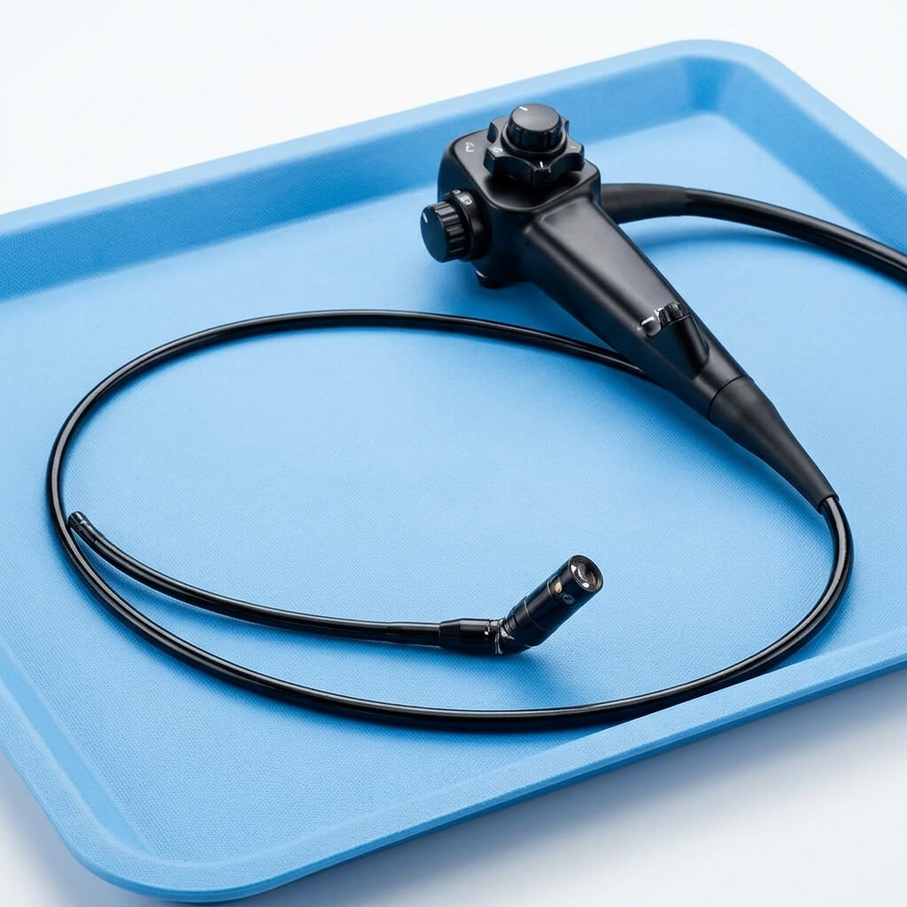
A medical fiberscope is a type of flexible endoscope designed for internal visualization.
Unlike a rigid endoscope, a flexible fiberscope can bend along its insertion path. This makes it useful for examining anatomical areas that are difficult to reach with a straight instrument.
Common terms used by hospitals, doctors and medical equipment buyers include:
- Medical fiberscope
- Medical fiber scope
- Medical fiberoptic scope
- Fiberoptic endoscope
- Fiber optic endoscope
- Flexible fiberscope
- Flexible fiberoptic endoscope
- Fiber endoscope
- Flexible medical endoscope
- Diagnostic fiberscope
- Fiberoptic medical endoscope
- Flexible optical endoscope
Although these terms are related, the exact design and intended use depend on the specific product.
2. Fiberscope vs Endoscope: What Is the Difference?
Endoscope is a broad term for an instrument used to look inside the body.
A fiberscope is an endoscope that uses optical fiber technology to transmit light and images.
Therefore:
All medical fiberscopes are endoscopic instruments, but not every endoscope is a fiberscope.
Modern medical endoscopy can use different image technologies.
For example, some flexible endoscopes use a fiber-optic image bundle, while video endoscopes use an electronic image sensor near the distal tip.
This distinction is important when a buyer is comparing:
- Fiberoptic endoscope
- Video endoscope
- Flexible endoscope
- Digital endoscope
- HD endoscope
The choice depends on the required imaging workflow, display system, application and budget.
3. How Does a Fiberoptic Endoscope Work?
The working principle of a medical fiberscope can be explained simply.
Step 1: Light Enters the Scope
A light source sends illumination through a light guide toward the distal tip.
Step 2: Light Reaches the Target Area
The distal end of the fiberscope illuminates the anatomical area being examined.
Step 3: The Lens Captures the Image
The objective lens collects reflected light from the tissue.
Step 4: Optical Fibers Transmit the Image
An organized bundle of optical fibers carries the image toward the proximal end.
Step 5: The Image Is Viewed
Depending on the equipment, the image may be viewed through:
- Eyepiece
- Camera adapter
- Monitor
- Video processor
This basic optical pathway allows a flexible instrument to transmit an image from a difficult-to-reach anatomical location.
Research on fiber-optic endoscopy describes how optical fibers can transmit illumination and images through a flexible instrument, while the distal optics and fiber bundle determine important aspects of the image.
4. What Is Fiber Optics in Medical Endoscopy?
Fiber optics refers to the transmission of light through very thin optical fibers.
In a fiberscope, many fibers are arranged into a bundle.
The optical fibers work by guiding light through repeated internal reflection.
This makes it possible to transmit illumination and image information through a narrow, flexible pathway.
Common SEO terms related to this technology include:
- Fiber optic technology
- Medical fiber optics
- Optical fiber endoscope
- Fiber optic medical instrument
- Fiberoptic imaging
- Fiber bundle endoscope
- Medical optical fibers
- Fiber optic imaging system
5. Main Components of a Medical Fiberscope
A medical fiberscope consists of several interconnected parts.
Understanding these components is useful for both medical users and purchasing teams.
1. Distal Tip
The distal tip is the end of the scope inserted toward the area being examined.
It may contain:
- Objective optics
- Illumination
- Working-channel opening
- Other functional components
2. Objective Lens
The objective lens collects light from the area being examined and directs it into the imaging system.
Its optical design contributes to:
- Field of view
- Image clarity
- Viewing direction
3. Fiber-Optic Image Bundle
This is the main optical image-transmission component of a traditional fiberscope.
The fiber bundle contains many optical fibers arranged to transmit an image from the distal end to the viewing end.
4. Light Guide
The light guide delivers illumination to the distal end of the scope.
Adequate illumination is essential for viewing internal anatomy.
5. Insertion Tube
The insertion tube is the flexible section introduced into the body.
Its design influences:
- Flexibility
- Outer diameter
- Working length
- Durability
6. Control Handle
The operator uses the handle to control the instrument.
Depending on the design, controls may include:
- Tip deflection
- Suction
- Air or water
- Working-channel functions
7. Deflection Mechanism
The distal tip of many flexible scopes can be moved using a control mechanism.
This allows the operator to change the viewing direction and navigate anatomical pathways.
8. Working Channel
Some medical fiberscopes include a working channel.
Depending on the intended application, it may be used for:
- Suction
- Irrigation
- Injection
- Brush sampling
- Biopsy instruments
- Other compatible accessories
A flexible bronchoscope, for example, may contain a working channel for suction and compatible diagnostic or therapeutic instruments.
9. Eyepiece or Camera Connection
Traditional fiberscopes can use an eyepiece for direct viewing.
Some systems can also connect to a camera, allowing the image to be displayed on a monitor.
6. Flexible Fiberscope vs Rigid Endoscope
One of the most common searches is:
Flexible endoscope vs rigid endoscope
The main difference is mechanical design.
| Feature | Flexible Fiberscope | Rigid Endoscope |
|---|---|---|
| Shaft | Flexible | Rigid |
| Navigation | Can follow curved pathways | Generally straight access |
| Tip movement | Often deflectable | Usually fixed viewing direction |
| Typical use | Airways and other curved anatomical routes | Procedures requiring a rigid optical path |
| Insertion | Flexible | Rigid |
A flexible design can be useful when the anatomical pathway is curved or difficult to access.
A rigid endoscope may be preferable when a stable straight optical path is required.
The appropriate type depends on the procedure.
7. Fiberscope vs Video Endoscope
This is another important distinction for medical equipment buyers.
Fiberoptic Endoscope
A traditional fiberscope uses an optical fiber bundle to transmit the image.
Typical components include:
- Fiber bundle
- Objective lens
- Eyepiece
- Light guide
Video Endoscope
A video endoscope generally uses an electronic imaging sensor near the distal end.
The image is then transmitted electronically to an image processor or display.
Simple Comparison
| Feature | Fiberoptic Endoscope | Video Endoscope |
|---|---|---|
| Image transmission | Optical fiber bundle | Electronic sensor |
| Viewing | Eyepiece or camera | Monitor |
| Image processing | More limited in traditional designs | Digital processing possible |
| Typical setup | Flexible scope + light source | Scope + camera/processor + monitor |
| Main technology | Fiber optics | Digital imaging |
The choice should depend on clinical workflow, image requirements, available infrastructure and purchasing budget.
8. What Is a Flexible Fiberoptic Endoscope Used For?
Medical fiberscopes can be used in several departments.
The specific indication always depends on the individual device.
Common areas include:
Respiratory Medicine
- Bronchoscopy
- Airway examination
- Respiratory tract visualization
ENT
- Nasal examination
- Nasopharyngeal examination
- Laryngeal examination
Urology
- Urinary tract examination
- Flexible cystoscopy
- Selected urological procedures
Gastroenterology
- Gastrointestinal examination
- Flexible endoscopy
Gynecology
- Selected gynecological endoscopic examinations
Anesthesia
- Difficult-airway assessment
- Fiberoptic intubation workflows
The next article in this series will examine these different medical fiberscope types and specialty applications in much greater detail.
9. Medical Fiberscope for Bronchoscopy
A fiberoptic bronchoscope is a flexible scope designed to visualize the airways.
Common search terms include:
- Fiberoptic bronchoscope
- Flexible bronchoscope
- Medical bronchoscope
- Flexible fiberoptic bronchoscope
- Bronchoscopy scope
- Bronchial fiberscope
- Airway fiberscope
A flexible bronchoscope can be introduced through the oral or nasal route and used to inspect the respiratory tract. NCBI’s current clinical reference describes flexible bronchoscopy as an established tool for diagnosing and managing lung disease and notes that flexible bronchoscopes incorporate fiber optics, imaging components and working channels.
Depending on the system and indication, bronchoscopy can involve:
- Airway inspection
- Secretions removal
- Bronchoalveolar lavage
- Brush sampling
- Biopsy
- Selected therapeutic procedures
10. Fiberoptic Scope for Airway Management
Another important application is airway management.
Searchers may use terms such as:
- Fiberoptic intubation scope
- Flexible intubation scope
- Fiberoptic airway scope
- Difficult airway fiberscope
- Airway management fiberscope
- Fiberoptic intubation device
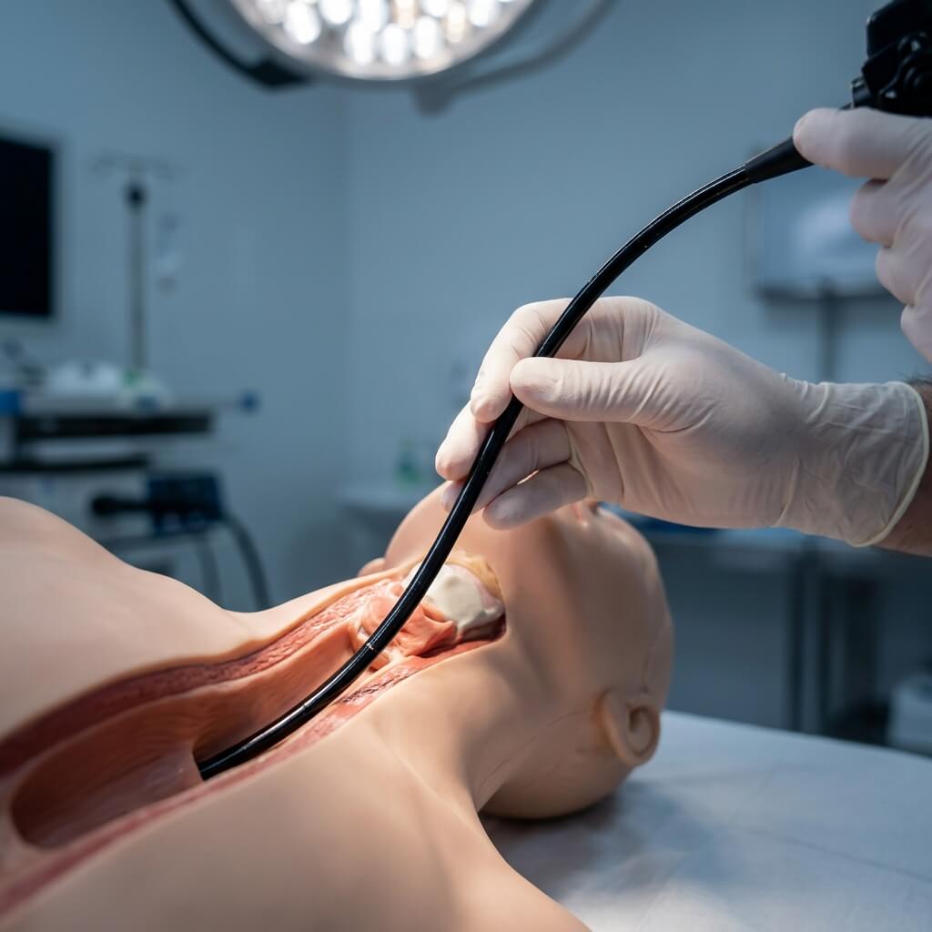
Flexible fiberoptic equipment can assist clinicians in selected difficult-airway workflows.
The slim, flexible insertion tube can help clinicians navigate the airway while viewing the anatomy.
The precise scope diameter, working length and channel configuration depend on the intended application.
11. Medical Fiberscope for ENT
ENT is another important application area.
Common terms include:
- ENT fiberscope
- ENT fiberoptic endoscope
- Flexible ENT endoscope
- Fiberoptic laryngoscope
- Flexible laryngoscope
- Nasopharyngoscope
- Flexible nasopharyngoscope
- Nasal fiberscope
- Throat fiberscope
Depending on the scope design, ENT fiberscopes may be used to examine:
- Nasal cavity
- Nasopharynx
- Pharynx
- Larynx
- Vocal-cord region
Flexible ENT examination is particularly useful when a flexible pathway is needed to reach anatomical areas that cannot be viewed easily with a straight instrument.
12. Medical Fiberscope for Urology
Flexible optical instruments are also used in urology.
Common keywords include:
- Urology fiberscope
- Flexible urology endoscope
- Fiberoptic cystoscope
- Flexible cystoscope
- Urinary tract fiberscope
- Bladder fiberscope
A flexible cystoscope is designed to follow the urinary tract and allow direct visual inspection of relevant anatomy.
For buyers, factors such as:
- Outer diameter
- Working length
- Deflection
- Optical performance
- Working channel
can be important when selecting a urology fiberscope.
13. Gastrointestinal Fiberscope
Flexible endoscopy has also been used in gastrointestinal medicine.
Related keywords include:
- Gastrointestinal fiberscope
- GI fiberscope
- Flexible gastroscope
- Fiberoptic gastroscope
- Gastrointestinal endoscope
- Flexible GI endoscope
The design of a gastrointestinal scope is different from a respiratory or ENT scope because it needs to navigate the gastrointestinal tract and may require different working-channel and control characteristics.
Specific applications should be confirmed against the intended device and regulatory documentation.
14. Medical Fiberscope for Gynecology
Flexible and specialized optical scopes can also be used for selected gynecological examinations.
Related keywords include:
- Gynecology fiberscope
- Gynecological endoscope
- Flexible gynecology scope
- Hysteroscope
- Fiberoptic hysteroscope
A hysteroscope is specifically designed to visualize the uterine cavity and is distinct from a general-purpose fiberscope.
This is an important distinction for purchasing teams:
The term “fiberscope” describes the imaging technology, while the clinical product name describes the intended application.
15. Pediatric and Small-Diameter Fiberscopes
Some medical procedures require a smaller insertion diameter.
This has created search demand around:
- Pediatric fiberscope
- Pediatric bronchoscope
- Small diameter fiberscope
- Small diameter endoscope
- Ultrathin fiberscope
- Mini fiberscope
- Small caliber flexible endoscope
For example, medical literature describes pediatric and ultrathin flexible bronchoscopes with smaller outer diameters and corresponding smaller working channels.
When selecting a small-diameter fiberscope, buyers need to balance:
Diameter + Image Quality + Working Channel + Flexibility + Durability
A smaller diameter can improve access, but may also affect available channel size and other design characteristics.
16. What Are the Advantages of a Medical Fiberscope?
Medical fiberscopes have several characteristics that make them useful in selected procedures.
Flexible Design
The scope can follow curved anatomical pathways.
Direct Visualization
The operator can see internal anatomy through the optical system.
Compact Distal Tip
The distal end can be designed for relatively narrow anatomical pathways.
Instrument Channel Options
Some designs provide channels for suction or compatible instruments.
Reusable Workflow
Some medical fiberscopes are reusable when designed and processed according to the manufacturer’s instructions.
17. What Are the Limitations of Fiberoptic Endoscopes?
Fiberscopes also have limitations.
Image Quality
Traditional fiber bundles have a different imaging structure from modern digital video sensors.
Fiber Damage
Individual optical fibers can become damaged over time.
This may produce:
- Dark dots
- Image artifacts
- Reduced image quality
Cleaning Requirements
Reusable flexible scopes require appropriate cleaning and reprocessing procedures.
Equipment Compatibility
A fiberscope may require a specific:
- Light source
- Camera
- Adapter
- Processor
- Connector
The complete system should therefore be evaluated rather than purchasing only the scope body.
18. Common Signs of a Damaged Fiberscope
Medical equipment users may notice:
- Dark spots in the image
- Reduced illumination
- Blurred image
- Broken or unstable image
- Reduced tip movement
- Cable damage
- Cracked insertion tube
- Working-channel problems
Dark spots can occur when individual fibers in an imaging bundle are damaged.
A fiberscope showing significant damage should be inspected according to the manufacturer’s service procedures before continued clinical use.
19. Medical Fiberscope Image Quality
Image quality depends on the complete optical system.
Important factors include:
- Fiber bundle quality
- Objective lens
- Illumination
- Field of view
- Resolution
- Optical transmission
- Distal-tip design
Buyers often search:
- Fiberoptic endoscope image quality
- Fiberscope resolution
- Fiber bundle resolution
- Medical endoscope image quality
- Flexible endoscope optics
However, comparing one specification alone can be misleading.
For example, fiber count, optical resolution, field of view and illumination should be considered together.
20. What Is the Field of View of a Fiberscope?
Field of view (FOV) describes how much of the scene can be seen through the scope.
Related keywords include:
- Fiberscope field of view
- Fiberoptic endoscope field of view
- Endoscope viewing angle
- Medical scope FOV
- Endoscope angle of view
A wider field of view can provide a larger visual area, while a narrower field may provide a different viewing perspective.
The appropriate FOV depends on the clinical application.
21. Fiberscope Diameter and Working Length
Two highly important specifications are:
Insertion tube diameter
and
Working length
The diameter affects accessibility.
The working length determines how far the instrument can reach.
Related search terms:
- Fiberscope diameter
- Flexible endoscope diameter
- Medical fiberscope size
- Fiberscope working length
- Flexible scope insertion length
- Small diameter endoscope
These specifications should always be selected according to the intended anatomy and procedure.
Detailed specification selection will be covered in Post 4.
22. Why Working Channels Matter
Some medical fiberscopes include a working channel.
The channel may allow compatible accessories to pass through the scope.
Possible functions include:
- Suction
- Fluid delivery
- Brush sampling
- Biopsy
- Other compatible instruments
Common search terms:
- Fiberscope working channel
- Flexible endoscope working channel
- Endoscope biopsy channel
- Endoscope suction channel
- Instrument channel endoscope
A larger working channel can provide compatibility with certain accessories, but it may also influence the overall dimensions and design of the scope.
23. What Is Tip Deflection?
Tip deflection describes how far the distal tip can be bent in different directions.
Common keywords:
- Fiberscope tip deflection
- Flexible endoscope tip movement
- Endoscope bending angle
- Flexible scope angulation
- Endoscope tip angulation
Deflection is important because it determines how effectively the operator can steer the scope.
For example, a flexible bronchoscope uses a control mechanism to steer the distal tip through the airway.
24. How to Choose a Medical Fiberscope?
A simple selection process is:
Step 1: Identify the Application
Is the scope for:
- Bronchoscopy?
- ENT?
- Urology?
- Gastroenterology?
- Airway management?
Step 2: Select the Appropriate Scope Type
Different applications require different designs.
Step 3: Check Diameter
Make sure the outer diameter is appropriate.
Step 4: Check Working Length
Confirm that the scope can reach the target area.
Step 5: Check Tip Deflection
Make sure the bending range fits the intended workflow.
Step 6: Check Working Channel
Confirm compatibility with required accessories.
Step 7: Check Imaging
Evaluate:
- Field of view
- Image clarity
- Optical system
- Illumination
Step 8: Check Compatibility
Confirm compatibility with:
- Light source
- Camera
- Processor
- Connector
- Accessories
25. Choosing a Medical Fiberscope Supplier
For hospitals and international medical equipment distributors, choosing a supplier involves more than comparing product prices.
Consider:
- Product specifications
- Application support
- Quality control
- Compatibility
- Documentation
- Warranty
- Technical support
- Spare parts
- International shipping
For a replacement fiberscope, compatibility should be confirmed before ordering.
For a new endoscopy department, it may be more appropriate to evaluate the complete system, including the scope, light source, camera, processor and display.
26. UMY Medical Equipment Co., Ltd. and Medical Endoscopy Solutions
UMY Medical Equipment Co., Ltd. provides medical equipment solutions for international healthcare customers.
The company’s current product portfolio includes medical endoscopy equipment and fiber-optic products. For example, UMY currently lists a Fiber Cystoscope, Nasopharyngoscope, Bronchoscope, Choledochoscope, and Hysteroscope product covering several endoscopic applications. The listed product information includes flexible designs, different insertion diameters, a 90° field of view and specified tip bending ranges, although exact specifications vary by configuration.
UMY also lists an endoscope camera system supporting applications including laparoscopy, hysteroscopy, ENT and fiberscope workflows, with camera, display, light-source and image-processing functions.
According to UMY Medical’s current company profile, the company serves healthcare entities across more than 168 countries and regions and offers a broad portfolio of medical devices and consumables, together with OEM/custom solutions and multilingual after-sales support.
For an international buyer, these capabilities can be relevant when a medical fiberscope purchase is part of a broader endoscopy or hospital equipment project.
27. Medical Fiberscope Buying Checklist
Before requesting a quotation, prepare:
Application
☐ Bronchoscopy
☐ ENT
☐ Urology
☐ Gastroenterology
☐ Gynecology
☐ Airway management
Scope
☐ Fiberscope type
☐ Outer diameter
☐ Working length
☐ Field of view
☐ Tip deflection
☐ Working channel
Imaging
☐ Fiber bundle / optical system
☐ Light source
☐ Camera compatibility
☐ Display requirements
Procurement
☐ Quantity
☐ Warranty
☐ Spare parts
☐ Documentation
☐ Certification
☐ Technical support
This information allows suppliers to provide a more accurate quotation.
28. Frequently Asked Questions About Medical Fiberscopes
What is a medical fiberscope?
A medical fiberscope is a flexible optical instrument used to visualize internal anatomy through a fiber-optic image-transmission system.
Is a fiberscope the same as an endoscope?
A fiberscope is a type of endoscope. “Endoscope” is the broader term.
What is a fiberoptic endoscope?
A fiberoptic endoscope uses optical fibers to transmit illumination and image information through the scope.
How does a fiberoptic endoscope work?
Light is delivered to the distal tip, reflected from the target anatomy, and collected by the optical system. The image is transmitted through the fiber bundle toward the viewing end.
What is a flexible fiberscope used for?
Applications include bronchoscopy, ENT, airway management, urology and other selected endoscopic examinations.
What is a fiberoptic bronchoscope?
It is a flexible medical scope designed to visualize the respiratory tract and airways.
What is a fiberscope used for in ENT?
Depending on the design, it may be used for visualization of the nose, nasopharynx, throat and laryngeal region.
What is a flexible cystoscope?
A flexible cystoscope is an endoscopic instrument designed for examination of the urinary tract and bladder.
What is the difference between a fiberscope and a videoscope?
A traditional fiberscope transmits images through an optical fiber bundle, while a videoscope generally uses an electronic image sensor and digital processing.
What is the advantage of a flexible fiberscope?
Its flexible insertion tube allows it to navigate curved anatomical pathways.
What is the working channel of an endoscope?
A working channel is an internal passage that can accommodate compatible instruments or provide suction, irrigation or other functions depending on the scope.
What does fiberscope diameter mean?
It refers to the outer diameter of the insertion tube. It is an important specification when evaluating access to a particular anatomical pathway.
What does fiberscope working length mean?
It is the usable length of the scope available for insertion toward the examination area.
What is tip deflection?
Tip deflection describes the ability of the distal end of a flexible scope to bend in different directions.
Can a medical fiberscope be connected to a camera?
Some fiberscopes can be connected to compatible camera adapters or camera systems. Compatibility should be confirmed with the manufacturer.
Can a fiberscope display images on a monitor?
Traditional fiberscopes can often be connected to an appropriate camera system, while some integrated systems are designed for direct monitor display.
How long does a medical fiberscope last?
Lifespan depends on construction, frequency of use, cleaning and reprocessing, storage and physical handling.
Can a damaged fiberscope be repaired?
Some types of damage may be serviceable, but the feasibility and cost depend on the scope design and the specific failure.
How do I choose a medical fiberscope?
Start with the clinical application, then compare diameter, working length, field of view, tip deflection, working channel, imaging characteristics, compatibility and supplier support.
Conclusion
A medical fiberscope is a flexible optical endoscopic instrument designed to provide direct visualization of internal anatomy.
Its basic system can be understood as:
Light Source → Fiber-Optic System → Distal Optics → Internal Anatomy → Image Transmission → Eyepiece or Camera
The main factors that distinguish one fiberscope from another include:
- Clinical application
- Outer diameter
- Working length
- Field of view
- Tip deflection
- Working channel
- Optical image quality
- Light-source compatibility
- Camera or processor compatibility
Fiberscopes remain relevant for selected medical applications because their flexible design can provide access to curved or difficult anatomical pathways. At the same time, buyers should understand the differences between fiberoptic endoscopes, video endoscopes, flexible endoscopes and rigid endoscopes before making a purchasing decision.
For hospitals, clinics and international medical equipment distributors, the right approach is to select the scope according to the intended clinical application first, then compare technical specifications and supplier support.