1. What Does “Slice Count” Mean in Computed Tomography?
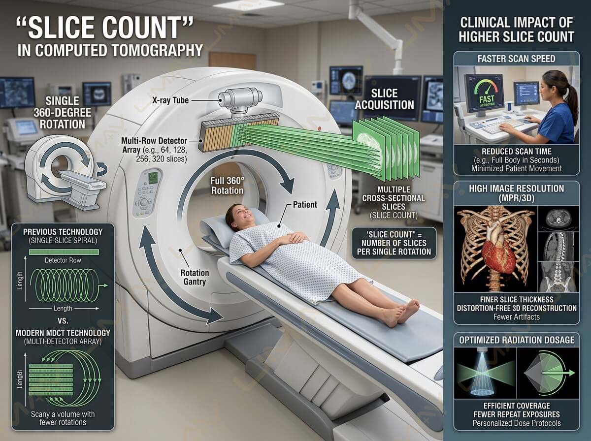
In Computed Tomography (CT), the term “slice count” refers to the number of two-dimensional cross-sectional image slices a scanner’s detector array can acquire in a single 360-degree rotation of the X-ray gantry.
When multi-detector CT (MDCT) technology replaced older single-slice spiral scanners, it transformed diagnostic imaging. Rather than using a single row of detectors, modern CT scanners feature multi-row detector arrays. As the X-ray tube rotates around the patient, these detector banks simultaneously record data across multiple channels.
A higher slice count directly impacts three core clinical factors:
-
Scan Speed: Capturing more physical area per gantry rotation reduces total scan time significantly.
-
Image Resolution: Finer slice thickness allows for thinner anatomical cross-sections, enabling 3D multiplanar reconstruction (MPR) without image distortion.
-
Radiation Efficiency: Faster coverage minimizes patient movement, reducing the need for repeat exposures and optimizing radiation dosage.
2. The Progression of Multi-Detector CT Architecture
16-Slice (Basic Routine Work)
    └──► 64-Slice (General Hospital Workhorse)
            └──► 128/256-Slice (Advanced Angiography & Oncology)
                    └──► 320-Slice (Ultra-Fast Whole-Organ Imaging)
Understanding how slice capabilities scale helps radiology directors and biomedical engineers align equipment capabilities with diagnostic requirements.
16-Slice CT Scanners: The Entry-Level Diagnostic Baseline
16-slice CT scanners are widely utilized in outpatient clinics, urgent care facilities, and community health centers. They offer reliable cross-sectional imaging for basic diagnostic needs.
-
Primary Use Cases: Routine brain scans, sinus and temporal bone imaging, basic chest and abdominal scans, non-complex trauma, and emergency orthopedic evaluations.
-
Key Advantages: Low initial capital expenditure, lower power requirements (often running on standard 50–60 kW generators), simple site installation, and modest maintenance costs.
-
Limitations: Slower acquisition times make cardiac coronary imaging or fast vascular tracking difficult.
64-Slice CT Scanners: The Global Clinical Workhorse
The 64-slice CT system is considered the industry standard for general acute care hospitals, trauma centers, and high-volume diagnostic labs.
-
Primary Use Cases: Full-body emergency trauma, pulmonary embolism protocols, high-resolution chest/abdomen/pelvis scans, basic oncology staging, and screening cardiac calcium scoring.
-
Key Advantages: Covers approximately 40 mm of anatomical distance per rotation. A standard chest scan can be completed in under 5 seconds, drastically reducing motion artifacts caused by patient breathing.
-
Limitations: Requires cardiac beta-blockers for high-heart-rate patients during advanced coronary angiography.
128-Slice and 256-Slice CT Scanners: High-Speed Advanced Diagnostics
128-slice and 256-slice systems represent advanced diagnostic technology, providing sub-millimeter spatial resolution and rapid gantry rotation speeds.
-
Primary Use Cases: Comprehensive coronary CT angiography (CCTA), neuro-vascular stroke imaging, pediatric imaging (minimizing sedations due to sub-second scanning), and dynamic tumor perfusion studies.
-
Key Advantages: Extremely high temporal resolution captures sharp images of moving structures like coronary arteries and beating heart valves.
-
Limitations: Higher initial purchasing cost, elevated X-ray tube replacement costs, and increased environmental HVAC requirements.
320-Slice CT Scanners: Ultra-Fast Whole-Organ Coverage
320-slice CT scanners represent high-end multi-detector technology. Featuring up to 16 cm of physical detector coverage in a single gantry turn, these systems can image entire organs instantaneously.
-
Primary Use Cases: Whole-brain dynamic perfusion for acute stroke, single-heartbeat complete cardiac angiography, dynamic joint kinematic studies, and specialized academic research.
-
Key Advantages: Captures the entire heart or brain in a single gantry rotation (approx. 0.25 to 0.28 seconds), completely eliminating helical step-and-shoot motion artifacts.
-
Limitations: Significant capital investment and specialized structural/electrical facility requirements.
3. Comprehensive Technical Specifications Comparison Matrix
| Technical Feature | 16-Slice CT | 64-Slice CT | 128-Slice CT | 320-Slice CT |
| Detector Coverage / Pass | ~20 mm | ~40 mm | ~80 mm | Up to 160 mm |
| Gantry Rotation Speed | 0.5s – 0.8s | 0.33s – 0.4s | 0.28s – 0.35s | 0.25s – 0.28s |
| Generator Power Rating | 40 kW – 50 kW | 60 kW – 80 kW | 80 kW – 100 kW | 100 kW – 120+ kW |
| Typical Slice Thickness | 0.625 mm – 1.25 mm | 0.5 mm – 0.625 mm | 0.33 mm – 0.5 mm | 0.25 mm – 0.5 mm |
| Cardiac Angiography Capability | Calcium Scoring Only | Standard (Requires ECG) | Advanced High Heart Rate | Gold Standard (1 Beat) |
| Average Patient Scan Time | 15 – 30 seconds | 5 – 10 seconds | 2 – 4 seconds | Under 1 second |
4. Key Performance Parameters: Resolution, Pitch, and Dose
When evaluating CT scanner architecture beyond slice numbers, three core technical parameters dictate image quality:
Spatial Resolution vs. Temporal Resolution
-
Spatial Resolution: The ability to distinguish two high-contrast objects close together (measured in line pairs per centimeter, lp/cm). High spatial resolution is critical for identifying tiny bone fractures, lung nodules, and fine vascular branches.
-
Temporal Resolution: The speed at which an image frame is acquired (measured in milliseconds). High temporal resolution is essential for cardiac imaging to freeze cardiac motion during fast heart rates.
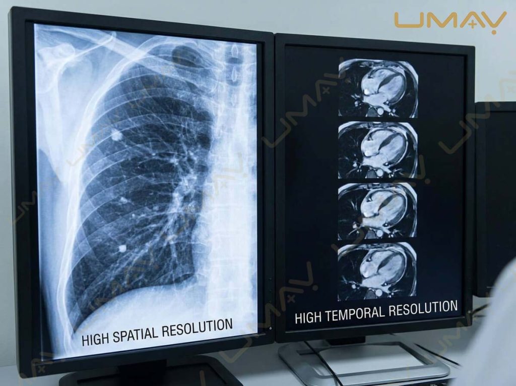
Diagnostic radiology workstation showing chest X-ray and cardiac imaging scans to compare spatial and temporal resolution performance.
5. Decision Framework: Selecting the Right Slice Count for Your Facility
To select the appropriate CT scanner slice count, facility management should evaluate four primary operational metrics:
                  ┌──────────────────────────────────────────┐
                  │    Step 1: Patient Volume & Turnaround   │
                  └────────────────────┬─────────────────────┘
                                       │
                                       ▼
                  ┌──────────────────────────────────────────┐
                  │    Step 2: Core Clinical Workload        │
                  └────────────────────┬─────────────────────┘
                                       │
                                       ▼
                  ┌──────────────────────────────────────────┐
                  │   Step 3: Facility Power & Space Audit   │
                  └────────────────────┬─────────────────────┘
                                       │
                                       ▼
                  ┌──────────────────────────────────────────┐
                  │  Step 4: Total Budget & Tube Lifecycle   │
                  └──────────────────────────────────────────┘
-
Patient Volume: Outpatient clinics imaging 5 to 10 routine cases per day can operate efficiently with a 16-slice scanner. High-volume emergency rooms handling 30+ trauma cases daily require a 64-slice or 128-slice scanner to ensure rapid patient throughput.
-
Clinical Specialization: If your facility specializes in cardiology, neurology, or pediatric care, a 128-slice or 320-slice system is necessary to handle non-invasive vascular studies and low-dose pediatric protocols.
-
Power Infrastructure: Higher slice count systems utilize powerful generators (80–120 kW) that require dedicated 3-phase 480V electrical connections, specialized line transformers, and high-capacity chillers.
-
Capital & Lifecycle Budget: Factor in future maintenance expenses. Higher slice count systems utilize larger X-ray tubes (7.0 to 9.0 MHU) which cost more to replace when tube wear reaches threshold levels.
6. Frequently Asked Questions (FAQ)
Q1: What is the main difference between a 16-slice and 64-slice CT scanner?
A 64-slice CT scanner covers twice the anatomical distance per rotation compared to a 16-slice machine and features faster rotation speeds. This enables 64-slice systems to complete full-body scans in under 10 seconds, making them suitable for emergency trauma and cardiac screening, whereas 16-slice machines are better suited for routine head, chest, and body imaging.
Q2: Is a higher slice count always better for general diagnostic clinics?
Not necessarily. While higher slice counts offer faster scan speeds and cardiac capabilities, they come with higher upfront purchasing costs, increased power requirements, and elevated tube replacement expenses. For general outpatient clinics that do not perform cardiac angiography, a 16-slice or mid-range 64-slice scanner often provides the best return on investment.
Q3: Can a 64-slice CT scanner perform full cardiac scans?
Yes, a 64-slice CT scanner can perform coronary CT angiography (CCTA). However, it usually requires prospective ECG gating and patient heart rate control (typically using beta-blockers) to keep the heart rate below 60–65 bpm during image acquisition.
Q4: How does slice count impact patient radiation dose?
Modern multi-detector CT scanners with high slice counts feature advanced iterative reconstruction and automated exposure control (AEC). Although high-slice scanners emit higher total energy per second, their rapid acquisition speed drastically reduces overall beam exposure time, resulting in lower total radiation doses per exam when optimized correctly.