What Does C-Arm Mean?
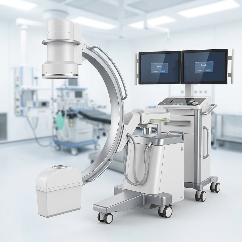
The term C-arm comes from the physical shape of the system.
The X-ray tube and image receptor are mounted on opposite sides of a C-shaped structure. This design lets the system move around the patient while maintaining alignment between the X-ray source and detector.
A simplified imaging path is:
X-ray Tube → Patient → Detector → Image Processing → Monitor
The C-arm structure allows the operator to change the imaging angle without repeatedly repositioning the patient.
That mobility is one of the main reasons C-arms are widely used in operating rooms.
Is a C-Arm an X-Ray Machine?
Yes.
A C-arm is a specialized X-ray imaging system, with many models designed specifically for fluoroscopic imaging.
The U.S. FDA classifies C-arm fluoroscopic X-ray systems within diagnostic X-ray equipment and describes them as systems in which the X-ray tube and image receptor are arranged so the beam direction can be changed relative to the patient.
The main difference is the intended workflow.
Conventional X-Ray
Usually focuses on:
- static radiographic images
- fixed-room examinations
- general diagnostic imaging
C-Arm
Mainly focuses on:
- intraoperative imaging
- fluoroscopy
- image-guided procedures
- mobile operation
- multiple projections during a procedure
Some C-arms can also provide radiographic exposures, depending on the system.
What Is C-Arm Fluoroscopy?
Fluoroscopy is an X-ray imaging method that produces a sequence of images so the medical team can observe anatomy or instruments during a procedure.
This is especially useful when imaging guidance is needed while the patient is already on the operating table.
A simplified workflow is:
X-ray generation → Patient → Detector → Image processing → Live or sequential display
The operator can reposition the C-arm to obtain different projections.
Fluoroscopy can therefore support procedures where the clinician needs imaging during rather than only before or after the intervention.
How Does a C-Arm Work?
The basic working principle is straightforward.
1. X-Rays Are Generated
The X-ray generator supplies power to the X-ray tube.
The tube produces an X-ray beam according to the selected operating settings.
2. The Beam Passes Through the Patient
Different tissues attenuate X-rays differently.
This creates an X-ray pattern that contains information about the anatomy and objects being imaged.
3. The Detector Captures the Signal
The image receptor receives the X-rays after they pass through the patient.
Depending on the C-arm design, this can be:
- flat panel detector
- image intensifier
4. The Signal Is Processed
The system converts and processes the detector signal into an image.
Digital image processing may adjust contrast, brightness, noise, edge information, and other image characteristics.
5. The Image Is Displayed
The processed image appears on the C-arm monitor or workstation.
6. The C-Arm Is Repositioned
The operator can change the angle or position of the C-arm to obtain additional views.
This is particularly useful during orthopedic, trauma, spine, and other procedures.
Main Components of a C-Arm
A C-arm is a complete imaging system rather than a single component.
X-Ray Tube
The X-ray tube creates the X-ray beam.
Relevant specifications can include:
- tube voltage
- tube current
- focal spot
- heat capacity
- exposure modes
The correct tube configuration depends on the intended application and system design.
X-Ray Generator
The generator supplies and controls power for X-ray production.
Depending on the model, the generator may support different fluoroscopy and radiography modes.
Image Detector
The detector receives X-rays and generates the image signal.
Two major configurations are:
Flat Panel Detector
and
Image Intensifier
The detector is one of the major components affecting the imaging architecture.
C-Shaped Arm
The C-arm mechanically connects and positions the X-ray tube and detector.
Its movement determines the available imaging angles and working range.
Collimator
The collimator shapes the X-ray beam and restricts the field to the required region.
Monitor
The monitor displays the fluoroscopic or radiographic image.
Control System
The operator uses the control panel or console to manage imaging functions and system settings.
Mobile Base
Many C-arms are mounted on wheels so that they can be moved between procedure rooms.
What Is a Mobile C-Arm?
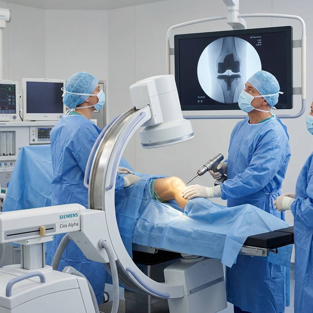
A mobile C-arm is designed to be transported between different locations rather than permanently installed in one imaging room.
A hospital may use a mobile C-arm in:
- operating rooms
- orthopedic rooms
- trauma areas
- procedure rooms
- emergency environments
Its main benefit is flexibility.
A single mobile C-arm can potentially support multiple compatible clinical areas according to the hospital’s workflow.
For this reason, buyers often search for:
- mobile C-arm
- mobile C-arm X-ray machine
- mobile fluoroscopy system
- portable C-arm
- surgical C-arm
- digital mobile C-arm
What Is a Digital C-Arm?
A digital C-arm uses digital image acquisition and computer-based image processing.
Digital systems can provide features such as:
- digital image display
- image storage
- image processing
- image review
- data export
- DICOM-related connectivity on supported systems
The exact capabilities vary between manufacturers and models.
When comparing digital C-arms, it is therefore more useful to check the actual specifications than to rely only on the term “digital.”
What Is an FPD C-Arm?
An FPD C-arm uses a flat panel detector as its image receptor.
FPD means Flat Panel Detector.
Compared with older image-intensifier designs, a flat-panel detector has a different physical and digital architecture and does not have the same image-intensifier shape.
Important FPD-related specifications can include:
- detector size
- active area
- pixel pitch
- resolution
- detector type
- image performance
- weight
The choice between different detector technologies should be based on the intended application and complete system specification.
UMY Medical currently lists an all-in-one mobile C-arm with FPD for orthopedic, urology, and gynecology applications.
What Is an Image Intensifier C-Arm?
An image intensifier C-arm uses an image intensifier as its image receptor.
Image intensifiers have been widely used in fluoroscopy systems.
When comparing image-intensifier and flat-panel systems, buyers may consider:
- image quality
- detector geometry
- system dimensions
- image distortion characteristics
- maintenance
- workflow
- replacement considerations
The exact differences depend on the system design.
What Is a Mini C-Arm?
A mini C-arm is a compact C-arm mainly associated with extremity imaging.
Common applications include:
- hand surgery
- wrist surgery
- foot surgery
- ankle surgery
- orthopedic procedures involving smaller anatomical regions
Its smaller design can make positioning easier when the imaging target is limited to an extremity.
A mini C-arm is therefore not simply a smaller version of every full-size C-arm feature set. Its main value is its suitability for specific procedures.
Mini C-Arm vs Full-Size C-Arm
The choice usually starts with the procedure.
Mini C-Arm
Often considered for:
- hands
- wrists
- feet
- ankles
- extremity procedures
Full-Size Mobile C-Arm
Can support a broader range of procedures, potentially including:
- orthopedics
- trauma
- spine
- urology
- general surgery
A hospital should select the system according to its actual procedural requirements.
Where Are C-Arms Used?
C-arms are most useful where intraoperative or procedural X-ray imaging is required.
Major applications include:
Orthopedic Surgery
C-arms may support:
- fracture fixation
- implant positioning
- screw placement
- bone alignment
- orthopedic procedures
Trauma
Mobile imaging can help provide intraoperative views during trauma and fracture procedures.
Spine Procedures
C-arms can provide imaging guidance during selected spinal and vertebral procedures.
Urology
Fluoroscopy can support selected urological procedures involving devices, positioning, or other image-guided steps.
Pain Management
C-arms can be used for selected fluoroscopy-guided procedures such as certain spinal and pain interventions.
Interventional Procedures
Some C-arms can support interventional applications, although complex procedures may require specialized equipment with additional imaging capabilities.
General Surgery
C-arms can provide intraoperative imaging when the surgical team needs X-ray guidance.
UMY Medical’s current C-arm range identifies applications including orthopedics, urology, gynecology, angiography, and medical imaging, depending on the specific model.
Why Do Hospitals Use C-Arms?
The main reason is intraoperative imaging.
A C-arm can bring X-ray imaging directly to the procedure instead of requiring the patient to be transferred to a separate radiography room for every image.
Potential workflow benefits include:
- imaging at the operating table
- multiple imaging angles
- intraoperative image review
- flexible positioning
- digital image storage on compatible systems
The exact benefit depends on the procedure and how the equipment is integrated into the operating workflow.
C-Arm for Orthopedic Surgery
Orthopedic procedures are among the most common applications for mobile C-arms.
During a procedure, clinicians may need to confirm:
- fracture alignment
- implant location
- screw placement
- bone position
- final fixation
The C-arm can be repositioned to provide different projections.
For orthopedic buyers, useful selection factors include:
- detector size
- image quality
- C-arm movement
- working range
- operating-room clearance
- image display
- workflow
C-Arm for Trauma Surgery
Trauma cases can require rapid imaging while the patient is already positioned for surgery.
A mobile C-arm can support procedures such as:
- fracture fixation
- trauma orthopedics
- implant placement
- intraoperative verification
Mobility is particularly useful when the same system needs to serve different operating rooms.
C-Arm for Spine Procedures
Spine procedures can require imaging from different angles.
A C-arm can support selected procedures where the surgical team needs intraoperative X-ray guidance.
For spine applications, buyers should consider:
- movement range
- detector field of view
- image quality
- operating-table compatibility
- positioning space
C-Arm for Urology
C-arm fluoroscopy can support various urological procedures.
Potential applications include:
- device positioning
- stone-related procedures
- stent-related procedures
- catheter guidance
- other fluoroscopy-guided interventions
The appropriate system depends on the exact procedure and required imaging workflow.
C-Arm for Pain Management
C-arm fluoroscopy can provide image guidance for selected pain-management procedures.
Depending on clinical practice, applications may include:
- spinal injections
- epidural procedures
- facet-related procedures
- other image-guided interventions
For these environments, compact size, positioning flexibility, and image display can be useful considerations.
C-Arm in the Operating Room
An operating room is a challenging environment for mobile imaging equipment.
The C-arm needs to work around:
- operating tables
- surgical lights
- anesthesia equipment
- staff
- other devices
Therefore, physical design is important.
Before purchasing, check:
system dimensions
C-arm depth
movement range
wheelbase
vertical travel
horizontal travel
monitor position
cable management
A good C-arm should be technically suitable and physically practical.
What Is C-Arm Movement?
C-arm movement allows the imaging system to obtain different projections.
Common movement specifications include:
- orbital movement
- vertical movement
- horizontal movement
- rotation
- angulation
The available range varies by model.
For procurement, movement should be evaluated based on the procedures rather than by comparing numbers alone.
A large movement range is useful only when the surgical workflow requires it.
What Is C-Arm Fluoroscopy Mode?
Fluoroscopy displays a sequence of X-ray images during the procedure.
Depending on the system, available modes may include:
- continuous fluoroscopy
- pulsed fluoroscopy
- digital fluoroscopy
- radiographic exposure
- image hold
- image sequence
Not every C-arm provides every mode.
For a purchase project, ask for the exact imaging modes included in the quoted configuration.
Continuous vs Pulsed Fluoroscopy
Continuous Fluoroscopy
X-ray exposure is provided continuously during the selected imaging period.
Pulsed Fluoroscopy
X-rays are produced in pulses.
Pulsed operation can help manage radiation output under appropriate settings, but actual radiation exposure depends on many factors, including:
- pulse rate
- exposure settings
- patient size
- procedure duration
- collimation
- operator technique
Therefore, the word “pulsed” alone should not be treated as a guarantee of a specific dose.
C-Arm Radiation Safety
C-arms use X-rays, so radiation protection must be part of normal operation.
Important practices include:
- appropriate shielding
- correct positioning
- collimation
- suitable exposure settings
- protective equipment
- staff training
- dose monitoring
Radiation safety should follow applicable regulations, institutional procedures, and the manufacturer’s instructions.
When comparing equipment, buyers should look at actual dose-related specifications and understand the test conditions rather than relying only on marketing descriptions.
What Is a C-Arm Generator?
The generator supplies electrical power to the X-ray tube.
Specifications can include:
- maximum power
- tube voltage range
- tube current range
- exposure modes
- pulse settings
The appropriate generator depends on:
- procedure type
- patient population
- imaging mode
- workload
- tube design
More generator power does not automatically mean a better C-arm.
What Is a C-Arm Detector?
The detector converts incoming X-ray information into an image signal.
A C-arm may use:
- flat panel detector
- image intensifier
For digital flat-panel systems, buyers can also compare:
- detector size
- pixel pitch
- active area
- image resolution
- sensitivity
- image processing
These specifications should be evaluated as a group.
What Is C-Arm Image Processing?
Image processing converts detector information into an image suitable for viewing.
Depending on the system, functions can include:
- brightness adjustment
- contrast adjustment
- edge enhancement
- noise reduction
- zoom
- rotation
- annotation
- measurement
- image storage
Software capability varies among manufacturers.
This is why buyers should request a software feature list rather than assuming all digital C-arms offer the same functions.
C-Arm Monitor and Workstation
The monitor and workstation are important parts of the user workflow.
A system may provide:
- one monitor
- dual monitors
- integrated display
- mobile monitor cart
- image review
- patient information
- image storage
- image export
Some digital systems can also support DICOM functions for hospital imaging workflows.
Before purchase, confirm exactly which features are included.
C-Arm and DICOM
DICOM is widely used for medical image communication and management.
A compatible digital C-arm may support functions such as:
- image export
- storage
- worklist
- PACS connection
However, DICOM capabilities vary.
Do not assume that a C-arm with “DICOM support” provides every DICOM service.
Ask the supplier for the exact supported functions.
Can a C-Arm Connect to PACS?
A compatible digital C-arm can connect to PACS through the appropriate software and network configuration.
A simplified workflow can be:
C-Arm → Workstation → DICOM → PACS
Before installation, the hospital IT team should verify:
- network requirements
- IP configuration
- DICOM services
- worklist
- PACS compatibility
- security requirements
C-Arm vs Conventional X-Ray Machine
| Feature | C-Arm | Conventional X-Ray |
|---|---|---|
| Main purpose | Intraoperative/procedural imaging | Diagnostic radiography |
| Mobility | Usually mobile | Often fixed or room-based |
| Fluoroscopy | Common | Usually not the main function |
| Surgical use | Designed for procedures | Limited |
| Imaging angles | Flexible | More fixed |
| Operating room | Common application | Not typically the main setting |
The two systems serve different roles.
A hospital may need both.
C-Arm vs Mobile X-Ray Machine
A mobile X-ray machine and a mobile C-arm are both movable X-ray systems, but their primary purposes differ.
Mobile X-Ray
Commonly used for:
- bedside radiography
- ICU
- emergency imaging
- ward imaging
Mobile C-Arm
Mainly used for:
- surgery
- fluoroscopy
- orthopedic procedures
- image-guided interventions
The C-arm’s defining advantage is its ability to position the X-ray tube and detector around the patient during a procedure.
What Should You Consider Before Buying a C-Arm?
Start with the intended procedures.
Then evaluate:
Clinical Requirements
- procedures
- anatomy
- imaging mode
- required field of view
Equipment
- detector
- generator
- tube
- movement
- monitor
Workflow
- operating-room space
- positioning
- image review
- storage
- PACS
Commercial
- price
- warranty
- service
- spare parts
- delivery
Regulatory
- applicable local requirements
- documentation
- registration
- certification
This process is more useful than choosing a C-arm from a specification list without defining the application.
UMY Medical C-Arm Solutions
UMY Medical Equipment Co., Ltd. is a Guangzhou-based medical equipment provider serving international healthcare markets.
According to the company’s current profile, UMY Medical was established in 2003 as Guangzhou Umy Medical Equipment Co., Ltd. and reports serving more than 1,100 healthcare entities in over 168 countries and regions, supported by more than 30 medical subsidiaries and 18 production bases. The company also states that its portfolio includes more than 1,500 types of medical devices and consumables.
For C-arm sourcing, UMY Medical currently lists several configurations, including:
- mobile C-arm fluoroscopy systems
- FPD C-arms
- high-frequency mobile C-arms
- portable high-frequency C-arms
- digital C-arm X-ray systems
The current product range identifies applications such as orthopedics, urology, gynecology, angiography, and medical imaging across different models.
For international buyers, the company’s broader medical equipment portfolio can also be relevant when a C-arm is part of a larger hospital or healthcare project. UMY Medical states that it supports hospitals, clinics, universities, veterinary institutions, and other healthcare customers, with international and multilingual after-sales support.
Why Supplier Capability Matters
A C-arm is not normally purchased as an isolated consumer product.
A hospital or distributor may also need:
- technical documentation
- product configuration
- installation guidance
- software information
- spare parts
- warranty
- training
- after-sales service
For an international project, logistics and communication can also become important.
This is why supplier evaluation should include both:
the C-arm itself
and:
the supplier’s ability to support the project.
What Information Should You Give a C-Arm Supplier?
For a more accurate quotation, provide:
Application:
Orthopedic / Trauma / Spine / Urology / Pain / Other
System Type:
Mini / Mobile / Full-Size / FPD
Detector:
FPD / Image Intensifier
Generator Requirement:
Based on application
Operating Room:
Available space and table configuration
Movement:
Required orbital / vertical / horizontal / rotation
Imaging:
Fluoroscopy / Radiography / Pulsed Mode
Digital Workflow:
DICOM / PACS / Image Storage
Quantity:
Number of units
Target Market:
Country and applicable requirements
The more specific the inquiry, the easier it is for a supplier to recommend a suitable configuration.
Frequently Asked Questions
What is a C-arm X-ray machine?
A C-arm is a mobile X-ray imaging system that uses a C-shaped structure to position the X-ray tube and image receptor around the patient for fluoroscopic and radiographic imaging.
Why is it called a C-arm?
It is called a C-arm because the main supporting structure is shaped approximately like the letter C.
What is a C-arm used for?
C-arms are commonly used for intraoperative and image-guided procedures, including orthopedic, trauma, spine, urology, pain-management, and other surgical applications.
Is a C-arm an X-ray machine?
Yes. A C-arm is a specialized X-ray imaging system designed primarily for fluoroscopic and procedural imaging.
What is C-arm fluoroscopy?
C-arm fluoroscopy is an X-ray imaging method that produces a sequence of images during a procedure.
How does a C-arm work?
The X-ray tube produces radiation, the beam passes through the patient, the detector captures the remaining X-rays, and the system processes and displays the resulting image.
What are the main components of a C-arm?
The main components include the X-ray tube, generator, detector, C-shaped arm, collimator, monitor, control system, and mobile base.
What is a mobile C-arm?
A mobile C-arm is a C-arm designed to move between operating rooms and other procedural locations.
What is a digital C-arm?
A digital C-arm uses digital image acquisition and computer-based image processing.
What is an FPD C-arm?
An FPD C-arm uses a flat panel detector as its X-ray image receptor.
What is an image intensifier C-arm?
It is a C-arm that uses an image intensifier as its image receptor.
What is a mini C-arm?
A mini C-arm is a compact C-arm commonly designed for extremity and orthopedic procedures.
What is the difference between a mini C-arm and a full-size C-arm?
Mini C-arms are generally focused on extremity imaging, while full-size systems can support a broader range of surgical procedures.
What is the difference between a C-arm and a mobile X-ray machine?
A mobile X-ray machine is mainly designed for portable radiography, while a C-arm is designed for fluoroscopy and intraoperative image guidance.
What is the difference between a C-arm and a fixed X-ray machine?
A fixed X-ray machine is generally installed in a dedicated radiography room, while a mobile C-arm is designed to move around the patient during surgical or procedural imaging.
What is a C-arm used for in orthopedic surgery?
It can provide intraoperative imaging for fracture fixation, implant positioning, screw placement, bone alignment, and other orthopedic procedures.
Can a C-arm be used for trauma surgery?
Yes. Mobile C-arms can provide imaging during trauma and fracture procedures.
Can a C-arm be used for spine surgery?
Yes. C-arms can support selected spine and vertebral procedures requiring intraoperative X-ray guidance.
Can a C-arm be used for urology?
Yes. C-arm fluoroscopy can support selected urological procedures.
Can a C-arm be used for pain management?
Yes. C-arm fluoroscopy can provide image guidance for selected pain-management procedures.
Can a C-arm be used for vascular procedures?
Some C-arms can support vascular or interventional applications, but more advanced procedures may require specialized imaging systems.
What is the difference between an FPD C-arm and an image intensifier C-arm?
The main difference is the detector technology. FPD systems use a flat panel detector, while image-intensifier systems use an image intensifier.
What is a C-arm detector?
It is the image receptor that captures X-rays after they pass through the patient.
What is C-arm generator power?
It describes the power capability of the X-ray generation system. The appropriate level depends on the intended procedure and system configuration.
What is C-arm orbital movement?
It is movement of the C-arm that changes the direction of the X-ray beam relative to the patient.
Why does C-arm movement matter?
Movement allows the operator to obtain different imaging projections during a procedure.
What is pulsed fluoroscopy?
Pulsed fluoroscopy generates X-rays in pulses rather than continuously.
Does pulsed fluoroscopy always mean lower radiation?
Not automatically. Actual radiation exposure depends on the system settings, patient, procedure, fluoroscopy time, collimation, and operator technique.
Can a C-arm connect to PACS?
A compatible digital system can connect to PACS using supported DICOM functions.
Does every C-arm support DICOM?
No. DICOM functions vary between models.
What is an FPD in a C-arm?
FPD stands for Flat Panel Detector. It is a digital X-ray image receptor used in compatible C-arm systems.
What size detector does a C-arm need?
The appropriate detector size depends on the procedure, anatomy, C-arm design, and required field of view.
What should I consider when buying a C-arm?
Start with the procedures, then evaluate system type, detector, generator, movement, imaging modes, operating-room space, software, connectivity, warranty, and service.
What should I ask a C-arm supplier?
Ask about the detector, generator, movement range, fluoroscopy modes, image processing, DICOM/PACS, compatibility, warranty, spare parts, installation, and technical support.
How do I request a C-arm quotation?
Provide the intended applications, preferred system type, detector technology, quantity, software requirements, target country, and any existing equipment that the C-arm needs to integrate with.
C-Arm Buying Checklist
Before placing an order, confirm:
Clinical
□ Intended procedures
□ Patient types
□ Required imaging modes
□ Field of view
System
□ Mobile or mini
□ FPD or image intensifier
□ Generator
□ X-ray tube
□ Collimator
Movement
□ Orbital
□ Vertical
□ Horizontal
□ Rotation
□ Angulation
Digital
□ Monitor
□ Workstation
□ Image processing
□ DICOM
□ PACS
□ Storage
Operating Room
□ Table compatibility
□ Floor space
□ C-arm clearance
□ Wheel movement
□ Cable management
Commercial
□ Price
□ Accessories
□ Warranty
□ Spare parts
□ Installation
□ Training
□ Technical support
Conclusion
A C-arm X-ray machine is a mobile imaging system designed primarily for fluoroscopy and intraoperative procedures.
Its basic structure is simple:
X-ray Tube → Patient → Detector → Image Processing → Monitor
The C-shaped arm gives the system its defining flexibility, allowing the X-ray tube and detector to move around the patient and provide different imaging projections.
C-arms are widely associated with:
- orthopedic surgery
- trauma
- spine procedures
- urology
- pain management
- interventional imaging
- general surgery
Depending on the model, a system may use an FPD or image intensifier, support fluoroscopy and radiography, and provide digital workflow features such as image storage and supported DICOM connectivity.
For buyers, however, the most important question is not simply which C-arm has the longest specification sheet.
Start with:
What procedures will the system support?
Then evaluate:
C-arm type → detector → generator → movement → imaging modes → operating-room fit → digital workflow → service
UMY Medical Equipment Co., Ltd. currently offers several mobile C-arm configurations and serves international healthcare customers across a broad medical equipment portfolio.
For hospitals, clinics, distributors, and healthcare projects, selecting the right C-arm means matching the equipment to the clinical application, operating environment, imaging requirements, and long-term support needs.
Start with the procedure. Choose the C-arm around the workflow.