1. Introduction: The Evolution of Point-of-Care Ultrasound (POCUS)
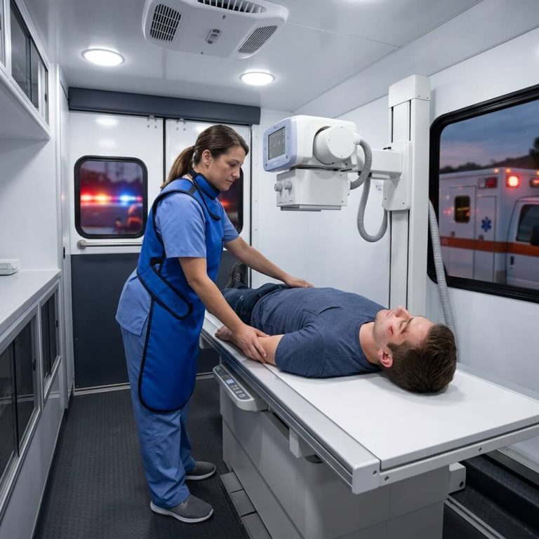
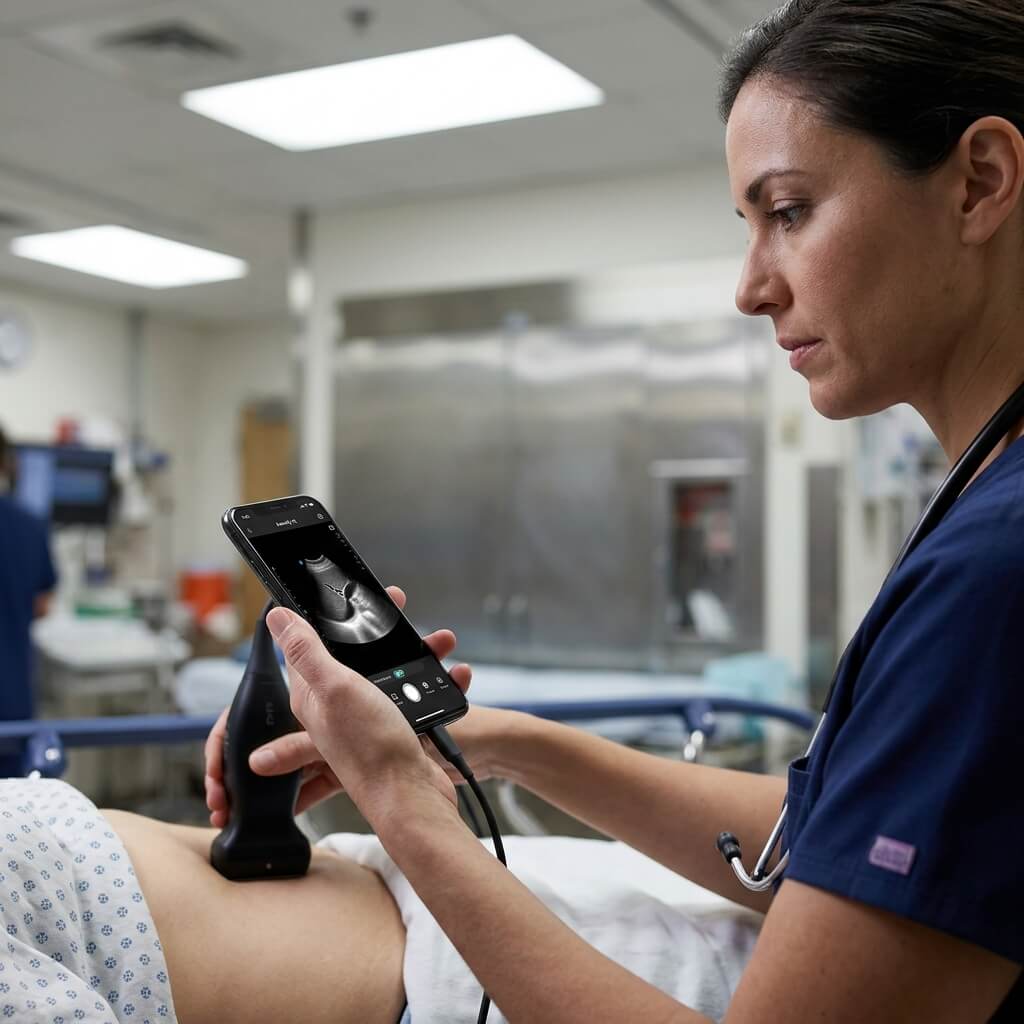
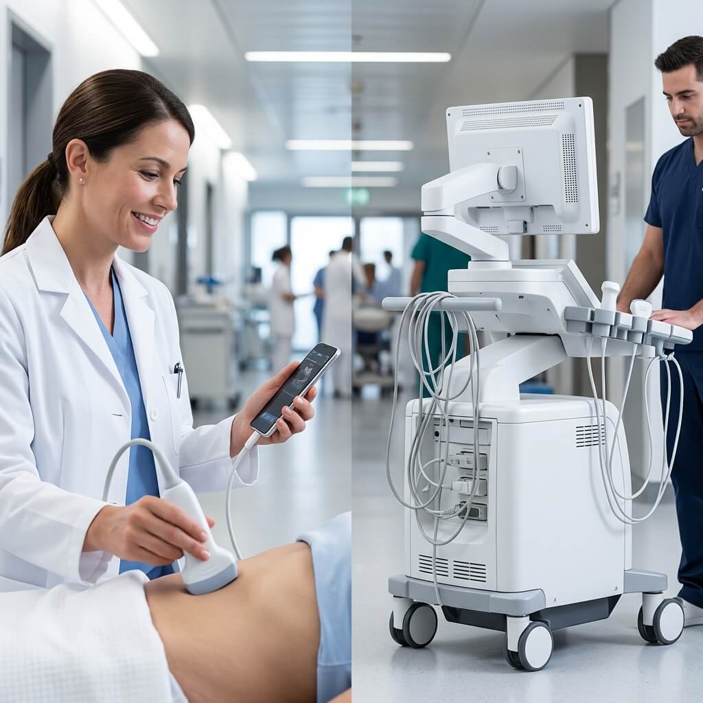
Point-of-care ultrasound (POCUS) has transformed bedside diagnostics across modern medicine. What once required a bulky 100-kilogram cart-based ultrasound console can now be accomplished with a pocket-sized handheld ultrasound scanner connected directly to a smartphone, tablet, or medical display.
                     POCUS PLATFORM EVOLUTION
  +-----------------------+     +-----------------------+     +-----------------------+
  |  Traditional Console  | --> |   Laptop Portable     | --> |   Hand-held Wireless  |
  |  Heavy, Stationed     |     |  10-15 lbs, Wheel Cart|     |  200-300g, Pocket Fit |
  +-----------------------+     +-----------------------+     +-----------------------+
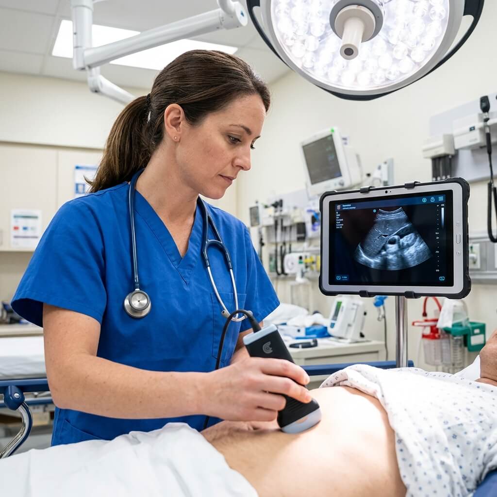
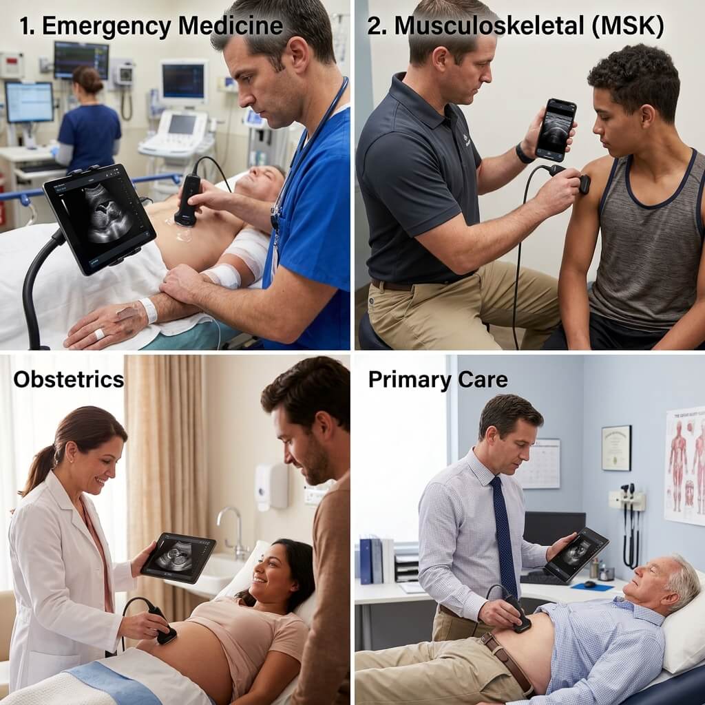
Handheld ultrasound devices empower emergency room physicians, general practitioners, anesthesiologists, vascular surgeons, and field medics to perform real-time diagnostic scans anywhere. Whether assessing internal bleeding during trauma triage, guiding needle placement for joint injections, or monitoring fetal biometry in remote clinics, pocket ultrasound probes deliver immediate clinical clarity.
However, with dozens of wireless ultrasound probes, pocket transducers, and smart ultrasound app platforms on the market, choosing the right system can be complex. This buyer’s guide outlines key hardware specifications, probe configurations, software workflows, and procurement criteria to help clinicians and healthcare buyers make informed decisions.
2. Core Transducer Architecture: Probe Types & Frequency Selection
Selecting the correct transducer frequency and acoustic design is crucial for ultrasound image quality. Different body tissues require specific acoustic wave penetration and spatial resolution.
                  TRANSDUCER FREQUENCY & DEPTH RELATIONSHIP
  +-------------------------------------------------------------------------+
  | High Frequency (7.5 - 18 MHz)  --> Shallow Depth (2 - 10 cm)  --> Linear  |
  | Mid Frequency  (2.5 - 6.0 MHz) --> Deep Depth    (9 - 30 cm)  --> Convex  |
  | Low Frequency  (2.0 - 4.0 MHz) --> Cardiac Deep (10 - 24 cm)  --> Phased  |
  +-------------------------------------------------------------------------+
Linear Transducers (7.5 MHz to 18 MHz)
-
Best Used For: Superficial structures, vascular access guidance, musculoskeletal (MSK) diagnostics, thyroid, breast, nerve blocks, and facial aesthetics.
-
Scanning Depth: Shallow depth range (typically 2 cm to 10 cm).
-
Key Benefit: High operating frequencies deliver fine spatial resolution, rendering clear tissue layers, small blood vessels, and needle trajectories.
Convex / Curved Array Transducers (2.0 MHz to 5.0 MHz)
-
Best Used For: Abdominal exams, liver and kidney screening, obstetrics and gynecology (OB/GYN), bladder volume measurements, and FAST trauma protocols.
-
Scanning Depth: Deep penetration range (typically 9 cm to 30 cm).
-
Key Benefit: The wide, curved sector field of view allows clinicians to visualize deep visceral organs through abdominal tissue barriers.
Phased Array Transducers (2.0 MHz to 4.0 MHz)
-
Best Used For: Bedside echocardiography, cardiac chamber assessment, stroke volume estimations, and intercostal lung imaging.
-
Scanning Depth: Deep thoracic penetration (10 cm to 24 cm).
-
Key Benefit: A compact acoustic footprint fits easily between rib spaces (intercostal windows) without rib shadowing blocking the cardiac view.
Next-Gen 3-in-1 Tri-Mode & Dual-Head Probes
To avoid purchasing and carrying multiple physical transducers, modern buyers increasingly choose 3-in-1 multipurpose handheld ultrasound probes.
-
Tri-Mode (3-in-1) Probes: Integrate convex, linear, and phased array capabilities into a single lightweight handle. Clinicians can switch software presets from abdominal to vascular or cardiac modes instantly without changing devices.
-
Dual-Head Probes: Feature a double-sided acoustic head (e.g., Convex on one end, Linear on the opposite end). Turning the probe upside down switches between deep abdominal scans and superficial vascular mapping.
3. Key Hardware Specifications & Selection Criteria
When evaluating technical spec sheets from handheld ultrasound suppliers, pay close attention to the following parameters:
                      ESSENTIAL HARDWARE EVALUATION
+---------------------------------------------------------------------------+
| 1. ELEMENT COUNT  --> 128, 192, or 256 Elements (Higher = Sharper Image)  |
| 2. CONNECTIVITY   --> 5GHz Wireless WiFi / Direct USB Type-C Cable        |
| 3. IMAGING MODES  --> B-Mode, M-Mode, Color Doppler, Power Doppler, PW    |
| 4. BATTERY & HEAT --> 3+ Hours Continuous Scan, Waterproof IP67 Casing    |
+---------------------------------------------------------------------------+
Transducer Element Count (64 vs. 128 vs. 192 vs. 256 Elements)
The element count refers to the number of individual piezoelectric crystal channels housed inside the probe head.
-
64 Elements: Entry-level or basic guidance probes. Suitable for simple line placement, but image resolution can be noisy.
-
128 Elements: The industry standard for high-performance point-of-care ultrasound. Offers balanced image clarity, gray-scale contrast, and tissue differentiation.
-
192 / 256 Elements: Premium diagnostic grade. Delivers crisp border definition, low speckle noise, and high detail required for nerve tracking, MSK tendon assessment, and micro-vascular flow analysis.
Wireless WiFi vs. Wired USB Connectivity
-
Wireless Handheld Probes: Connect to iOS or Android smart devices via an onboard high-speed 5GHz WiFi hot-spot. Wireless probes offer complete freedom of movement without cables interfering with sterile surgical fields or emergency procedures.
-
Wired USB-C Probes: Connect directly to phones, tablets, or Windows laptops via standard USB Type-C cables. Wired probes eliminate potential wireless interference in crowded hospital environments and draw power directly from the host display device.
Imaging Modes & Feature Set
A professional handheld diagnostic ultrasound must support a complete suite of ultrasound display modes:
-
B-Mode (Brightness Mode): Standard 2D grayscale anatomical imaging.
-
M-Mode (Motion Mode): Tracks movement over time, essential for fetal heart rate assessment and cardiac valve motion.
-
Color Doppler: Visualizes blood flow direction and velocity inside arteries and veins.
-
Power Doppler (PDI): Highly sensitive to low-velocity blood flow, ideal for detecting inflammation, tumor vascularity, and small organ perfusion.
-
Pulsed Wave (PW) Doppler: Measures precise hemodynamic flow velocities and spectral waveforms across vascular structures.
Battery Endurance, Thermal Management & IP Rating
-
Continuous Operating Time: Look for integrated lithium-ion batteries that provide at least 2.0 to 4.0 hours of active continuous scanning. Fast USB-C or wireless induction charging docks ensure the probe is ready for emergency shifts.
-
Passive Heat Dissipation: High-element probes produce heat during extended Color Doppler sessions. Quality probes use lightweight magnesium or aluminum alloy housings to dissipate heat safely without built-in cooling fans that draw dust.
-
IP Ingress Protection: Choose probes rated at IP67 or IP68 waterproof protection. Fully submersible probes can be easily disinfected, wiped down, or chemically sterilized between patients.
4. Software Compatibility, DICOM PACS & Subscription Models
Hardware is only half the equation. The operating app and software integration define daily workflow efficiency.
                      SOFTWARE WORKFLOW INTEGRATION
+---------------------------------------------------------------------------+
| PROBE SENSORS --> Mobile Imaging App (iOS / Android / Windows)            |
|                        |                                                  |
|                        v                                                  |
| ONBOARD TOOLS --> Auto-Measurements (EF, Bladder, Vascular Indices)       |
|                        |                                                  |
|                        v                                                  |
| NETWORKING    --> Encrypted Wireless DICOM Export to Hospital PACS / Cloud|
+---------------------------------------------------------------------------+
App Ecosystem & Device Compatibility
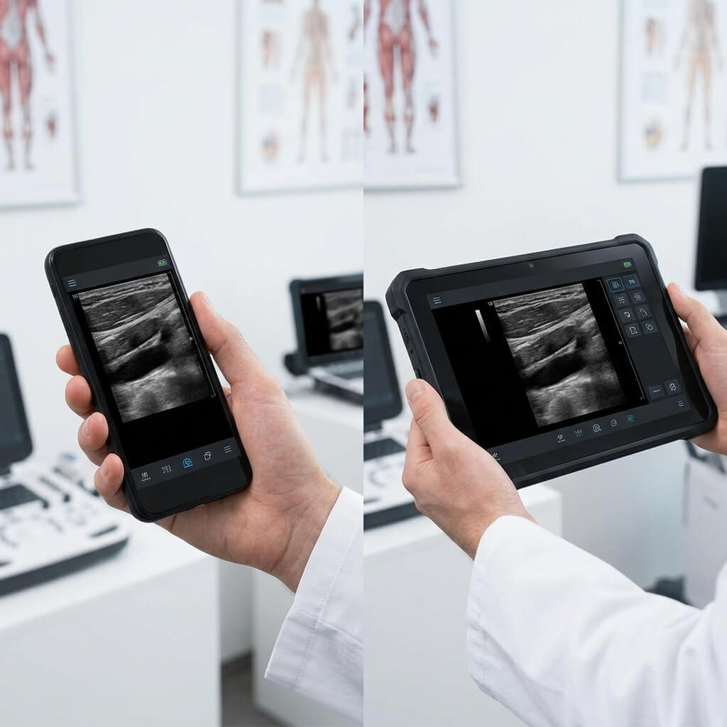
Ensure the manufacturer’s application installs smoothly across multiple platforms, including iOS (iPhone/iPad), Android (tablets/phones), and Windows OS. The user interface should feature intuitive touch-screen controls for gain adjustment, depth tuning, focus positioning, freeze-frame, and image annotation.
DICOM PACS & EHR Integration
In hospital settings, diagnostic images and patient reports must seamlessly export to local or cloud-based PACS (Picture Archiving and Communication System) and Electronic Health Record (EHR) networks. Look for native DICOM 3.0 compatibility, enabling structured report exports, store-and-forward image transmission, and worklist management via secure encrypted WiFi.
Watch Out for Hidden Software Subscriptions
Some global brand handheld ultrasound vendors sell hardware at a lower initial price point but require mandatory annual software subscription fees to unlock basic scanning modes, Color Doppler features, or DICOM exports.
For public health institutions, medical distributors, and private practices, choosing a supplier with an open, subscription-free software policy saves thousands of dollars in long-term operating costs.
5. Handheld Ultrasound Technical Comparison Matrix
To assist procurement managers, the table below compares key structural configurations commonly found across the market:
| Feature Specification | Standard Wireless Single-Head | High-Frequency Linear MSK | Multi-Head Dual-End Probe | Tri-Mode 3-in-1 Transducer |
| Primary Clinical Use | General Abdominal / OB | Vascular, MSK, Nerve | Abdominal + Vascular | Full-Body POCUS (ER/ICU) |
| Probe Architecture | Single Convex or Linear | High-Density Linear | Double-Sided Head | Convex + Linear + Phased |
| Frequency Range | 2.5 MHz – 5.0 MHz | 7.5 MHz – 18.0 MHz | 3.5 MHz / 10.0 MHz | 2.5 MHz – 12.0 MHz |
| Piezo Element Count | 64 or 128 Elements | 128 or 192 Elements | 128 Elements per head | 128 or 192 Elements |
| Scan Depth Range | 90 mm – 305 mm | 20 mm – 100 mm | 20 mm – 220 mm | 20 mm – 305 mm |
| Display Modes | B, M, Color, PW | B, M, Color, PDI, PW | B, M, Color, PDI, PW | B, M, Color, PDI, PW |
| Connectivity Type | 5GHz WiFi / USB-C | 5GHz WiFi / USB-C | 5GHz WiFi Wireless | 5GHz WiFi Wireless |
| Average Device Weight | 220g – 260g | 200g – 250g | 250g – 280g | 250g – 300g |
6. Why UMY Medical Equipment Co Ltd Leads Handheld Ultrasound Manufacturing
When sourcing medical equipment for hospitals, private clinics, or international distribution networks, partnering with an established direct-factory manufacturer ensures consistent quality and long-term technical support.
                      UMY MEDICAL MANUFACTURING STANDARDS
                                       |
       +-------------------------------+-------------------------------+
       |                               |                               |
       v                               v                               v
[ ISO 13485 & CE Directives ]   [ High-Element Crystal Array ]  [ Open DICOM & No Subscriptions ]
 Full Quality Management System   128 / 192 Element High Res      Zero Paywalls, Lifetime Updates
Direct Factory Excellence & Global Credibility
-
ISO 13485 & CE Quality Certification: UMY Medical Equipment Co Ltd manufactures pocket ultrasound scanners in ISO 13485 certified facilities, adhering to strict international medical device manufacturing directives and CE safety standards.
-
Advanced 128 & 192 Element Micro-Array Technology: UMY Medical utilizes high-density piezoelectric crystal arrays, delivering clear grayscale contrast, sensitive Color Doppler flow detection, and deep signal penetration rivaling conventional console units.
-
Subscription-Free, Open System Policy: Unlike brand vendors that charge monthly software fees, UMY Medical provides lifetime app updates, open DICOM 3.0 export capabilities, and full mode access without hidden paywalls.
-
OEM / ODM Customization & B2B Solutions: As a trusted original equipment manufacturer, UMY Medical supports global distributors and hospital chains with custom brand logo integration, software language localization, specialized probe presets, and factory-direct pricing.
Frequently Asked Questions (FAQ)
Q1: Can a handheld ultrasound machine replace a traditional hospital cart system?
Handheld ultrasound systems excel at point-of-care screening, needle guidance, emergency triage, and bedside diagnostics. While premium cart-based machines are still used for complex cardiology studies and deep radiological evaluations, handheld units handle over 80% of routine clinical diagnostic tasks faster and at a fraction of the cost.
Q2: How long does the battery last on a wireless handheld ultrasound probe?
Most wireless handheld ultrasound probes feature lithium-ion batteries providing 2 to 4 hours of active continuous scanning. In typical clinical workflows (where individual scans take 3 to 5 minutes), a single charge easily lasts an entire clinical shift.
Q3: What is the difference between B-Mode and Color Doppler mode on a pocket scanner?
B-Mode (Brightness Mode) provides a 2D grayscale anatomical image of organs and tissues. Color Doppler overlays real-time color signals onto the 2D image to indicate the direction and velocity of blood flow through vessels, heart valves, or vascular lesions.
Q4: Are handheld ultrasound scanners waterproof and easy to sterilize?
Yes. Quality handheld scanners built by manufacturers like UMY Medical feature IP67 or IP68 ingress protection. The seamless casing allows full immersion in disinfectant solutions or wipe-downs with medical-grade germicidal sprays without damaging internal electronics.
Q5: How do I export diagnostic images from a handheld probe to a hospital PACS network?
Modern handheld ultrasound apps connect directly to local hospital WiFi networks. Clinicians select DICOM export to send patient images, loop recordings, and measurement reports directly to the facility’s PACS server or cloud storage with a single tap.