Image Processing Software: The Brain Behind Every Digital X-Ray System
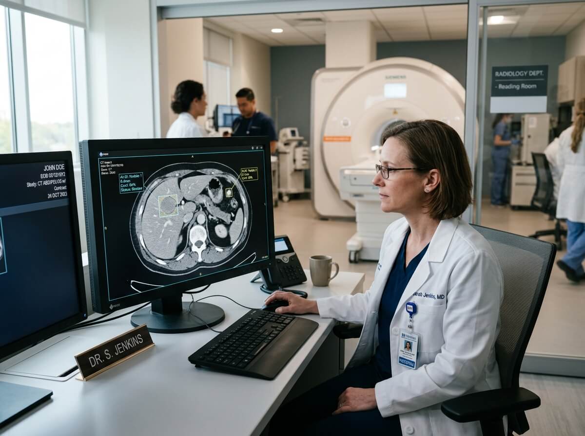
While the X-ray generator and flat panel detector capture the image, it is the image acquisition software and image processing algorithms that transform raw data into clear, diagnostic-quality images.
Modern digital radiography software automatically optimizes image quality within seconds, allowing radiographers and physicians to make faster and more accurate clinical decisions.
When evaluating a digital X-ray system, buyers should not only compare hardware specifications but also assess the capabilities of the software platform.
Key software features include:
- Automatic image stitching
- Bone suppression technology
- Multi-frequency image enhancement
- Intelligent noise reduction
- Dynamic range optimization
- Automatic exposure detection
- Anatomical image optimization
- AI-assisted positioning
- Image annotation
- Measurement tools
- Report generation
- Image export and sharing
A user-friendly software interface reduces training time and improves workflow, especially in busy radiology departments.
Artificial Intelligence Is Changing Digital Radiography
Artificial Intelligence (AI) is becoming an increasingly valuable tool in medical imaging.
Although AI does not replace radiologists, it helps improve efficiency by assisting with image analysis and workflow management.
Many modern DR systems now include AI-powered functions such as:
- Automatic patient positioning guidance
- Intelligent exposure parameter recommendations
- Bone suppression imaging
- Chest abnormality detection
- Pneumothorax screening
- Fracture detection assistance
- Image quality assessment
- Automatic anatomy recognition
These technologies reduce repeat examinations, improve consistency, and support faster diagnosis.
Hospitals investing in AI-ready imaging systems are better prepared for future developments in digital healthcare.
Understanding PACS, RIS, HIS, and DICOM Integration
A high-quality digital X-ray machine should integrate seamlessly with existing hospital information systems.
Without proper integration, image management becomes inefficient and increases the risk of workflow errors.
What Is DICOM?
DICOM (Digital Imaging and Communications in Medicine) is the international standard for storing, transmitting, and displaying medical images.
Almost every modern digital radiography system supports DICOM communication.
DICOM allows hospitals to:
- Store diagnostic images
- Transfer images between departments
- Share examinations with specialists
- Enable tele-radiology services
- Archive patient records securely
Compatibility with DICOM is essential for hospitals planning long-term digital transformation.
PACS (Picture Archiving and Communication System)
PACS eliminates the need for printed films by storing images electronically.
Benefits include:
- Instant image retrieval
- Secure long-term storage
- Remote image review
- Improved collaboration
- Reduced film costs
- Faster diagnosis
Cloud-based PACS solutions further improve accessibility for multi-site healthcare organizations.
RIS (Radiology Information System)
RIS manages the administrative side of radiology departments.
Typical functions include:
- Patient scheduling
- Examination management
- Report tracking
- Radiologist workload management
- Billing support
- Department statistics
When integrated with a DR system, RIS helps improve workflow efficiency and reduces administrative errors.
HIS (Hospital Information System)
The Hospital Information System connects all departments within a healthcare facility.
A digital X-ray machine that integrates with HIS enables automatic sharing of patient demographics, examination requests, and imaging reports, reducing duplicate data entry and improving operational efficiency.
Radiation Safety and Dose Management
Patient safety is one of the most important considerations when selecting an X-ray system.
Modern digital radiography equipment is designed to produce excellent diagnostic images while minimizing radiation exposure.
Radiation dose management should always follow the ALARA Principle — As Low As Reasonably Achievable.
Hospitals can reduce unnecessary exposure through:
- High-efficiency flat panel detectors
- Automatic exposure control (AEC)
- Intelligent image processing
- Proper patient positioning
- Routine equipment calibration
- Staff training
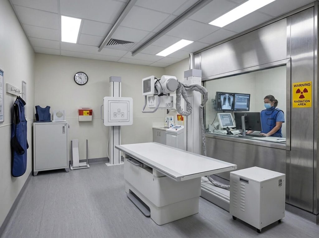
A lower radiation dose benefits both patients and healthcare professionals, particularly in pediatric imaging and high-volume departments.
Radiation Protection Equipment
Every radiology department should be equipped with appropriate radiation protection accessories.
Common protective equipment includes:
- Lead aprons
- Thyroid shields
- Lead gloves
- Lead glasses
- Mobile lead barriers
- Radiation warning signs
- Personal dosimeters
These accessories help protect healthcare workers from unnecessary exposure during daily clinical practice.
X-Ray Room Planning and Installation Requirements
Purchasing a digital X-ray machine also requires careful planning of the installation environment.
Proper room design ensures safe operation, efficient workflow, and compliance with local regulations.
Before installation, hospitals should evaluate:
- Available room dimensions
- Ceiling height
- Floor load capacity
- Electrical supply
- Grounding system
- Network infrastructure
- Air conditioning
- Lighting conditions
- Radiation shielding requirements
A well-designed radiology room improves patient comfort while allowing radiographers to work more efficiently.
Electrical and Network Requirements
Most digital radiography systems require a stable electrical supply and reliable network connection.
Typical installation requirements include:
- Stable power voltage
- Independent electrical circuit
- UPS (Uninterruptible Power Supply)
- Gigabit Ethernet connection
- Hospital network access
- PACS connectivity
- Internet access for remote service
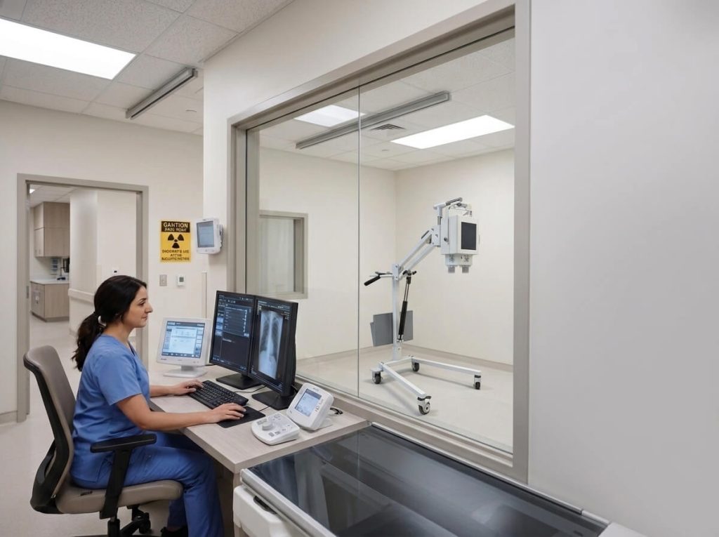
Proper infrastructure reduces downtime and simplifies future software upgrades.
Daily Maintenance Best Practices
Routine maintenance extends equipment life while maintaining image quality.
Daily maintenance should include:
- Cleaning detector surfaces
- Inspecting cables
- Checking tube movement
- Verifying workstation operation
- Reviewing system error logs
- Performing detector calibration if required
- Cleaning touch screens and keyboards
- Inspecting emergency stop functions
These simple procedures help reduce unexpected equipment failures.
Preventive Maintenance
In addition to daily inspections, manufacturers typically recommend scheduled preventive maintenance.
Preventive maintenance often includes:
- Generator testing
- Tube performance evaluation
- Detector calibration
- Mechanical alignment
- Software updates
- Safety inspections
- Image quality testing
Regular preventive maintenance minimizes downtime and protects the hospital’s investment.
Common X-Ray Machine Problems
Even high-quality systems may occasionally experience technical issues.
Some common problems include:
- Blurry images
- Detector communication failure
- Exposure errors
- Tube overheating
- Image artifacts
- Calibration errors
- Software connection issues
- PACS communication failure
Working with an experienced manufacturer that provides remote technical support and readily available spare parts can significantly reduce service interruptions.
Choosing a System That Supports Future Growth
Medical imaging technology continues to evolve rapidly.
When purchasing a new DR system, hospitals should consider not only current requirements but also future expansion.
Look for systems that support:
- AI software upgrades
- Additional detector compatibility
- Cloud PACS integration
- Remote diagnostics
- Multi-workstation networking
- Future software licensing
- Expansion to multiple imaging rooms
A scalable digital radiography system protects your investment and ensures your imaging department can adapt to future clinical and technological developments.