The Technological Evolution of Bone Densitometry
Selecting the right bone densitometer requires evaluating beam geometry, diagnostic accuracy, radiation output, and software capabilities. While Dual-Energy X-Ray Absorptiometry (DXA / DEXA) remains the recognized gold standard for bone mineral density (BMD) measurement, alternative modalities like Quantitative Ultrasound (QUS) and Radiofrequency Echographic Multi Spectrometry (REMS) serve distinct roles in clinical screening and point-of-care settings.
For radiologists, medical directors, and B2B procurement officers, understanding the technical specifications and clinical tradeoffs among these technologies ensures optimal capital deployment and patient care.
+-----------------------------------------------------------------------------------+
|                     BONE DENSITOMETRY TECHNOLOGY LANDSCAPE                        |
+-----------------------------------------------------------------------------------+
| IONIZING RADIATION MODALITIES       | NON-IONIZING MODALITIES                     |
| - Pencil-Beam Central DXA           | - Quantitative Ultrasound (QUS)             |
| - Fan-Beam Central DXA              | - Radiofrequency Echographic Multi          |
| - Narrow-Angle Fan-Beam DXA         |   Spectrometry (REMS)                       |
| - Peripheral DXA (pDXA)             | - Peripheral QUS (pQUS)                     |
+-----------------------------------------------------------------------------------+
1. DXA Beam Technologies: Pencil Beam vs. Fan Beam vs. Narrow-Angle Fan Beam
Central DXA scanners utilize different X-ray beam configurations, directly impacting scan duration, spatial resolution, and radiation dosage.
                   DXA BEAM GEOMETRY ARCHITECTURE
                                  |
     +----------------------------+----------------------------+
     |                            |                            |
Pencil Beam                  Fan Beam                     Narrow-Angle Fan Beam
- Single X-ray point source  - Wide fan-shaped beam       - Multi-detector linear array
- Pin-point detector array   - Multi-element detector row - Short focal distance
- Scan time: 2–5 minutes     - Scan time: 10–30 seconds   - Reduced geometric parallax
- Lowest radiation dose      - Highest spatial resolution - Balanced speed & precision
Pencil-Beam DXA Technology
-
Operational Mechanism: A single X-ray beam pair moves synchronously with a single detector across the target area in a raster (zigzag) pattern.
-
Key Advantages: Ultra-low radiation dose and minimal geometric distortion (magnification error).
-
Limitations: Slower scan speeds (2 to 5 minutes per site) and lower spatial resolution compared to modern fan-beam units.
-
Best Suited For: Low-to-medium throughput clinics prioritizing minimal radiation exposure and lower hardware purchase costs.
Fan-Beam DXA Technology
-
Operational Mechanism: Uses a wide, fan-shaped X-ray beam coupled with a multi-element linear detector array to acquire an entire anatomical line in a single sweep.
-
Key Advantages: Extremely fast scan acquisition (10 to 30 seconds), high-definition imaging, and superior edge detection for automated region-of-interest (ROI) placement.
-
Limitations: Higher radiation output and slight geometric parallax distortion at the edges of the image area, requiring software correction.
-
Best Suited For: High-volume diagnostic imaging centers, tertiary hospitals, and sports science facilities performing advanced body composition scans.
Narrow-Angle Fan-Beam DXA Technology
-
Operational Mechanism: Combines multiple narrow fan-beam sweeps with intermediate detector arrays, capturing anatomical sites with reduced angular dispersion.
-
Key Advantages: Significantly reduces magnification distortion compared to wide fan-beam systems while maintaining rapid scan times (under 1 minute).
-
Limitations: Moderately higher cost than basic pencil-beam systems.
-
Best Suited For: Mid-to-high volume orthopedic and endocrinology practices requiring a balance of speed, precision, and geometric accuracy.
2. Non-Ionizing Alternatives: Quantitative Ultrasound (QUS) & REMS Technology
For screening programs, pediatric care, and settings where ionizing radiation is restricted or impractical, ultrasound-based technologies provide valuable diagnostic data.
Quantitative Ultrasound (QUS)
-
Technology Overview: Measures the attenuation and velocity of high-frequency sound waves through peripheral bone structures (most commonly the calcaneus/heel).
-
Primary Metrics:
-
Speed of Sound : Reflects bone density and structural elasticity.
-
Broadband Ultrasound Attenuation : Measures ultrasound energy loss absorbed by trabecular bone structure.
-
Stiffness Index (SI) / Quantitative Ultrasound Index (QUI): Derived composite metrics used to calculate clinical T-scores for pre-screening.
-
-
Clinical Application: Community health screenings, pharmacy health checks, and initial triage before ordering central DXA scans.
Radiofrequency Echographic Multi Spectrometry (REMS)
-
Technology Overview: Uses high-frequency ultrasound signals directed at central sites (lumbar spine and proximal femur).
-
Mechanism: Analyzes unfiltered raw radiofrequency (RF) signals acquired during an echographic scan, comparing spectral signatures against normative database models to determine bone density.
-
Clinical Application: Radiation-free central bone density assessment suitable for pregnant women, pediatric patients, and frequent therapeutic monitoring.
3. Trabecular Bone Score (TBS) & Advanced Software Modules
Modern bone densitometers rely on specialized software algorithms to enhance fracture prediction beyond basic bone mineral density.
                    ADVANCED DXA DIAGNOSTIC SOFTWARE
                                   |
       +---------------------------+---------------------------+
       |                                                       |
Trabecular Bone Score (TBS)                   Vertebral Fracture Assessment (VFA)
- Evaluates bone microarchitecture            - High-resolution lateral spine scan
- Textural analysis from spine DXA            - Identifies asymptomatic vertebral collapses
- Independent predictor of fracture risk      - Replaces standard lateral spine X-rays
-
Trabecular Bone Score (TBS): An analytical software module that assesses grey-level spatial variations from standard lumbar spine DXA images. Higher TBS values indicate stronger, dense microarchitecture; lower values indicate degraded, fragile bone structure—even in patients with normal T-scores.
-
Vertebral Fracture Assessment (VFA): A low-dose lateral imaging scan performed on central DXA machines to detect silent vertebral fractures, critical for accurate clinical risk staging.
-
Atypical Femur Fracture (AFF) Assessment: High-speed lateral femur scanning to identify periosteal thickening and early stress fractures caused by long-term bisphosphonate therapy.
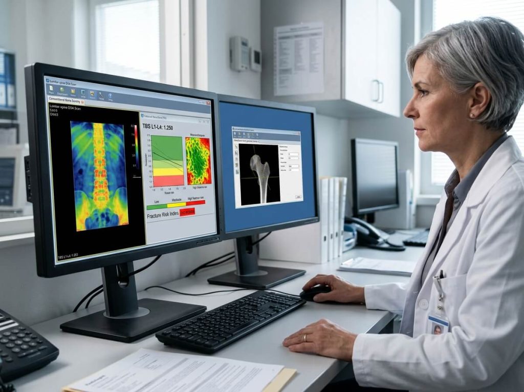
Female radiologist studying spine bone density scans and Trabecular Bone Score analysis on medical workstation monitors.
4. Technical Specification & Feature Comparison Matrix
| Specification / Feature | Pencil-Beam DXA | Fan-Beam DXA | Quantitative Ultrasound (QUS) | REMS Technology |
| Energy Source | Dual X-Ray | Dual X-Ray | High-Frequency Ultrasound | Raw Radiofrequency Ultrasound |
| Ionizing Radiation | Yes (Ultra-Low) | Yes (Low) | None (0 Radiation) | None (0 Radiation) |
| Scan Time (Spine/Hip) | 2 to 5 minutes | 10 to 30 seconds | 15 to 30 seconds (Heel) | 1 to 2 minutes |
| Anatomical Sites | Hip, Spine, Forearm | Hip, Spine, Forearm, Whole Body | Calcaneus (Heel), Radius | Lumbar Spine, Femoral Neck |
| Diagnostic Gold Standard | Yes (WHO Criteria) | Yes (WHO Criteria) | Screening Tool Only | Emerging Central Alternative |
| Spatial Resolution | Moderate | High / HD | Low | High (Signal-based) |
| TBS Compatibility | Yes | Yes | No | Integrated Equivalent |
| Body Composition | Basic | Advanced HD Mapping | Not Available | Not Available |
5. Procurement Selection Protocol for Clinical Buyers
When evaluating technical quotes and specification sheets from bone densitometer manufacturers, use this decision framework:
-
Patient Volume & Throughput: Select Fan-Beam systems if your facility scans more than 15 to 20 patients per day. Select Pencil-Beam or Narrow-Angle Fan-Beam for lower-volume practices.
-
Clinical Specialty Focus:
-
Rheumatology & Endocrinology: Prioritize Fan-Beam DXA equipped with TBS (Trabecular Bone Score) and VFA software.
-
Pediatric & Obstetric Care: Consider REMS or QUS systems to eliminate ionizing radiation risks.
-
Bariatrics & Sports Medicine: Look for central DXA tables with high weight capacities and full-body composition analysis capabilities.
-
-
Software & PACS Licensing: Ensure all advanced software add-ons (TBS, VFA, Advanced Hip Assessment, DICOM structured reporting) are explicitly listed in vendor contracts.
6. Frequently Asked Questions (FAQ)
Q1: What is the main clinical advantage of adding Trabecular Bone Score (TBS) software to a central DXA machine?
BMD measurements alone can miss up to 50% of individuals at risk for fractures because bone mass does not capture bone microarchitecture. TBS provides an independent evaluation of bone quality, significantly improving fracture risk predictions when combined with standard T-scores.
Q2: Can a REMS scan be used for official World Health Organization (WHO) osteoporosis staging?
While the WHO gold standard diagnostic criteria were historically established using central DXA, clinical validation studies show strong correlation between REMS T-scores and DXA T-scores at central sites. Many European clinical guidelines accept REMS as an accurate alternative, particularly when radiation exposure must be avoided.
Q3: Does a fan-beam DXA scanner require dedicated room shielding?
Because fan-beam DXA scanners deliver a slightly higher scatter dose than pencil-beam units, local regulations may require minor shielding or spatial clearance (typically maintaining operator distance of ≥ 2 meters or using a protective shield barrier). Always verify local radiation safety regulations before installation.