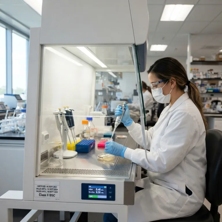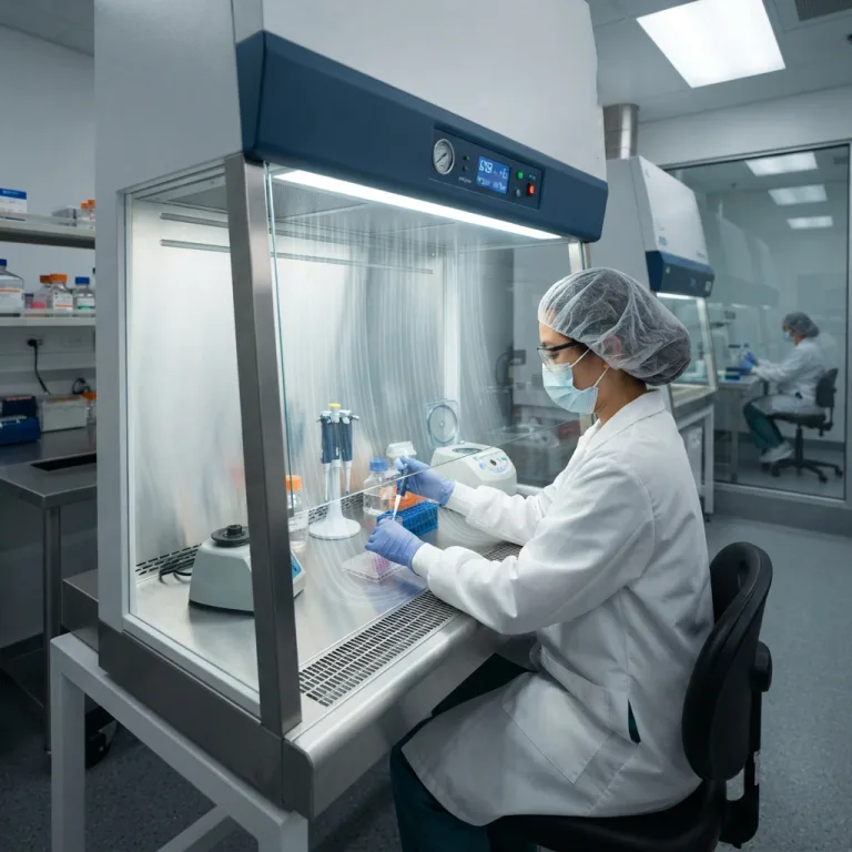
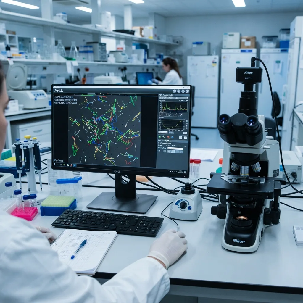
The 30% Subjectivity Crisis in Male Infertility Diagnostics
Place two experienced clinical embryologists in front of a dual-headed manual microscope looking at the exact same drop of liquefied human semen, and their visual estimates of progressive sperm motility will frequently differ by 30% to 40%.
This is not a failure of clinical training; it is a physiological limitation of the human visual cortex. When looking into a microscope field containing 60 to 100 spermatozoa swimming simultaneously in criss-crossing directions at speeds between 5 and 80 micrometers per second, the human eye is naturally drawn to rapid movement. This cognitive bias—known as visual motion capture—causes technicians to systematically overestimate progressive motility while losing track of slower or stationary cells in the background.
When evaluating sperm concentration on a manual hemocytometer grid or scoring abnormal head shapes under oil immersion, similar human fatigue and subjective threshold drift occur. A borderline oval sperm head classified as “normal” by a hospital laboratory at 8:00 AM may be scored as “abnormal” by the same technician at 4:00 PM.
In an era when global male sperm counts have declined significantly over the past four decades—and where male factor infertility accounts for roughly 40% to 50% of all reproductive challenges among couples—fertility clinics, urology departments, and hospital andrology laboratories can no longer rely on subjective visual guesswork. Whether a couple is guided toward timed natural conception, Intrauterine Insemination (IUI), conventional In Vitro Fertilization (IVF), or Intracytoplasmic Sperm Injection (ICSI) depends directly on the precision of the semen analysis report.
Computer-Assisted Semen Analysis (CASA)—commonly referred to as an automated sperm analyzer—solves this diagnostic crisis by replacing human visual estimation with high-speed digital micrography and multi-object computer vision tracking.
1. How a CASA Sperm Analyzer Works: From Live Video to Multi-Target Tracking
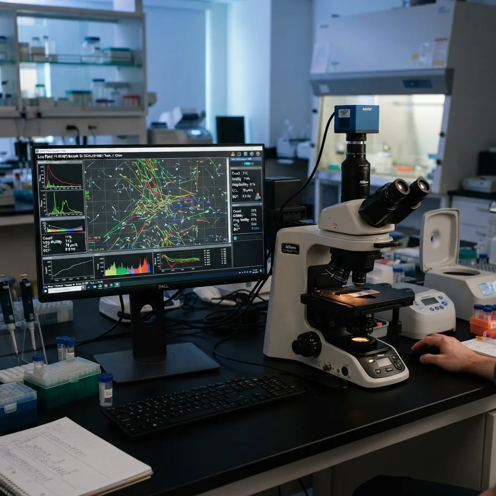
A modern CASA workstation integrates specialized illumination, optical magnification, high-speed digital photography, and algorithmic image processing to convert a live microscopic sample into quantitative clinical data within seconds.
Plaintext
                    THE 4-STAGE CASA ANALYTICAL ENGINE
+-------------------------------------------------------------------------+
| STAGE 1: HIGH-SPEED OPTICAL ACQUISITION                                 |
| - Phase-contrast optics illuminate transparent live sperm without stain |
| - High-speed camera records 30 to 60+ frames per second (fps)           |
|                                   |                                     |
|                                   v                                     |
| STAGE 2: HEAD CENTROID SEGMENTATION                                     |
| - Computer vision isolates bright phase-halo sperm heads from debris    |
| - Verifies target area (e.g., 5 to 20 square micrometers) & tail presence|
|                                   |                                     |
|                                   v                                     |
| STAGE 3: MULTI-FRAME TRAJECTORY RECONSTRUCTION                          |
| - Connects X-Y coordinates across 30–60 sequential video frames         |
| - Collision-resolution algorithms track crossing paths without swapping |
|                                   |                                     |
|                                   v                                     |
| STAGE 4: KINEMATIC & MORPHOLOGICAL CLASSIFICATION                       |
| - Color-codes tracks (Red = Rapid A, Green = Slow B, Blue = Non-Prog C) |
| - Computes WHO concentration, total count, kinematics & Kruger metrics  |
+-------------------------------------------------------------------------+
Step 1: Stain-Free Live Capture via Phase-Contrast Micrography
Chemical stains kill living spermatozoa immediately, meaning motility must be evaluated in native, unstained semen. However, unstained spermatozoa are nearly transparent under standard brightfield light.
-
CASA systems utilize phase-contrast optics (or specialized darkfield illumination) that convert subtle shifts in light phase passing through the dense sperm nucleus and delicate flagellar tail into high-contrast brightness variations.
-
Under negative or positive phase contrast, the sperm head stands out with a crisp, bright halo against a dark background, allowing computer vision software to lock onto the exact geometric center (centroid) of every cell.
Step 2: Frame-by-Frame Trajectory Reconstruction
As the motorized or manual microscope stage moves across 5 to 10 standardized fields of view inside a precision counting chamber, a high-speed CMOS digital camera captures continuous video sequences at 30 to 60 frames per second (fps) for 0.5 to 1.0 second per field:
-
Debris Rejection: In native semen, static particulate matter, prostatic concretions, epithelial cells, and round cells share the visual field with spermatozoa. Advanced CASA algorithms verify both head optical density and characteristic tail motion or flagellar presence, preventing immobile uric acid crystals or cellular debris from being miscounted as dead Grade D sperm (which would falsely lower the patient’s reported motility percentage).
-
Predictive Collision Tracking: When two rapidly swimming spermatozoa cross paths in the same optical plane, primitive software can confuse their identities. Modern CASA systems apply Kalman filtering and predictive velocity vector algorithms—using each cell’s momentum, swimming angle, and head orientation prior to intersection to maintain accurate individual trajectory lines after they cross.
2. Mastering WHO Semen Analysis Standards: 5th Edition vs. 6th Edition
The global clinical benchmark for evaluating male fertility is the World Health Organization (WHO) Laboratory Manual for the Examination and Processing of Human Semen. When procuring or operating an automated sperm analyzer, understanding how the software implements both the WHO 5th Edition (2010) and the updated WHO 6th Edition (2021) guidelines is essential.
Plaintext
               CRITICAL SHIFT IN WHO MOTILITY CLASSIFICATION
+-------------------------------------------------------------------------+
| WHO 5TH EDITION (2010): 3-Tier Simplified System                        |
| - Pooled all forward-moving sperm into one "Progressive Motility (PR)"  |
| - Hid the clinical distinction between sluggish and fast swimmers       |
|                                                                         |
| WHO 6TH EDITION (2021): Return of the 4-Tier Clinical Velocity System   |
| - Grade A (Rapid Progressive): Velocity >= 25 micrometers per second    |
| - Grade B (Slow Progressive):  Velocity 5 to < 25 micrometers per second|
| - Grade C (Non-Progressive):   Velocity < 5 micrometers/sec (In-place)  |
| - Grade D (Immotile):          Zero movement (Static / Dead or Ciliary) |
+-------------------------------------------------------------------------+
Why the WHO 6th Edition Reinstated Grade A vs. Grade B Motility
In the 2010 WHO 5th Edition, the committee combined all forward-moving sperm into a single category called “Progressive Motility (PR)” because human technicians could not reliably distinguish fast vs. slow swimming speed by eye.
However, a decade of clinical IVF and IUI outcomes proved that swimming velocity matters immensely: a patient whose progressive sperm are all swimming sluggishly at 8 micrometers per second (Grade B) has a drastically lower natural conception and Intrauterine Insemination (IUI) success rate than a patient with abundant Grade A rapid progressive sperm swimming at 25 micrometers per second or faster (roughly equivalent to 5 sperm head lengths per second).
Because automated CASA systems measure exact micrometer-per-second velocities effortlessly, the WHO 6th Edition officially restored the 4-tier motility classification and explicitly highlighted CASA technology for objective velocity grading.
WHO Lower Fifth Percentile Reference Limits & Clinical Nomenclature
The table below outlines the baseline WHO reference cutoffs (representing the 5th percentile of fertile men whose partners conceived within 12 months) and the diagnostic terminology automatically flagged by CASA software:
| Semen Parameter | WHO Reference Lower Limit | Pathological Condition Flagged Below Limit | Clinical Definition of Pathology |
| Ejaculate Volume | 1.4 mL – 1.5 mL | Hypospermia (or Aspermia if 0 mL) | Low seminal vesicle fluid output, partial ejaculatory duct obstruction, or retrograde ejaculation. |
| Sperm Concentration | 15 – 16 million / mL | Oligozoospermia (Severe if < 5M/mL) | Reduced spermatogenesis in the seminiferous tubules of the testes. |
| Total Sperm Number | 39 million / ejaculate | Oligozoospermia (Azoospermia if 0) | Reflects total testicular output and ductal patency per ejaculation. |
| Total Motility (A+B+C) | 40% – 42% motile | Asthenozoospermia | Impaired flagellar mitochondrial ATP production, varicocele, antisperm antibodies, or oxidative stress. |
| Progressive Motility (A+B) | 30% – 32% progressive | Asthenozoospermia | Inability of sperm to ascend through cervical mucus and the uterotubal junction. |
| Normal Morphology | 4% normal forms | Teratozoospermia | Defective spermiogenesis affecting head acrosome, midpiece, or tail structure. |
| Sperm Vitality | 54% – 58% membrane-intact | Necrozoospermia | High percentage of structurally dead sperm due to epididymal pathology or toxins. |
| Peroxidase Leukocytes | < 1.0 million / mL | Leukocytospermia (Pyospermia) | Male accessory gland infection (prostatitis, epididymitis) generating reactive oxygen species. |
(Note: When Oligozoospermia, Asthenozoospermia, and Teratozoospermia occur simultaneously in the same patient, the CASA workstation flags Oligoasthenoteratozoospermia [OAT Syndrome], the most common clinical indication for IVF/ICSI).
3. Beyond the Human Eye: CASA Kinematic Trajectory Parameters Explained
The greatest diagnostic advantage of a CASA sperm analyzer over manual microscopy is its ability to map sperm kinematics—the precise physics and geometry of how each spermatozoon propels itself through fluid.
As a spermatozoon swims forward, its flagellar tail beats back and forth, causing the sperm head to oscillate from side to side across an average central path. A CASA system reconstructs this exact path at 60 frames per second and calculates eight core kinematic metrics:
Plaintext
                  ANATOMY OF A SPERM KINEMATIC TRAJECTORY
+-------------------------------------------------------------------------+
|   Start (A)                                                   End (B)   |
|      o ---/\----/\----/\----/\----/\----/\----/\----/\----/\---> (O)    |
|                                                                         |
| 1. VCL (Curvilinear Velocity): Total actual zig-zag distance / time     |
|    (Follows the /\/\/\ head oscillations; always the highest velocity)  |
|                                                                         |
| 2. VSL (Straight-Line Velocity): Direct crow-flies distance A to B /time|
|    (Measures net forward progression from first frame to last frame)    |
|                                                                         |
| 3. VAP (Average Path Velocity): Smoothed central midline path / time    |
|    (Mathematically smooths out the side-to-side head wobble)            |
|                                                                         |
| 4. ALH (Amplitude of Lateral Head): Width of head swing from midline    |
| 5. BCF (Beat-Cross Frequency): How many times/sec head crosses midline  |
+-------------------------------------------------------------------------+
The Three Velocity Metrics (Measured in Micrometers per Second)
-
VCL (Curvilinear Velocity): The actual point-to-point speed of the sperm head along its true, undulating two-dimensional track. VCL reflects the raw mechanical vigor and mitochondrial energy output of the cell.
-
VSL (Straight-Line Velocity): The net straight-line speed measured directly from the very first point where the sperm was detected to the very last point at the end of the video capture. VSL reflects net directional progress.
-
VAP (Average Path Velocity): The speed of the sperm head along a mathematically smoothed five-point moving average path.
The Three Trajectory Ratios (Expressed as Percentages)
-
LIN (Linearity = VSL divided by VCL): Measures how straight the overall swimming track is. A sperm swimming in a tight circle has a high VCL (it is moving fast) but a near-zero VSL and very low Linearity.
-
STR (Straightness = VSL divided by VAP): Measures the directional conformity of the smoothed average path.
-
WOB (Wobble = VAP divided by VCL): Measures the degree of side-to-side oscillation of the actual head path relative to its average trajectory.
Head Oscillation & Flagellar Drive Metrics
-
ALH (Amplitude of Lateral Head Displacement, in Micrometers): Measures the physical width of the sperm head’s side-to-side swing as it progresses forward.
-
BCF (Beat-Cross Frequency, in Hertz): Counts how many times per second the oscillating actual path of the sperm head crosses over its smoothed average path. BCF provides a direct proxy for flagellar tail beat frequency.
Clinical Application: Detecting “Hyperactivated Motility” for IVF
Why do fertility specialists care about ALH, VCL, and Linearity? Because of a critical biological phenomenon called capacitation and hyperactivation.
-
In the fresh ejaculate, healthy sperm swim in relatively straight, efficient lines (high VSL, high Linearity, moderate ALH) to travel up the female cervical canal.
-
However, once spermatozoa reach the fallopian tube and undergo capacitation near the egg, they must switch into Hyperactivated Motility—a frantic, high-amplitude whiplash swimming pattern needed to mechanically bore through the sticky cumulus oophorus cells and dense zona pellucida shell surrounding the human oocyte.
-
CASA Hyperactivation Criteria: A hyperactivated spermatozoon exhibits very high VCL , wide lateral head swing , and low Linearity. An automated CASA system quantifies the exact percentage of hyperactivated sperm after capacitation washing. If a patient has normal baseline motility but fails to achieve hyperactivation, natural fertilization and conventional IVF often fail—alerting the embryologist to proceed directly to ICSI (Intracytoplasmic Sperm Injection), where a single sperm is injected directly into the egg, sparing the couple a failed IVF cycle.
4. Automated Strict Kruger Morphology: Sub-Micrometer Biometrics
Even if a patient has 50 million rapidly swimming sperm per milliliter, fertilization will fail if those spermatozoa are structurally deformed and lack the enzymatic machinery to bind to the oocyte.
Following Tygerberg Strict Kruger Criteria (adopted by the WHO), a spermatozoon is only classified as morphologically normal if all four anatomical regions—the head, acrosome, neck/midpiece, and tail—fall within strict sub-micrometer biometric tolerances simultaneously.
Plaintext
               STRICT KRUGER MORPHOLOGY BIOMETRIC STANDARDS
+-------------------------------------------------------------------------+
| 1. HEAD & ACROSOME (Smooth Oval Contour)                                |
|    - Length: 3.7 to 4.7 μm | Width: 2.5 to 3.2 μm | L/W Ratio: 1.3–1.8  |
|    - Acrosomal Cap: Distinct anterior region covering 40%–70% of head   |
|    - Vacuoles: <= 2 small vacuoles occupying < 20% of head area         |
|                                                                         |
| 2. NECK & MIDPIECE (Mitochondrial Engine)                               |
|    - Slender, axially attached (~4.0 to 5.0 μm long, < 1.0 μm wide)     |
|    - Cytoplasmic Droplet: Must be < 1/3 of normal head size             |
|                                                                         |
| 3. PRINCIPAL TAIL (Flagellar Propulsor)                                 |
|    - Uniform thinner caliber (~45 μm long, ~10x head length)            |
|    - Uncoiled, free of sharp hairpin bends (which indicate tail break)  |
+-------------------------------------------------------------------------+
How Automated CASA Morphology Eliminates Human Eyeball Drift
On a stained semen smear (prepared with Diff-Quik, Papanicolaou, or SpermBlue differential stains), the CASA morphology module captures high-magnification brightfield images and applies automated edge-detection segmentation to measure over 20 biometric parameters on 200+ individual cells:
-
Head Defects: Automatically flags pyriform (pear-shaped), tapered (cigar-shaped, common in varicocele), amorphous, microcephalic (pinhead), macrocephalic (giant multi-nucleated), double-headed, and globozoospermic (round heads completely lacking an acrosome cap, which cannot fertilize an egg without ICSI and artificial oocyte activation).
-
Neck & Midpiece Defects: Measures insertion angle asymmetry, bent necks, thickened mitochondrial sheaths, and retained excess residual cytoplasm (ERC) droplets that generate destructive reactive oxygen species (ROS).
-
Tail Defects: Identifies short tails, stump tails, duplicate tails, and coiled tails (associated with epididymal osmotic dysfunction or infection).
-
Automated Teratozoospermia Index (TZI) & Multiple Anomalies Index (MAI): Because a single abnormal spermatozoon can exhibit up to four concurrent defects (head + midpiece + tail + cytoplasmic droplet), the CASA software automatically calculates the TZI (average number of defects per abnormal sperm, ranging from 1.00 to 4.00). A TZI exceeding 1.60 to 1.80 strongly correlates with impaired in-vivo fertilization potential.
5. Advanced Functional Modules: Vitality, DNA Fragmentation (DFI) & Round Cells
Approximately 15% of infertile men exhibit a “normal” routine semen analysis (normozoospermia) yet remain unable to achieve a viable pregnancy or suffer recurrent early miscarriage. Modern CASA platforms bridge this gap by automating three specialized functional assays on the same optical workstation.
Plaintext
                  ADVANCED FUNCTIONAL ANDROLOGY MODULES
                                    |
     +------------------------------+------------------------------+
     |                              |                              |
     v                              v                              v
[ SPERM VITALITY ]          [ DNA FRAGMENTATION ]          [ LEUKOCYTE vs GERM ]
- Eosin-Nigrosin Stain      - SCD "Halo Test" Assay        - Peroxidase Staining
- Live = White Unstained    - Intact DNA = Large Halo      - Brown = WBC Neutrophil
- Dead = Pink/Red Stained   - Broken DNA = No/Tiny Halo    - Unstained = Spermatid
A. Automated Sperm Vitality (Live vs. Dead Immotile Sperm)
When a patient’s motility is severely depressed (total motility below 40%), the clinician must know whether the stationary Grade D sperm are structurally dead (Necrozoospermia) or alive with paralyzed tails (such as in Kartagener syndrome / primary ciliary dyskinesia).
-
Automated Eosin-Nigrosin Colorimetric Scoring: Live spermatozoa possess intact plasma membranes that exclude eosin dye, remaining white against the dark purple nigrosin background. Dead spermatozoa have damaged, permeable membranes that absorb eosin, staining bright pink or red. The CASA color camera counts 200 stained vs. unstained heads automatically in under 30 seconds, identifying viable live immobile sperm that can still be safely selected for ICSI.
B. Automated Sperm DNA Fragmentation Index (DFI / SCD Halo Assay)
Even a fast-swimming, normal-looking spermatozoon can carry shattered genetic material inside its nucleus—caused by varicocele heat stress, smoking, chemotherapy, advanced paternal age, or genital tract infection. While an egg can sometimes be fertilized by a DNA-damaged sperm, embryonic development frequently arrests at the blastocyst stage or results in first-trimester miscarriage.
-
Sperm Chromatin Dispersion (SCD / Halo Test) Automation: Treated spermatozoa are embedded in an agarose microgel on a slide, incubated with an acid denaturation solution and lysis buffer to remove nuclear proteins, and stained.
-
Optical Halo Measurement: Spermatozoa with intact, healthy double-stranded DNA disperse massive radiating loops of chromatin around the head (forming a large, bright Halo). Spermatozoa with fragmented single- or double-stranded DNA breaks produce a tiny halo or no halo at all.
-
The CASA DFI module automatically measures the precise ratio of halo thickness to nuclear core diameter across 300 to 500 cells and computes the DNA Fragmentation Index (% DFI):
-
DFI < 15%: Excellent fertility potential.
-
DFI 15% – 25%: Moderate DNA damage; lifestyle intervention and antioxidant therapy indicated.
-
DFI > 25% – 30%: High DNA fragmentation; reduced success with natural conception or IUI; strong clinical indication for IVF/ICSI or testicular sperm aspiration.
-
C. Differentiating Round Cells: Leukocytes vs. Immature Germ Cells
Unstained semen frequently contains “round cells” lacking tails. Under manual brightfield microscopy, harmless immature spermatogenic cells (spermatids and spermatocytes sloughed from the testes) look nearly identical to inflammatory polymorphonuclear leukocytes (WBCs).
-
Using the WHO ortho-toluidine or benzidine peroxidase staining protocol, the CASA workstation automatically distinguishes peroxidase-positive brown neutrophils (Leukocytospermia ≥ 1.0 million/mL, indicating prostatitis or accessory gland infection requiring antibiotic therapy) from peroxidase-negative pink/unstained immature germ cells.
6. Elevating Andrology & IVF Workflows: The UMY Medical Advantage
In reproductive medicine centers, urology clinics, and hospital pathology departments, the sperm analyzer plays a dual role: diagnostic screening and therapeutic sperm preparation QC. Before performing Intrauterine Insemination (IUI) or IVF, embryologists wash raw semen using density-gradient centrifugation or swim-up techniques to isolate the cleanest, fastest-moving spermatozoa. Running a quick 30-second pre-wash and post-wash CASA check verifies the exact Total Motile Sperm Count (TMSC) recovered—where a post-wash TMSC exceeding 5 to 10 million progressive sperm predicts optimal IUI pregnancy rates.
To make this advanced computer-vision technology accessible without the prohibitive capital costs and recurring software fees of legacy specialty vendors, UMY Medical Equipment Co Ltd engineers precision automated sperm analyzers tailored for modern fertility centers and clinical laboratories worldwide.
Plaintext
                  UMY MEDICAL CASA WORKSTATION ADVANTAGES
                                     |
     +-------------------------------+-------------------------------+
     |                               |                               |
     v                               v                               v
[ High-Speed Computer Vision ]  [ Built-In WHO 5th & 6th ]     [ Complete Clinical Suite ]
 60 fps Anti-Collision Tracking  Instant Grade A/B/C/D Switch   Motility, Kruger Morphology,
 Phase-Contrast Optical Clarity  Full VCL/VSL/ALH Kinematics    Vitality & DFI Halo Modules
Why Fertility Clinics and B2B Distributors Partner with UMY Medical:
-
Multi-Frame Anti-Collision Tracking Precision: UMY Medical CASA workstations pair high-contrast phase-contrast optical systems with high-frame-rate digital cameras and intelligent trajectory reconstruction software, accurately tracking dense concentrations of spermatozoa while filtering out seminal debris and round cells.
-
One-Click WHO 5th & 6th Edition Compliance: Clinicians can toggle seamlessly between WHO 5th Edition (3-tier PR/NP/IM) and WHO 6th Edition (4-tier Rapid Grade A, Slow Grade B, Non-Progressive Grade C, Immotile Grade D) reporting standards, complete with full kinematic scattergrams (VCL, VSL, VAP, ALH, BCF, LIN, STR, WOB) and visual trajectory color overlays.
-
All-in-One Functional Andrology Platform: Beyond baseline motility and concentration, UMY Medical software integrates automated Strict Kruger Morphology, Eosin-Nigrosin Vitality, Sperm DNA Fragmentation (SCD Halo), and Peroxidase Leukocyte quantification into a single unified workstation—generating comprehensive, image-rich diagnostic reports with real video clip archiving.
-
Certified Quality & Open Hospital IT Integration: Manufactured under strict ISO 13485 quality management systems and carrying CE compliance, UMY Medical sperm analyzers feature subscription-free software and bidirectional LIS/HIS connectivity, allowing seamless electronic reporting and direct factory wholesale value for global B2B medical distributors.
Frequently Asked Questions (FAQ)
Q1: Why is sexual abstinence time (2 to 7 days) critical before a CASA sperm test?
The WHO mandates a strict sexual abstinence window of 2 to 7 days (ideally 3 to 5 days) prior to semen collection. If abstinence is shorter than 48 hours, sperm concentration and total ejaculate volume may appear falsely low because epididymal reserves have not fully replenished. Conversely, if abstinence exceeds 7 days, older spermatozoa accumulate in the epididymis and undergo senescence and oxidative damage—resulting in a deceptively high sperm count paired with falsely depressed progressive motility (asthenozoospermia) and elevated Sperm DNA Fragmentation (DFI).
Q2: How does a CASA analyzer handle extremely low sperm counts (Severe Oligozoospermia or Post-Vasectomy Checks)?
In severe oligozoospermia (under 1 to 2 million sperm/mL), cryptozoospermia, or post-vasectomy clearance checks, standard 5-field automated scanning may only capture a handful of cells. Modern CASA systems feature a dedicated “Low-Concentration / Post-Vasectomy Extended Scan Mode” that automatically captures 20 to 50+ optical fields across the entire counting chamber, flagging every single motile or immotile spermatozoon on screen for rapid operator confirmation.
Q3: What is the difference between Total Motility, Progressive Motility, and Total Motile Sperm Count (TMSC)?
-
Total Motility (Grades A + B + C): The percentage of all sperm showing any movement at all, including cells twitching in place.
-
Progressive Motility (Grades A + B): The percentage of sperm swimming actively forward along a trajectory.
-
Total Motile Sperm Count (TMSC): Calculated automatically by the CASA software as Ejaculate Volume (mL)× Sperm Concentration (millions/mL) $\times$ Progressive Motility (%). TMSC is considered by reproductive endocrinologists to be the single most predictive composite number for guiding couples toward natural conception, IUI, IVF, or ICSI.
Q4: Why can a patient have 5% normal Kruger morphology and still be classified as fertile?
Under the Tygerberg Strict Kruger Criteria adopted by the WHO, even highly fertile men whose partners conceived naturally within 12 months exhibit a vast majority of pleomorphic sperm shapes, placing the lower reference cutoff at just 4.0% morphologically normal forms. This occurs because strict criteria apply rigid sub-micrometer measurements learned from analyzing the select subpopulations of elite sperm capable of penetrating human cervical mucus and binding to the zona pellucida. A score of 4% or higher is classified as normal, whereas 0% to 3% indicates Teratozoospermia.
Q5: Can the same CASA workstation be configured for veterinary and livestock artificial insemination (AI) centers?
Yes. Advanced CASA platforms feature customizable multi-species biometric and velocity profiles. Because bull, boar, stallion, ram, canine, and poultry spermatozoa differ significantly from human sperm in head dimensions (for example, boar and bull sperm heads are much larger and flatter than human oval heads) and swim at much higher curvilinear velocities (often exceeding 150 to 250 micrometers per second), dedicated veterinary software modules adjust the particle size gates, frame acquisition rates, and dose-calculation formulas for commercial livestock breeding centers.