
1. Executive Overview: Incorporating Bone Mineral Density Testing into Clinical Practice
Bone mineral density (BMD) testing is a fundamental component of preventative healthcare, orthopedic evaluation, endocrinology, and rheumatology. Bone densitometers measure bone mineral content to diagnose osteoporosis, assess osteopenia, predict future fracture risk, and monitor patient response to therapeutic interventions.
For hospital procurement teams, clinic directors, and medical equipment distributors, investing in bone densitometry technology requires balancing diagnostic accuracy, patient throughput, physical clinic footprint, and long-term operating costs.
+-----------------------------------------------------------------------------------+
|                        CLINICAL BONE DENSITY TESTING WORKFLOW                     |
+-----------------------------------------------------------------------------------+
|  1. Patient Triage & History | Risk factor screening (FRAX), height/weight check  |
|  2. Modality Selection       | Central DXA (Hip/Spine) or Peripheral (Heel/Arm)  |
|  3. Scan Execution & QC      | Laser positioning, daily phantom calibration check |
|  4. Diagnostic Output        | T-Score, Z-Score, & Body Composition Analysis      |
+-----------------------------------------------------------------------------------+
2. Primary Technologies: Dual-Energy X-Ray Absorptiometry (DXA) vs. Quantitative Ultrasound (QUS)
Modern bone densitometry relies on two main technology standards, each suited for specific clinical roles.
Dual-Energy X-Ray Absorptiometry (DXA / DEXA)
-
Working Principle: Passes two distinct X-ray energy beams through the body. Soft tissue and bone absorb the beams differently; subtracting soft tissue absorption yields an exact bone mineral density reading
-
Clinical Role: Recognized globally by the World Health Organization (WHO) as the gold standard for diagnosing osteoporosis and monitoring changes over time.
-
Primary Scan Sites: Lumbar spine, proximal femur (total hip and femoral neck), and dual forearm.
Quantitative Ultrasound (QUS)
-
Working Principle: Uses high-frequency sound waves to measure Speed of Sound (SOS) and Broadband Ultrasound Attenuation (BUA) through peripheral bone structures.
-
Clinical Role: Radiation-free initial screening tool used to identify individuals at risk who require follow-up central DXA testing.
-
Primary Scan Sites: Calcaneus (heel), radius (forearm), and phalanges (fingers).
3. Central DXA vs. Peripheral Densitometers (pDXA & pQUS)
Categorizing machines by form factor helps practices match equipment with their spatial limitations and patient volume.
                        BONE DENSITOMETER CLASSIFICATION
                                       |
       +-------------------------------+-------------------------------+
       |                                                               |
Central / Full-Body Systems                      Peripheral Screening Units
- Tabletop design for full-body bed             - Compact desktop or mobile cart
- Scans Hip, Spine, & Whole Body                - Scans Heel, Forearm, or Finger
- Gold Standard Osteoporosis Diagnosis          - Low-Cost Prescreening & Community Drives
Central / Full-Body DXA Systems
-
Design & Footprint: Full-length padded examination table with an overhead scanning arm. Requires a dedicated room.
-
Key Features: High-speed fan-beam or pencil-beam X-ray acquisition, automated region-of-interest (ROI) placement, advanced body composition software (fat mass, lean mass, visceral adipose tissue), and Vertebral Fracture Assessment (VFA).
-
Target Users: Hospitals, diagnostic imaging centers, rheumatology practices, and metabolic bone disease clinics.
Peripheral Bone Densitometers (pDXA & pQUS)
-
Design & Footprint: Portable desktop units or small wheeled carts requiring zero dedicated room buildouts.
-
Key Features: Fast scan times (15 to 60 seconds), plug-and-play setup, minimal operator training requirements, and low capital cost.
-
Target Users: General optometry/primary care practices, mobile health units, pharmacies, and occupational health clinics.
4. Key Clinical Outputs: T-Scores, Z-Scores & Advanced Software
Understanding diagnostic output software ensures buyers select equipment that meets local medical billing and clinical reporting requirements.
-
T-Score: Compares a patient’s bone density to a healthy 30-year-old adult reference population. Used for diagnosing osteoporosis in postmenopausal women and men aged 50 and older.
-
Z-Score: Compares bone density to an age-, gender-, and ethnicity-matched reference population. Used for pediatric patients, premenopausal women, and younger men.
-
FRAX (Fracture Risk Assessment Tool): Integrated software calculating a patient’s 10-year probability of major osteoporotic or hip fracture.
-
Advanced Body Composition: Measures whole-body fat percentage, lean muscle mass balance, and visceral fat, popular in sports medicine, wellness, and bariatric programs.
5. Physical, Electrical & Regulatory Siting Requirements
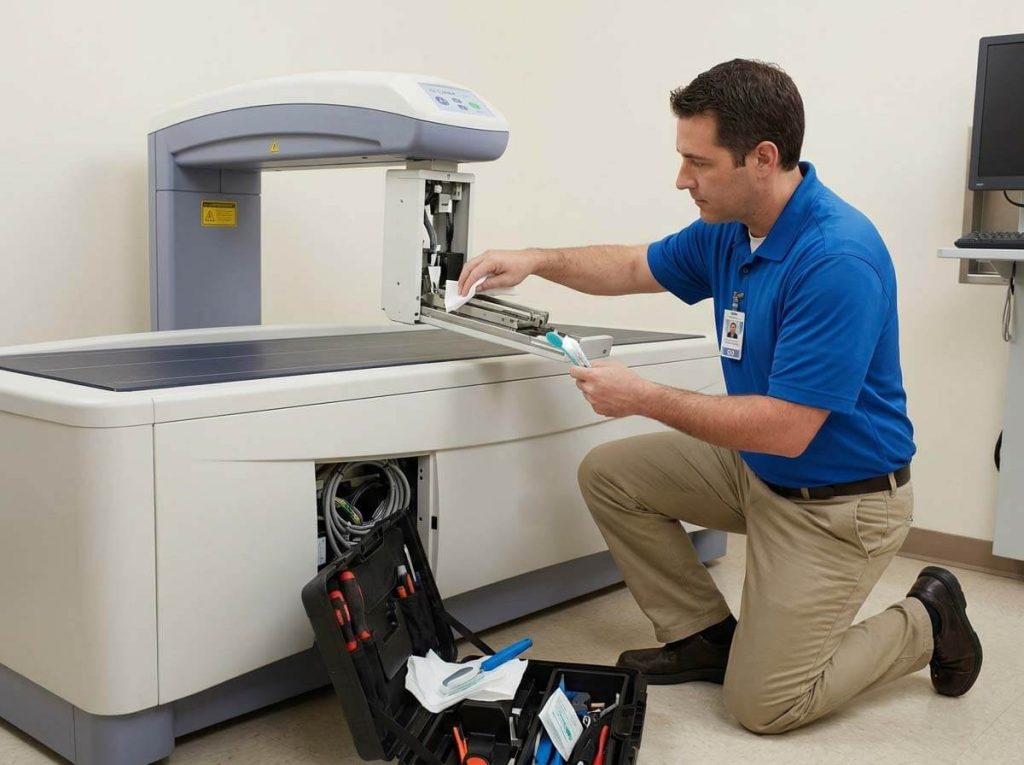
Installing a bone densitometer requires basic facility planning to comply with local radiation safety and operational standards.
+-----------------------------------------------------------------------------------+
|                        FACILITY & SITING CHECKLIST                                |
+-----------------------------------------------------------------------------------+
| Power Supply         | Dedicated 100–240 V AC, 50/60 Hz grounded circuit         |
| Climate Control      | Temperature: 18°C–25°C; Humidity: 20%–80% non-condensing |
| Floor Load & Level   | Level floor supporting 150–350 kg total system weight      |
| Radiation Shielding  | Standard drywall usually sufficient (low scatter dose)    |
+-----------------------------------------------------------------------------------+
-
Radiation Safety: DXA scanners emit extremely low scatter radiation doses (often equivalent to less than a daily background radiation exposure). In many jurisdictions, standard drywall is sufficient without lead shielding, though local radiation protection advisor (RPA) sign-off is required.
-
Network Integration: Ensure the system includes DICOM 3.0 export options for sending reports directly to PACS and local Electronic Medical Record (EMR) systems.
6. Technical Comparison Matrix of Primary Densitometer Types
| Feature / Metric | Central Fan-Beam DXA | Central Pencil-Beam DXA | Peripheral DXA (pDXA) | Quantitative Ultrasound (QUS) |
| Primary Clinical Site | Hip, Spine, Whole Body | Hip, Spine, Whole Body | Forearm, Heel | Calcaneus (Heel) |
| Diagnostic Status | Gold Standard (WHO) | Gold Standard (WHO) | Screening / Baseline | Screening / Triage |
| Radiation Exposure | Very Low | Ultra-Low | Extremely Low | Zero (Ionizing Radiation Free) |
| Scan Speed | 10 to 30 seconds | 1 to 3 minutes | 30 to 60 seconds | 15 to 30 seconds |
| Body Composition | Full Advanced Features | Basic / Optional | Not Available | Not Available |
| Space Footprint | Large Dedicated Room | Large Dedicated Room | Tabletop / Compact Cart | Desktop Portable |
| Relative Equipment Cost | Moderate to High | Moderate | Low to Moderate | Low |
7. B2B Procurement & Supplier Evaluation Checklist
When sourcing a bone densitometer from international manufacturers or distributors, evaluate suppliers using these criteria:
-
Daily Phantom Quality Control: Ensure the machine includes an automated calibration phantom (e.g. spine phantom) to perform daily precision and drift checks required by international standards (ISCD).
-
Regulatory Certifications: Verify ISO 13485 manufacturing quality compliance, CE marks under EU Medical Device Regulation (MDR), and FDA 510(k) clearance where applicable.
-
Software Upgrades & Licensing: Confirm whether software licenses (body composition, VFA, FRAX, pediatric modules) are bundled or sold separately.
-
Service Level Agreement (SLA): Check the availability of regional field engineers, replacement X-ray tube turnarounds, and remote diagnostic capabilities.
8. Frequently Asked Questions (FAQ)
Q1: What is the main operational difference between pencil-beam and fan-beam DXA scanners?
Pencil-beam systems use a single collimated X-ray beam moving in a raster pattern, offering low radiation exposure but longer scan times. Fan-beam systems use a wide-angle X-ray beam with multi-element detectors, scanning patients significantly faster with higher image resolution.
Q2: Can a peripheral ultrasound bone densitometer replace a central DXA scanner?
No. Quantitative Ultrasound (QUS) is an effective, portable, radiation-free screening tool for identifying patients with potential bone loss, but official diagnosis of osteoporosis and drug treatment monitoring must be confirmed with a central DXA scan of the hip and spine.
Q3: How long does a standard bone densitometer system last?
With regular preventive maintenance, daily phantom calibration checks, and timely software updates, a high-quality central DXA machine typically has a operational lifespan of 8 to 12 years.