The Most Avoided Bench in the Clinical Laboratory
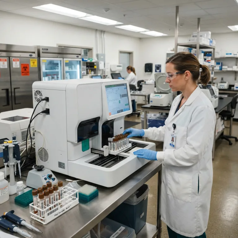
Walk into almost any hospital pathology department or outpatient diagnostic laboratory, and you will quickly discover which workstation medical technologists dread the most: the manual routine stool microscopy bench.
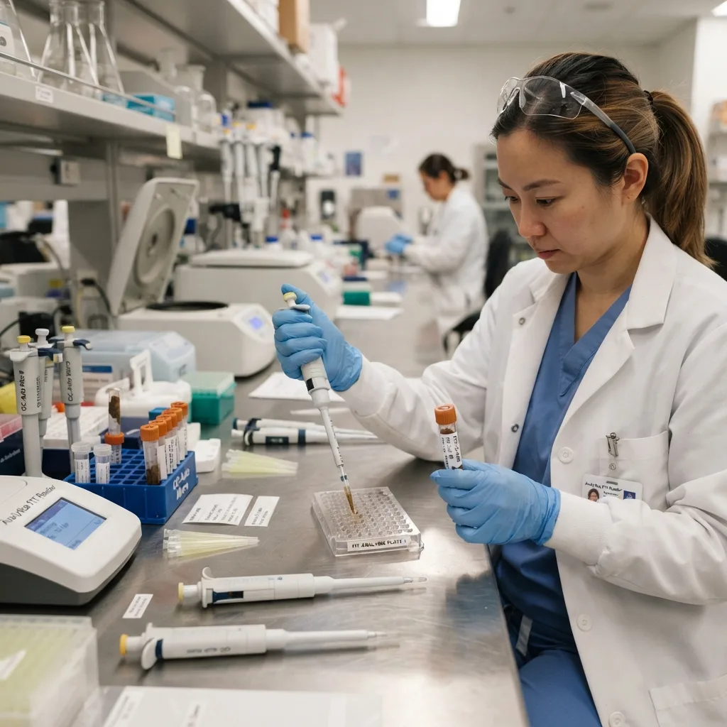
For over a century, clinical coprology (fecal analysis) remained frozen in time while hematology, biochemistry, and urinalysis underwent total robotic transformation. In the traditional manual workflow, a technician must uncap puluhan open plastic stool containers every morning, dip a wooden applicator stick into raw fecal matter, emulsify a tiny 2-milligram speck into a drop of saline or Lugol’s iodine on a glass microscope slide, drop a fragile glass coverslip on top, and lean over an optical microscope to scan through dense clouds of undigested plant fibers and bacterial debris.
This archaic manual process creates two severe problems for modern healthcare institutions:
-
Unacceptable Occupational Biosafety Hazards: Uncapping raw stool cups and vigorous manual stirring generate invisible biohazard aerosols containing viable Salmonella, Shigella, enterohemorrhagic E. coli, Clostridioides difficile spores, norovirus, hepatitis A, and infectious protozoan cysts. Even under a fume hood, laboratory personnel face constant mucosal exposure and olfactory exhaustion, driving high staff turnover among young laboratory graduates.
-
Massive Sampling Bias and False-Negative Diagnoses: An average bowel movement weighs between 100 and 250 grams, yet a traditional direct wet-mount smear examines only 2 to 5 milligrams of stool—less than 0.005% of the specimen. If a patient has an early bleeding colonic polyp or an intermittent parasitic infection where helminth eggs are unevenly distributed in the fecal bolus, dipping a wooden stick into a single random spot misses the pathology up to 30% to 40% of the time. Furthermore, if the technician smears the slide too thickly, fibrous debris hides translucent protozoan cysts; if smeared too thinly, scarce formed elements are washed off the edge of the glass.
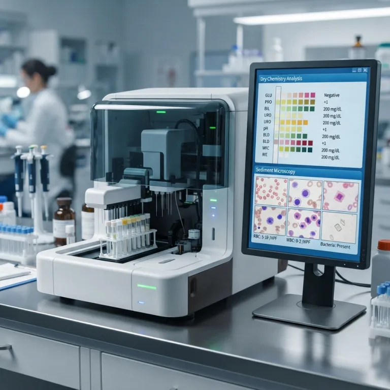
The rapid global shift toward the automated fecal analyzer is driven by the urgent clinical need to close this diagnostic gap—turning an unpleasant, hazardous, and highly subjective manual task into a standardized, closed-tube, high-precision diagnostic science.
1. Macroscopic Physical Standardization: Color, Consistency, and the Bristol Stool Scale
Before any chemical or microscopic evaluation takes place, the physical characteristics of a fecal specimen provide vital clinical clues regarding gastrointestinal transit time, biliary patency, and mucosal integrity.
In manual laboratories, recording stool color and consistency relies on quick visual glances that vary wildly between technicians. Automated fecal workstations standardize this step by positioning a high-resolution RGB spectral camera above the specimen entry port to photograph and classify the physical traits of the native sample prior to dilution:
Plaintext
                  CLINICAL SIGNIFICANCE OF MACROSCOPIC TRAITS
+-------------------------------------------------------------------------+
| STOOL COLOR SPECTRUM:                                                   |
| - Normal Brown      --> Healthy stercobilin pigment from bile metabolism|
| - Pitch Black/Tarry --> Upper GI bleeding (Melena:acid-oxidized hematin)|
| - Bright Red/Maroon --> Lower GI bleeding (Hematochezia: colon/rectum)  |
| - Pale Clay / White --> Acholic stool (Biliary obstruction / gallstones)|
| - Pea-Soup Green    --> Rapid intestinal transit(Unconverted biliverdin)|
|                                                                         |
| CONSISTENCY & PATHOLOGY:                                                |
| - Bristol Types 1–2 --> Hard,lumpy (Slow colonic transit / Constipation)|
| - Bristol Types 3–4 --> Smooth sausage (Ideal physiological baseline)   |
| - Bristol Types 5–7 --> Mushy to watery (Malabsorption / Secretory)     |
| - Rice-Water Stool  --> Severe secretory diarrhea (Vibrio cholerae)     |
| - Mucoid / Purulent --> Invasive bacterial colitis or ulcerative colitis|
+-------------------------------------------------------------------------+
By embedding an objective photograph of the raw specimen alongside automated Bristol Stool Chart grading (Types 1 through 7) directly into the Laboratory Information System (LIS) report, gastroenterologists and pediatricians receive verifiable visual evidence of mucus strings, visible blood streaks, or watery secretory output without relying on vague patient descriptions.
2. Fecal Occult Blood Testing (FOB / FIT): Overcoming the “Hook Effect” in Colorectal Cancer Screening
Colorectal cancer (CRC) is the second leading cause of cancer-related mortality worldwide, yet it has a five-year survival rate exceeding 90% when detected at an early, localized stage or intercepted as a precancerous adenomatous polyp. Because early colonic neoplasms and gastrointestinal ulcers bleed intermittently in microscopic quantities invisible to the naked eye, Fecal Occult Blood Testing (FOBT) serves as the frontline non-invasive screening tool globally.
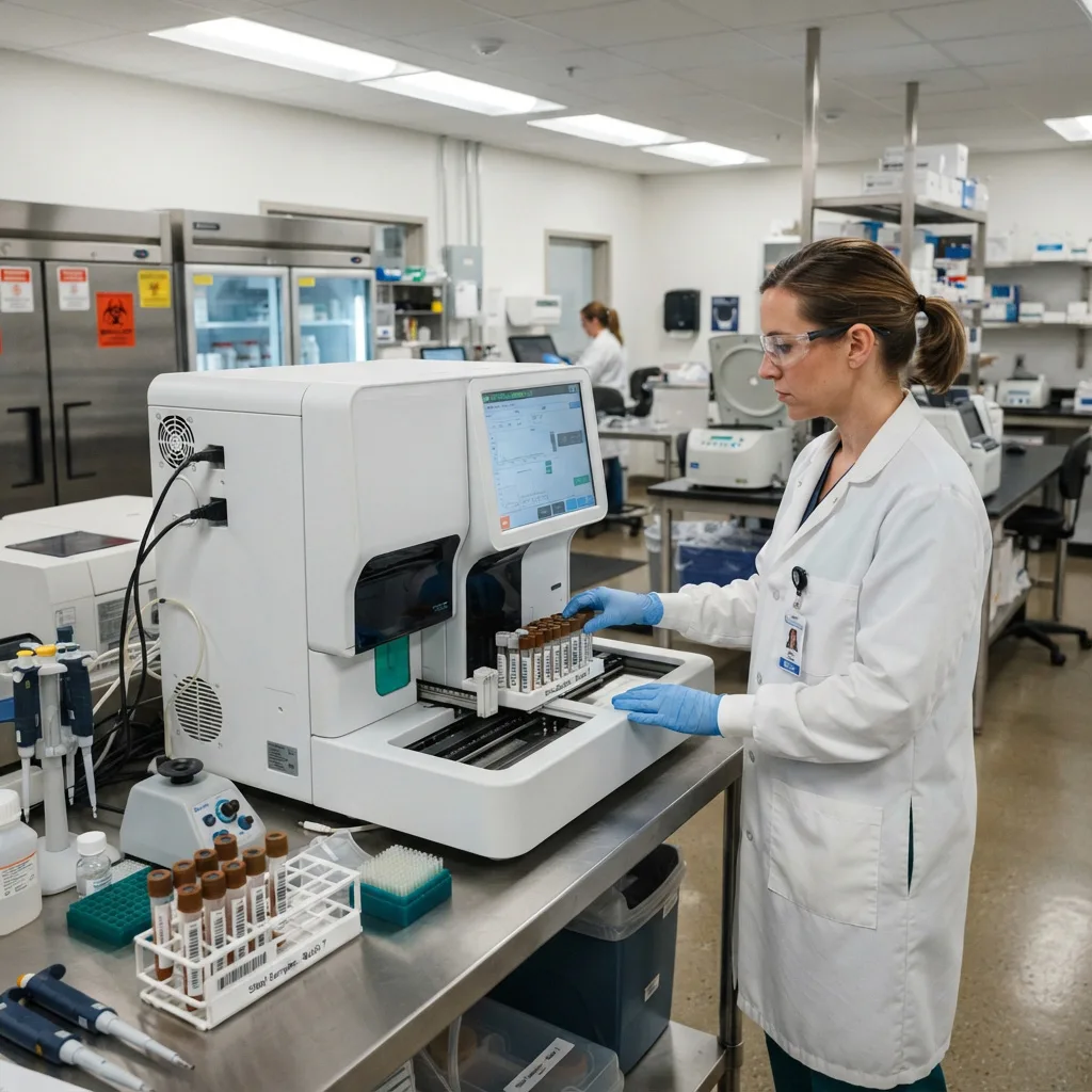
Historically, laboratories had to choose between two flawed standalone methods: Chemical FOBT (Guaiac / Pyramidon) or Immunochemical FOBT (FIT). Modern automated fecal analyzers resolve the limitations of both by running Dual-Method Occult Blood Testing simultaneously.
Plaintext
         WHY DUAL-METHOD OCCULT BLOOD TESTING PREVENTS MISDIAGNOSIS
+-------------------------------------------------------------------------+
| SCENARIO A: Upper GI Bleeding (Gastric Ulcer / Gastritis)               |
| - Stomach acid and pepsin digest the globin protein of hemoglobin.      |
| - Standalone FIT (Anti-Human Globin) ---> FALSE NEGATIVE                |
| - Dual Method (Chemical Heme + Transferrin) ---> TRUE POSITIVE          |
|                                                                         |
| SCENARIO B: Massive Lower GI Hemorrhage (Advanced Colonic Tumor)        |
| - Excessive free hemoglobin floods the antibody binding sites.          |
| - Standalone FIT (Prozone / Hook Effect) ---> FALSE NEGATIVE            |
| - Dual Method (Chemical FOB + Optical Alert) ---> TRUE POSITIVE         |
+-------------------------------------------------------------------------+
A. The Limitations of Chemical FOBT (cFOBT / gFOBT) Alone
Chemical occult blood tests rely on the pseudoperoxidase activity of the iron-containing heme moiety of hemoglobin to oxidize a chromogen (such as guaiac resin or tetramethylbenzidine) in the presence of hydrogen peroxide, producing a blue-green color shift.
-
Strength: Because the iron-porphyrin heme ring resists degradation by gastric hydrochloric acid and digestive proteases, chemical tests can detect bleeding originating high in the esophagus, stomach, or duodenum.
-
Weakness: Low specificity. Dietary animal hemoglobin from rare red meat (beef, lamb, pork), plant peroxidases from raw vegetables (horseradish, turnips, broccoli, cauliflower), and gastrointestinal irritation from NSAIDs cause high rates of false-positive results unless patients adhere to strict 3-day dietary restrictions. Conversely, high dietary Vitamin C (ascorbic acid greater than 250 mg/day) acts as a strong reducing agent that blocks oxidation, causing false-negative results.
B. The Limitations of Immunochemical Testing (FIT / iFOBT) Alone
Fecal Immunochemical Tests utilize monoclonal or polyclonal antibodies engineered to bind specifically to the intact globin protein chains of human hemoglobin.
-
Strength: Zero cross-reactivity with animal meat or vegetable peroxidases (no dietary restrictions required) and extreme sensitivity to trace lower gastrointestinal bleeding from colorectal adenomas and carcinomas.
-
Weakness 1 (Upper GI Blindness): If bleeding occurs in the stomach or upper small bowel, digestive proteolytic enzymes cleave the human globin protein before it reaches the rectum, rendering standard anti-hemoglobin FIT negative.
-
Weakness 2 (The High-Dose “Hook Effect” / Prozone Phenomenon): In lateral-flow immunochromatographic cassettes, a fixed concentration of gold-conjugated antibodies awaits the sample. If a patient has heavy lower gastrointestinal bleeding (exceeding 1,000 to 2,000 micrograms of hemoglobin per gram of stool), the massive excess of free hemoglobin saturates both the mobile gold-labeled antibodies and the immobilized capture antibodies on the test line independently. Prevented from forming the required “antibody-antigen-antibody sandwich,” the test line fails to appear—yielding a dangerous false-negative result in a heavily bleeding patient.
C. How Automated Dual-Method Testing + Transferrin Solves Both
Advanced automated fecal analyzers eliminate these diagnostic blind spots through two integrated protocols:
-
Simultaneous Chemical + Immunochemical Testing: By assaying the fecal suspension with both an immunochemical FIT cassette and a chemical heme channel (or comparing FIT against microscopic RBC counts and physical stool color), any sample with massive bleeding that triggers an immunological prozone hook effect will light up strongly positive on the chemical channel, alerting the software to flag a high-concentration hook effect.
-
Combined Hemoglobin + Transferrin (Hb/Tf) Dual Dipsticks: Transferrin is an iron-binding plasma glycoprotein present in blood that is significantly more stable against gastric acid and digestive bacterial degradation than hemoglobin. By incorporating dual-line immunochromatographic cards that detect both Human Hemoglobin (Hb) and Human Transferrin (Tf), automated analyzers capture both upper and lower gastrointestinal tract bleeding with high immunological specificity.
| Diagnostic Feature | Standalone Chemical FOBT | Standalone Immunochemical (FIT) | Automated Dual-Method (FIT + Hb/Tf + Chem) |
| Target Molecule | Heme iron ring (Peroxidase) | Intact human globin protein | Human Globin + Transferrin + Heme |
| Dietary Restrictions | Mandatory 3-day meat/veggie ban | None required | None (Software resolves discrepancies) |
| Vitamin C Interference | High risk of false negatives | Immune to Vitamin C | Immune on FIT/Tf channels |
| Lower GI Sensitivity (CRC) | Moderate (45% – 65%) | High (80% – 95%) | Maximum (Over 95% combined sensitivity) |
| Upper GI Bleeding Detection | Moderate | Poor (Globin digested by pepsin) | High (Via acid-stable Transferrin & Heme) |
| Heavy Bleed “Hook Effect” | Resistant (Stays strongly positive) | Vulnerable to false-negative prozone | Eliminated (Chemical channel catches excess) |
3. Automated Parasitology: Catching Helminth Ova and Protozoan Cysts
Intestinal parasitic infections affect hundreds of millions of individuals globally, causing iron-deficiency anemia, pediatric growth stunting, chronic malabsorption, and biliary obstruction. Yet clinical parasitology is notoriously difficult to master manually because parasite shedding is cyclical and morphological identification requires years of microscopic training.
Automated fecal analyzers revolutionize parasite detection through large-volume homogenization, selective micro-filtration, automated Lugol’s iodine staining, and deep-learning morphological recognition.
Plaintext
                  CLINICAL ATLAS OF FECAL PARASITE TARGETS
+-------------------------------------------------------------------------+
| 1. NEMATODES (Roundworms)                                               |
|    - Ascaris lumbricoides (Mammillated fertile & decorticated ova)      |
|    - Trichuris trichiura (Whipworm: barrel-shaped with bipolar plugs)   |
|    - Hookworm (Ancylostoma / Necator:thin hyaline shell, cleaved morula)|
|    - Enterobius vermicularis (Pinworm: flattened D-shaped translucent)  |
|                                                                         |
| 2. TREMATODES (Flukes) & CESTODES (Tapeworms)                           |
|    - Clonorchis sinensis (Liver fluke: tiny lightbulb shape + operculum)|
|    - Schistosoma japonicum / mansoni (Lateral or terminal spine ova)    |
|    - Taenia spp. (Thick radially striated embryophore with 6 hooklets)  |
|    - Hymenolepis nana (Dwarf tapeworm:polar filaments in inner membrane)|
|                                                                         |
| 3. INTESTINAL PROTOZOA (Cysts & Trophozoites)                           |
|    - Giardia duodenalis (lamblia) (Oval quadrinucleate cysts)           |
|    - Entamoeba histolytica / coli (1-to-8 nuclei + chromatoid bars)     |
|    - Blastocystis hominis (Vacuolar ring forms)                         |
+-------------------------------------------------------------------------+
Why Automated Analyzers Outperform Manual Smears in Parasitology
-
100-Fold Increase in Examined Sample Mass: Instead of picking up a 2-milligram speck on a toothpick, the sealed collection wand scoops a standardized 300-to-500-milligram core sample. Internal vortex mixing and multi-stage filtration strip away bulky undigested vegetable fibers and coarse fecal detritus while concentrating microscopic ova and cysts (ranging from 8 micrometers to 150 micrometers in size) into a clean optical counting chamber.
-
Automated On-Board Lugol’s Iodine Staining: Translucent protozoan cysts—such as Giardia lamblia and Entamoeba histolytica—are nearly invisible in plain saline brightfield microscopy because their refractive index matches the surrounding liquid. Automated analyzers feature a dedicated iodine staining channel that mixes diluted Lugol’s iodine into the suspension, staining glycogen vacuoles reddish-brown and highlighting internal nuclear karyosomes, axonemes, and median bodies in crisp contrast.
-
AI-Assisted Morphological Atlas: A technician who rarely sees tropical flukes like Clonorchis sinensis (measuring just 27 by 15 micrometers) or Fasciolopsis buski can easily overlook them or confuse pollen grains and plant hair cells with worm eggs. The analyzer’s convolutional neural network scans dozens of high-power fields at multiple focal depths, crops every suspected ovum or cyst, and presents them in a high-definition gallery on screen for instant confirmation.
4. Cellular Formed Elements & Digestive Function Markers
Beyond occult blood and parasites, microscopic examination of formed elements in stool allows clinicians to differentiate immediately between invasive inflammatory diarrhea and non-inflammatory secretory or malabsorptive diarrhea.
Plaintext
               DIFFERENTIAL DIAGNOSIS VIA FECAL MICROSCOPY
+-------------------------------------------------------------------------+
| INVASIVE / INFLAMMATORY ENTERITIS    | NON-INFLAMMATORY / MALABSORPTIVE |
+--------------------------------------+----------------------------------+
| - Abundant Fecal Leukocytes (WBCs)   | - Zero or scarce Fecal WBCs      |
| - Intact Red Blood Cells (RBCs)      | - Abundant Neutral Fat Globules  |
| - Macrophages & Pyocytes (Pus cells) | - Undigested Striated Muscle     |
| - Causes: Shigella, Campylobacter,   | - Starch Granules (Amylorrhea)   |
|   Invasive E. coli, Salmonella,      | - Causes: Viral enteritis,       |
|   Ulcerative Colitis, Crohn's Flare  |  Pancreatic insufficiency, Celiac|
+-------------------------------------------------------------------------+
Key Microscopic Formed Elements Quantified by Automated Systems
-
Fecal Leukocytes (WBCs) & Pyocytes: Healthy stool contains virtually zero white blood cells. Finding sheets of polymorphonuclear neutrophils or degenerated pus cells confirms that the colonic mucosa has been breached—pointing directly toward invasive bacterial dysentery (Shigella, Salmonella, Campylobacter, enteroinvasive E. coli) or an acute flare of Inflammatory Bowel Disease (Ulcerative Colitis or Crohn’s disease).
-
Fecal Erythrocytes (RBCs): Intact red blood cells only survive in stool if bleeding originates in the lower colon, sigmoid, rectum, or anal canal (as upper GI blood cells are digested into hematin before excretion).
-
Charcot-Leyden Crystals: Slender, hexagonal, double-pointed needle crystals formed from the breakdown of eosinophil granules (galectin-10 protein). Their presence in stool is a classic microscopic hallmark of eosinophilic gastroenteritis, allergic colitis, or invasive parasitic infections such as amoebiasis and trichuriasis.
-
Fat Globules (Steatorrhea) & Sudan III Staining: Rounded, highly refractive droplets of neutral triglycerides and fatty acids. When paired with automated Sudan III lipophilic staining (which turns fat droplets bright orange-red), elevated fecal fat globules indicate exocrine pancreatic insufficiency (chronic pancreatitis, cystic fibrosis), biliary obstruction, or small-bowel celiac malabsorption.
-
Undigested Striated Muscle Fibers (Creatorrhea) & Starch Granules: Finding rectangular muscle fibers with preserved transverse striations or iodine-stained blue-black starch granules signals rapid intestinal transit or deficiency of pancreatic trypsin and amylase.
-
Fungal Spores & Pseudohyphae (Candida): While trace budding yeast cells can be normal commensals, dense clusters of Candida blastospores sprouting elongated pseudohyphae alert clinicians to fungal dysbiosis—particularly critical in ICU patients receiving broad-spectrum intravenous antibiotics, chemotherapy patients, and premature neonates.
5. Multi-Card Immunochromatography: Enteric Viruses, H. pylori, and IBD Biomarkers
Modern automated fecal analyzers are no longer limited to wet microscopy and occult blood. By incorporating multi-slot colloidal gold immunochromatographic cassette carousels, a single fecal suspension tube can be automatically pipetted onto up to 4 to 8 different rapid diagnostic test cards simultaneously.
Plaintext
                  AUTOMATED MULTI-CARD IMMUNOCHROMATOGRAPHY
+-------------------------------------------------------------------------+
|                  [ Single Diluted Fecal Sample Tube ]                   |
|                                    |                                    |
|      +------------------+----------+----------+------------------+      |
|      |                  |                     |                  |      |
|      v                  v                     v                  v      |
| [ ONCOLOGY ]    [ PEDIATRIC VIRAL ]   [ GASTRIC ULCER ]  [ IBD vs IBS ] |
| - Hb (FIT)      - Rotavirus Group A   - H. pylori Ag     - Calprotectin |
| - Transferrin   - Adenovirus 40/41    - C. difficile     - Lactoferrin  |
|                 - Norovirus GI/GII      GDH + Toxin A/B                 |
+-------------------------------------------------------------------------+
1. Pediatric Infectious Diarrhea Triage (Rotavirus, Adenovirus, Norovirus)
In pediatric emergency departments, acute gastroenteritis is a leading cause of severe dehydration. However, prescribing antibiotics for a viral infection destroys normal gut flora and fuels antimicrobial resistance.
-
Automated fecal analyzers run combination Rotavirus Group A and Enteric Adenovirus (Serotypes 40/41) cassettes alongside routine microscopy.
-
Delivering an objective viral antigen confirmation alongside a negative fecal leukocyte count within 15 minutes gives pediatricians the confidence to withhold unnecessary antibiotics and focus on oral or intravenous rehydration therapy.
2. Non-Invasive Helicobacter pylori Stool Antigen Testing (HpSA)
Helicobacter pylori colonizes the gastric mucosa, causing chronic active gastritis, peptic ulcer disease, and gastric adenocarcinoma.
-
Unlike blood antibody serology—which remains positive for months or years after an infection has been cured—the fecal H. pylori antigen test detects active bacterial shedding directly in the stool.
-
Automated analyzers read monoclonal H. pylori antigen cassettes both for initial diagnosis in dyspeptic patients and for confirming bacterial eradication 4 weeks after completing antibiotic triple or quadruple therapy.
3. Differentiating IBD from IBS: Fecal Calprotectin & Lactoferrin
Millions of patients present to gastroenterology clinics with chronic abdominal cramping and altered bowel habits. Clinicians must distinguish between Irritable Bowel Syndrome (IBS)—a functional disorder with no mucosal damage—and Inflammatory Bowel Disease (IBD: Crohn’s disease and Ulcerative Colitis), which causes destructive ulcers requiring immunosuppressant therapy and colonoscopy.
-
Fecal Calprotectin: A calcium- and zinc-binding protein accounting for 60% of the cytosolic protein in human neutrophils. When intestinal inflammation occurs, neutrophils migrate into the gut lumen and release calprotectin, which resists bacterial degradation for up to 7 days at room temperature.
-
Fecal Lactoferrin: An iron-binding glycoprotein secreted by activated polymorphonuclear leukocytes.
-
Automated optical reading of semi-quantitative or quantitative Calprotectin and Lactoferrin cassettes provides an accurate non-invasive biomarker cutoff: patients with low calprotectin (less than 50 micrograms per gram) can safely avoid invasive diagnostic colonoscopies, while elevated levels prioritize urgent endoscopic evaluation and monitor mucosal healing during biologic therapy.
6. Clinical Impact: How Automation Elevates Hospital Diagnostic Quality
Transitioning from manual wooden-stick smears to an automated fecal analyzer delivers measurable clinical and operational gains across the entire hospital ecosystem:
| Clinical & Operational Metric | Traditional Manual Wet-Mount Bench | Automated Fecal Analyzer Workflow |
| Laboratory Biosafety Level | High Risk (Open cups, aerosols, direct odor) | Fully Sealed (Zero operator contact with raw stool) |
| Sample Mass Examined | 2 to 5 mg (Extreme random sampling error) | 300 to 500 mg (Homogenized & micro-filtered) |
| Parasite Ova Detection Rate | Baseline (Misses low-burden infections) | Increased by 30% to 50%+ via concentration |
| Occult Blood Reliability | Visual subjective color check on open bench | Automated optical reader + Dual Hb/Transferrin |
| Visual Result Traceability | Zero (Slide discarded immediately into trash) | Permanent HD digital photo archive in LIS/HIS |
| Turnaround Time (TAT) | Slow batching; bottlenecks in morning rush | Rapid continuous walk-away processing |
| Laboratory Accreditation | Difficult to standardize inter-tech QC | ISO 15189 compliant with liquid/image QC logs |
Frequently Asked Questions (FAQ)
Q1: Why is a positive chemical FOBT paired with a negative immunochemical FIT clinically important?
When a specimen tests negative on an immunochemical FIT channel but strongly positive on a chemical occult blood channel, three clinical possibilities exist: (1) Upper gastrointestinal bleeding (such as a bleeding gastric or duodenal ulcer) where digestive enzymes degraded the globin protein before excretion while leaving the iron heme ring intact; (2) Massive lower gastrointestinal bleeding triggering an immunological prozone “hook effect” that saturated the FIT antibodies; or (3) Dietary peroxidase interference from red meat consumption. Automated analyzers flag this exact pattern so clinicians can investigate upper GI sources or perform a 1:10 sample dilution to unmask a hook-effect hemorrhage.
Q2: How long can a stool sample sit before being tested on an automated fecal analyzer?
For standard helminth ova, protozoan cysts, and fecal calprotectin, samples collected in sealed preservative or diluent tubes remain stable for several hours. However, if the clinician suspects motile protozoan trophozoites (such as Entamoeba histolytica or Giardia trophozoites in acute watery diarrhea), the specimen should be processed within 30 to 60 minutes of passage, as fragile trophozoites lose motility and lyse rapidly once the sample cools to room temperature. Similarly, hemoglobin degrades slowly in warm environments, making rapid automated processing essential for accurate FIT quantification.
Q3: Why can’t fecal leukocytes alone replace a Fecal Calprotectin test for IBD?
Intact fecal leukocytes (WBCs) are fragile and begin to degranulate and lyse within hours of passing through the intestinal lumen, catalyzed by bacterial proteases and osmotic shifts. Consequently, a manual or microscopic WBC count can appear falsely low if transport is delayed. Fecal Calprotectin, released inside those neutrophils, is remarkably resistant to enzymatic proteolysis and remains stable in stool for days, providing a reliable quantitative measure of total neutrophil migration into the gut even when intact cell walls have lysed.
Q4: How does automated iodine staining improve protozoan detection without destroying other tests?
In manual microscopy, adding Lugol’s iodine directly to a single wet mount kills motile trophozoites (preventing observation of their characteristic movement) and can obscure the refractile appearance of unstained fat droplets or certain crystals. Automated fecal analyzers solve this by splitting the aspirated filtrate into parallel optical channels or sequential imaging passes: first capturing native brightfield frames to evaluate natural color, RBCs, WBCs, and motile organisms, and then injecting Lugol’s iodine or Sudan III stain into a secondary sub-volume to highlight protozoan cyst nuclei and glycogen vacuoles.
Q5: Does an automated fecal analyzer require patients to collect stool differently?
In many outpatient workflows, patients or ward nurses can either use a dedicated sealed quantitative sampling tube (featuring a grooved collection wand held inside a pre-filled buffer vial that goes directly onto the analyzer rack) or submit a standard clinic stool cup that a lab technician samples once using a snap-shut filtration tube. Both pathways eliminate open-slide smearing and keep the biological material hermetically contained inside the analyzer through disposal.