The Clinical Shift Toward Bedside Imaging
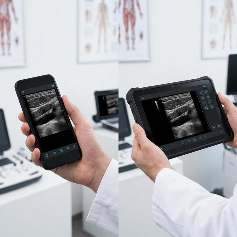
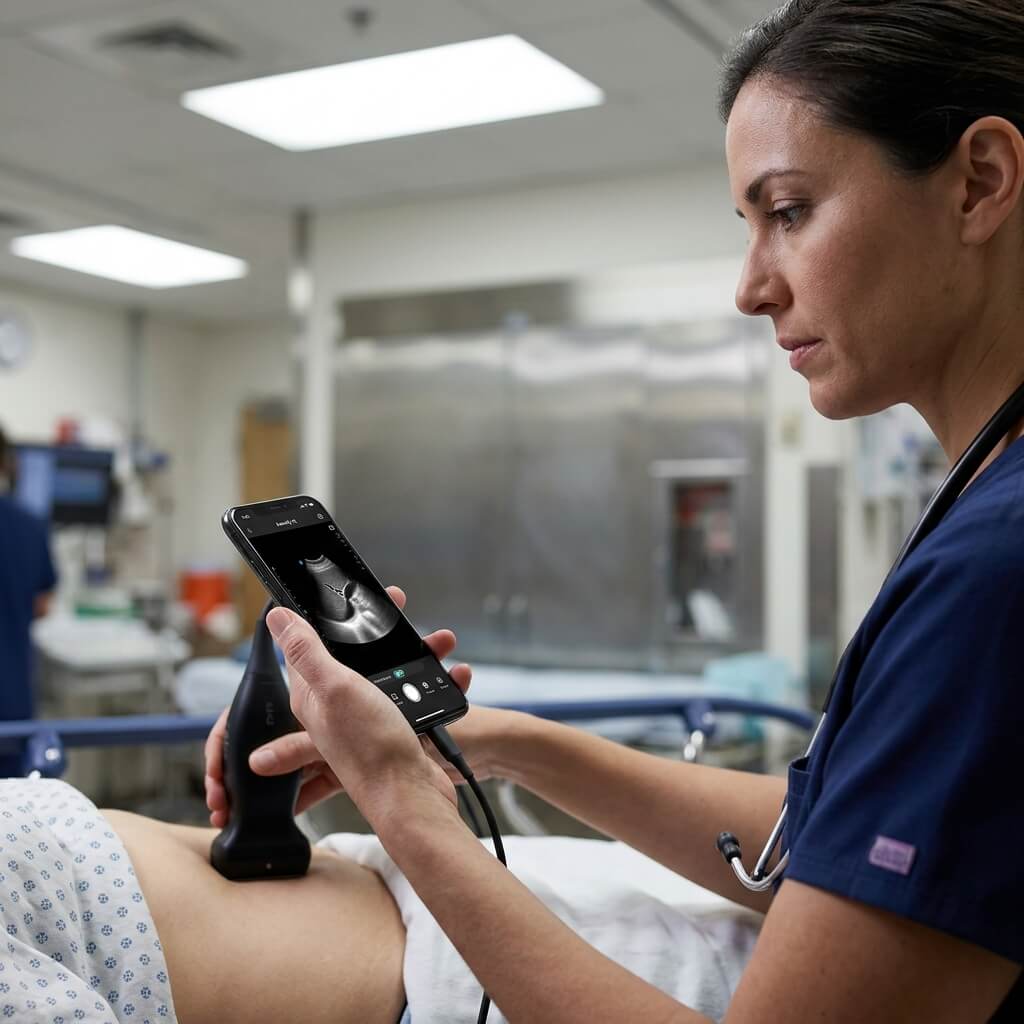
Point-of-care ultrasound (POCUS) is no longer confined to specialized radiology departments. Across emergency rooms, intensive care units, outpatient clinics, and rural health stations, handheld ultrasound devices have become an essential diagnostic extension of the physical examination—often referred to as the “modern visual stethoscope.”
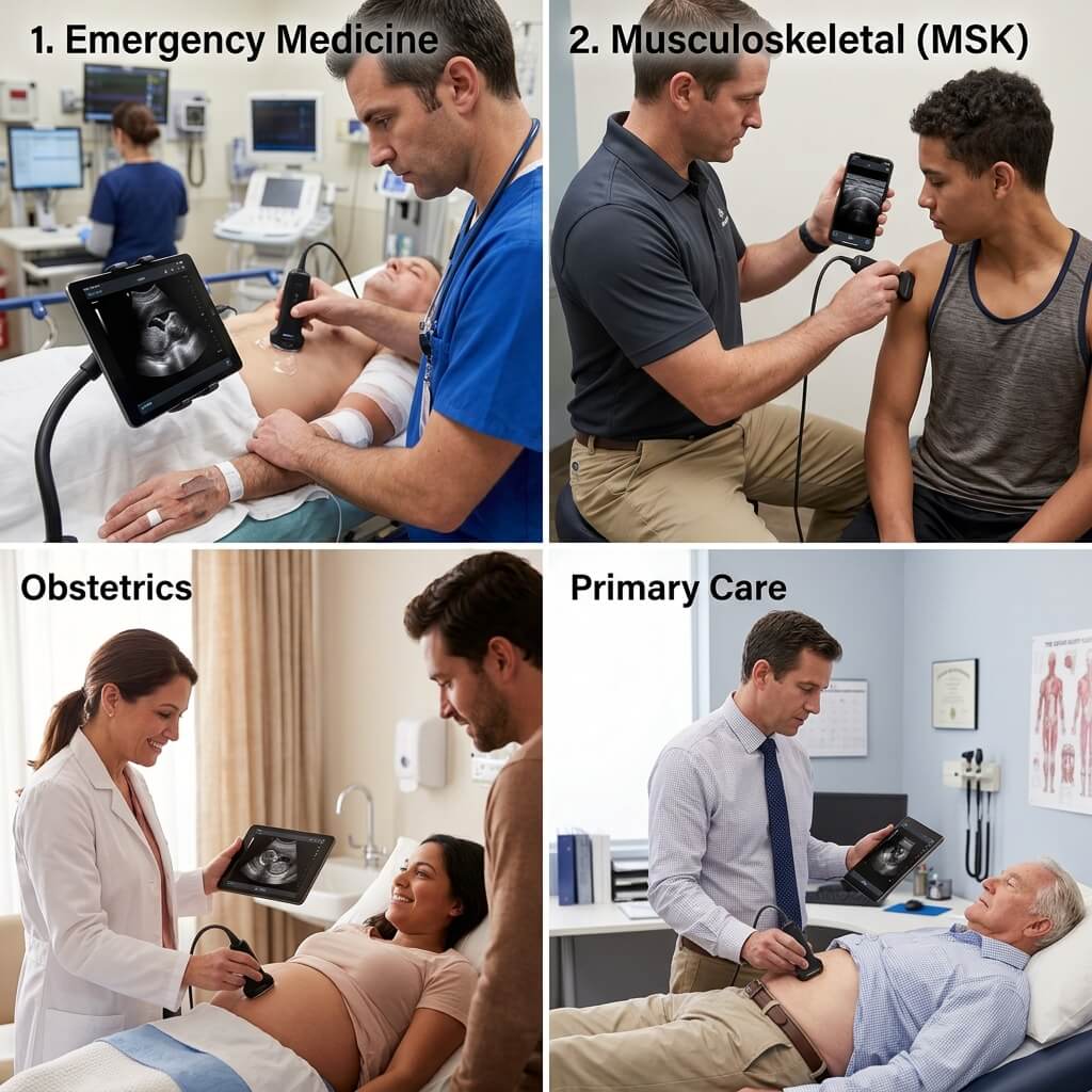
                       CLINICAL SPECTRUM OF POCUS
  +-------------------------------------------------------------------+
  | Emergency & Trauma  --> eFAST, Pneumothorax, Cardiac Arrest       |
  | Musculoskeletal     --> Tendon Tears, Joint Injections, Nerve     |
  | Obstetrics / GYN    --> Fetal Viability, Amniotic Fluid, Ectopic   |
  | Vascular & Anesthesia--> Central Lines, Nerve Blocks, DVT Scan    |
  | Primary Care        --> AAA Screening, Gallbladder, Bladder Scan  |
  +-------------------------------------------------------------------+
By bringing immediate, non-invasive imaging directly to the patient’s bedside, clinicians can establish rapid diagnoses, shorten emergency department stay times, minimize patient transport risks, and significantly improve first-pass needle placement accuracy.
Below is an in-depth breakdown of how pocket ultrasound scanners elevate clinical care across major medical disciplines.
2. Emergency Medicine, Trauma Triage & Critical Care
In emergency and trauma environments, every second counts. Handheld ultrasound scanners allow trauma teams to conduct rapid triage protocol assessments without waiting for a fixed console machine or moving unstable patients to a CT scanner.
                    eFAST TRAUMA DIAGNOSTIC PROTOCOL
+-------------------------------------------------------------------------+
| 1. RUQ (Morison's Pouch)  --> Detects free fluid in hepatoronal space.  |
| 2. LUQ (Splenorenal Recess)--> Detects perisplenic free fluid           |
| 3. Pelvis (Suprapubic View)--> Detects free fluid in rectovesical pouch |
| 4. Cardiac (Subxiphoid)   --> Evaluates pericardial effusion/tamponade. |
| 5. Anterior Thoracic View --> Rules out pneumothorax (sliding sign)     |
+-------------------------------------------------------------------------+
The eFAST Protocol (Extended Focused Assessment with Sonography for Trauma)
Using a convex or phased array handheld transducer, emergency physicians execute the eFAST protocol within minutes to detect life-threatening internal bleeding:
-
Abdominal Free Fluid: Rapidly visualizes blood accumulation in Morison’s pouch (Right Upper Quadrant), the splenorenal recess (Left Upper Quadrant), and the pelvis.
-
Pericardial Tamponade: Evaluates the pericardial space for fluid collection that restricts cardiac pumping during blunt chest trauma.
-
Pneumothorax Detection: Uses high-frequency linear scanning to observe pleural lung sliding. The absence of lung sliding or the presence of a “barcode sign” on M-mode confirms a pneumothorax.
Critical Care & Resuscitation
-
Rapid Cardiac Assessment: Phased array handheld probes allow intensivists to evaluate left ventricular ejection fraction (EF), assess right ventricular strain in suspected pulmonary embolism, and guide fluid resuscitation by measuring Inferior Vena Cava (IVC) diameter and respiratory collapse.
-
Acute Respiratory Failure (BLUE Protocol): Differentiates cardiogenic pulmonary edema (characterized by lung B-lines) from pneumonia, COPD, or pulmonary embolism at the bedside.
3. Musculoskeletal (MSK), Sports Medicine & Facial Aesthetics
High-frequency linear handheld ultrasound probes (7.5 MHz to 18 MHz) have revolutionized outpatient MSK care and minimally invasive aesthetic procedures by providing ultra-clear visualization of superficial anatomical structures.
                  SUPERFICIAL MSK & AESTHETIC DIAGNOSTICS
+---------------------------------------------------------------------+
| TENDONS & LIGAMENTS --> Rotator cuff, Achilles tendon, sprains      |
| JOINTS & BURSAE     --> Joint effusions, bursitis, gouty tophi      |
| NERVE ENTRAPMENT    --> Median nerve swelling (Carpal Tunnel)       |
| FACIAL AESTHETICS   --> Vascular mapping prior to hyaluronic filler |
+---------------------------------------------------------------------+
Sports Medicine & Orthopedics
-
Tendon & Ligament Evaluation: Instantly identifies full-thickness vs. partial-thickness rotator cuff tears, Achilles tendonitis, plantar fasciitis, and ligamentous sprains. Dynamic real-time ultrasound allows clinicians to flex joints during the examination to observe tissue movement.
-
Injections & Aspiration Guidance: Ultrasound guidance ensures 100% accurate needle placement into intra-articular spaces (glenohumeral joint, knee joint, hip) for corticosteroid, hyaluronic acid, or platelet-rich plasma (PRP) therapies while avoiding surrounding nerves and vessels.
Facial Aesthetics & Vascular Mapping
-
Filler Safety: In cosmetic dermatology, high-frequency linear pocket probes allow injectors to map the course of the facial artery and angular artery prior to hyaluronic acid dermal filler injections, dramatically reducing the risk of accidental vascular occlusion and necrosis.
-
Complication Management: Enables real-time guided injection of hyaluronidase to dissolve misplaced or intravascular filler material.
4. Obstetrics & Gynecology (OB/GYN) & Maternal Health
Access to diagnostic imaging during pregnancy is vital for reducing maternal and neonatal mortality. Pocket ultrasound transducers empower obstetricians, midwives, and rural healthcare workers to provide essential maternal screening anywhere.
                    OBSTETRIC SCREENING WORKFLOW
+----------------------------------------------------------------------+
| FIRST TRIMESTER  --> Confirm intra-uterine pregnancy & fetal heart   |
| SECOND/THIRD     --> Measure BPD, FL, AC for gestational age & weight|
| PLACENTAL/FLUID  --> Assess placental location & Amniotic Fluid Index|
| EMERGENCY SCREEN --> Rule out ectopic pregnancy & placental abruption|
+----------------------------------------------------------------------+
Key Obstetric Applications
-
First-Trimester Assessment: Confirms intrauterine pregnancy, visualizes the gestational sac and yolk sac, measures Crown-Rump Length (CRL) for accurate pregnancy dating, and confirms fetal cardiac activity using M-mode.
-
Growth & Biometry Monitoring: Measures Biparietal Diameter (BPD), Femur Length (FL), and Abdominal Circumference (AC) to assess fetal growth trajectory and estimated fetal weight (EFW).
-
Placental & Amniotic Fluid Evaluation: Checks for placenta previa (placenta covering the cervical os) and measures the Amniotic Fluid Index (AFI) to screen for oligohydramnios or polyhydramnios.
-
Ectopic Pregnancy Screening: In acute pelvic pain, bedside scanning helps clinicians rule out ectopic pregnancies in the adnexa or fallopian tubes.
5. Vascular Access, Regional Anesthesia & Interventional Procedures
Point-of-care ultrasound has become the global standard of care for needle-guided vascular and anesthetic procedures, turning blind landmark techniques into safe, visually guided interventions.
               ULTRASOUND-GUIDED NEEDLE INTERVENTIONS
+---------------------------------------------------------------------+
| CENTRAL VENOUS ACCESS --> Internal Jugular & Subclavian Vein lines  |
| PERIPHERAL LINES      --> Deep brachial/basilic vein cannulation    |
| REGIONAL ANESTHESIA   --> Brachial plexus, femoral & TAP blocks     |
| VASCULAR SCREENING    --> Deep Vein Thrombosis (DVT) compression    |
+---------------------------------------------------------------------+
Vascular Access
-
Central Venous Catheterization (CVC): Real-time linear ultrasound visualization of the Internal Jugular Vein (IJV), Femoral Vein, or Subclavian Vein reduces arterial puncture rates, hematoma formation, and pneumothorax complications.
-
Difficult Peripheral IV Access: Allows nurses and paramedics to cannulate deep brachial or basilic veins in obese, edematous, or oncology patients with poor superficial veins.
Regional Anesthesia & Pain Management
-
Peripheral Nerve Blocks: Anesthesiologists visualize local nerve plexus bundles (brachial plexus, femoral nerve, sciatic nerve, fascia iliaca, TAP block) and monitor the real-time spread of local anesthetic fluid around the target nerve sheath.
-
Deep Vein Thrombosis (DVT) Screening: Enables 2-point or 3-point compression ultrasound of the femoral and popliteal veins to diagnose acute lower-extremity deep vein thrombosis at the bedside.
6. Primary Care, Internal Medicine & Outpatient Screening
For family physicians, general practitioners, and outpatient internists, handheld ultrasound acts as a diagnostic accelerator, eliminating unnecessary hospital referrals and providing immediate answers during routine physical exams.
                    PRIMARY CARE OUTPATIENT SCREENING
+---------------------------------------------------------------------+
| ABDOMINAL  --> Gallstones, cholecystitis, hepatic steatosis (fatty) |
| RENAL/BLAD --> Hydronephrosis, renal calculi, post-void urine vol   |
| CARDIAC    --> Screening for Abdominal Aortic Aneurysm (AAA)        |
| LUNG/THORAX--> Pleural effusion, pneumonia consolidation detection  |
+---------------------------------------------------------------------+
Primary Care Diagnostic Use Cases
-
Abdominal Aortic Aneurysm (AAA) Screening: Rapid measurement of the abdominal aorta diameter in high-risk patients (smokers, elderly males) screens for life-threatening aneurysms.
-
Gallbladder & Liver Evaluation: Identifies gallstones (cholelithiasis), gallbladder wall thickening, acoustic shadowing, and fatty liver disease during routine abdominal check-ups.
-
Renal & Bladder Assessment: Evaluates flank pain for hydronephrosis (kidney swelling caused by urinary obstruction) and measures post-void residual (PVR) bladder volume using automated measurement tools.
7. Master Matrix: Clinical Presets vs. Probe Types & Frequencies
The matrix below illustrates the recommended probe selection, frequency range, and clinical software presets across primary medical specialties:
| Medical Specialty | Primary Clinical Indication | Recommended Transducer Type | Optimal Frequency | Essential Software Preset |
| Emergency Medicine | eFAST Trauma, Free Fluid | Convex / 3-in-1 Probe | 2.5 MHz – 5.0 MHz | Abdomen / Trauma Preset |
| Cardiology / ICU | Ejection Fraction, IVC Collapse | Phased Array / 3-in-1 Probe | 2.0 MHz – 4.0 MHz | Cardiac Preset |
| MSK / Orthopedics | Tendon Tear, Joint Injection | High-Frequency Linear | 7.5 MHz – 14.0 MHz | MSK / Small Parts Preset |
| Facial Aesthetics | Vascular Mapping, Filler Check | Ultra-High Linear Probe | 10.0 MHz – 18.0 MHz | Aesthetics / Vascular Preset |
| OB / GYN | Gestational Age, Fetal Heart | Convex / Dual-Head Probe | 2.5 MHz – 5.0 MHz | Obstetrics / Pelvic Preset |
| Vascular / Anesthesia | Central Line, Nerve Block | Linear Transducer | 6.0 MHz – 12.0 MHz | Vascular / Nerve Preset |
| Urology / Nephrology | Bladder Volume, Hydronephrosis | Convex Transducer | 3.0 MHz – 5.0 MHz | Urology / Renal Preset |
8. Why UMY Medical Equipment Co Ltd Empowers Clinical Excellence
Selecting handheld diagnostic equipment that performs consistently across diverse medical specialties requires precision acoustic engineering, robust physical construction, and specialized image processing software.
                 UMY MEDICAL CLINICAL SOLUTIONS
                               |
     +-------------------------+-------------------------+
     |                         |                         |
     v                         v                         v
[ Specialized Clinical Presets ] [ High-Purity Transducers ] [ Durable Medical Casing ]
 One-Touch Optimization Algorithms 128 / 192 Element Crisp Images IP67 Waterproof & Submersible
Engineering Advantages for Healthcare Providers:
-
One-Touch Optimized Clinical Presets: UMY Medical Equipment Co Ltd equips its handheld devices with pre-calibrated algorithmic presets for Abdomen, Cardiac, MSK, Vascular, Urology, OB/GYN, and Small Parts. Clinicians can switch specialties with a single tap.
-
128 & 192-Element Crystal Clarity: High-density piezoceramic crystal arrays deliver sharp tissue contrast, high frame rates, and deep acoustic penetration, making subtle clinical findings easily recognizable.
-
Ruggedized Medical Casing: Designed for demanding emergency rooms, field hospitals, and mobile clinics, UMY Medical handheld probes feature IP67-rated waterproof housing, allowing full submersion sterilization and impact resistance.
-
Factory-Direct Value for Health Systems: By working directly with UMY Medical’s direct-factory manufacturing facility, hospitals, clinics, and government health agencies secure high-performance diagnostic tools without distributor markups.
Frequently Asked Questions (FAQ)
Q1: Is specialized training required to use a handheld ultrasound scanner?
Yes. While handheld ultrasound devices feature user-friendly touch interfaces and automated presets, proper diagnostic interpretation requires foundational POCUS training. Many clinical societies (ACEP, AIUM, WINFOCUS) offer certified courses in bedside ultrasound application.
Q2: How does a handheld ultrasound assist in needle-guided nerve blocks?
By placing a linear high-frequency probe over the targeted anatomic area, the clinician visualizes the nerve bundle, surrounding blood vessels, and the incoming needle tip in real time. This ensures local anesthetic is delivered precisely into the fascial plane surrounding the nerve without puncturing the nerve tissue or nearby blood vessels.
Q3: Can handheld ultrasound scanners detect lung conditions like pneumonia or COVID-19?
Yes. Lung ultrasound using convex or linear probes can identify A-lines (normal air-filled lung), B-lines (interstitial edema or fluid accumulation), pleural thickening, and subpleural consolidations indicative of pneumonia or severe respiratory infections.
Q4: Are handheld ultrasound probes suitable for low-resource or off-grid field clinics?
Absoluely. Handheld ultrasound scanners are ideal for remote clinics, disaster response teams, and mobile health vehicles. Their lightweight design, long battery runtime, USB/wireless charging options, and resistance to environmental dust and moisture make them superior to bulky console units in field conditions.
Q5: How can medical institutions test UMY Medical handheld devices for their clinical teams?
Healthcare directors, hospital procurement officers, and medical distributors can contact UMY Medical Equipment Co Ltd through their online sales portal to request technical specifications, clinical sample images, demonstration videos, and OEM partnership terms.