
Clinical Fundamentals: What Makes Arthrogram MRI Indispensable?
In modern musculoskeletal (MSK) diagnostics, precision is paramount. While conventional Magnetic Resonance Imaging (MRI) serves as the cornerstone for general soft-tissue evaluation, precise diagnostic demands—particularly regarding subtle intra-articular pathologies, micro-labral tears, and postoperative cartilage integrity—frequently require higher diagnostic specificity. This requirement has established Arthrogram MRI (Direct MR Arthrography) as the clinical gold standard for intricate joint imaging.
An Arthrogram MRI is a specialized two-stage diagnostic procedure combining fluoroscopic or ultrasound-guided intra-articular injection of a diluted contrast agent directly into the joint capsule, followed by high-resolution T1-weighted fat-suppressed MR scanning.
-
Joint Capsule Distension: Fluid instillation expands the joint cavity, physically separating adjacent anatomic structures and highlighting subtle defects.
-
High Contrast-to-Noise Ratio (CNR): Delineates contrast penetrating into subtle torn fibrocartilage.
-
Superior Diagnostic Sensitivity: Significantly outperforms standard non-contrast MRI in detecting ALPSA, SLAP lesions, and hip femoroacetabular impingement (FAI).
B2B Technical & Performance Comparison Matrix
| Diagnostic Metric / Feature | Conventional Non-Contrast MRI (1.5T / 3.0T) | Direct Arthrogram MRI (Contrast-Enhanced) |
| Primary Target Pathology | General ligamentous injury, bone marrow edema, muscle strain | Subtle labral tears (Glenoid/Acetabular), partial rotator cuff tears, postoperative joint recurrent tears |
| Sensitivity for Hip/Shoulder Labrum | Moderate (~65% – 78%) | Exceptional (~90% – 98%) |
| Auxiliary Equipment Needed | MRI Scanner, standard receiving coils | MRI Scanner + Guidance Unit (Ultrasound / C-Arm) + Injection Kits + Dedicated MSK Coils |
| Procurement Capital Complexity | Single modality investment | Multi-modality integration (Radiology suite + Interventional capabilities) |
| Patient Throughput Rate | Higher (approx. 20–30 mins per scan) | Moderate (requires pre-scan injection procedure + scan time) |
Key Infrastructure & Equipment Investment Requirements
To successfully establish a high-throughput Arthrogram MRI diagnostic workflow, healthcare providers must evaluate three essential hardware components:
-
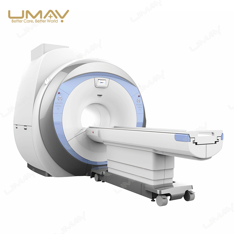
High-Field MRI Scanner Specifications: While 1.5T scanners remain global workhorses, 3.0T MRI systems offer superior signal-to-noise ratios (SNR), enabling thinner slice thicknesses critical for subtle MSK diagnostics.
-
Precision Guidance Modalities (Ultrasound / C-Arm Fluoroscopy): Direct arthrography requires accurate intra-articular needle placement. Integrating high-resolution diagnostic ultrasound units minimizes fluoroscopy radiation exposure and speeds up patient workflow.
-
Dedicated MSK Surface Coils: Equipping radiology suites with specialized phased-array coils ensures targeted signal acquisition and maximum clinical clarity.
Upgrade Your Diagnostic Capabilities with UMY Medical
Navigating medical imaging procurement requires choosing equipment engineered for long-term reliability, clinical clarity, and manageable maintenance overhead. At UMY Medical,we specialize in delivering comprehensive imaging and diagnostic solutions tailored for international hospitals, private clinics, and medical distributors.
From advanced digital radiography and specialized diagnostic imaging to comprehensive medical equipment integration, UMY Medical provides end-to-end support—including global logistics, regulatory compliance documentation, OEM options, and dedicated technical training.
Strategic B2B Procurement Advice
Compared to major tier-one players like GE Healthcare, Mindray, SonoScape, or United Imaging, hospital procurement teams should focus on:
-
Total Cost of Ownership (TCO): Balancing initial CapEx against maintenance, helium refills, and coil warranties.
-
Tiered Equipment Strategy: Pairing high-performance diagnostic ultrasound units for primary needle placement guidance alongside dependable 1.5T/3.0T MRI platforms yields optimal financial ROI.
Frequently Asked Questions (FAQ)
-
Q1: Is an Arthrogram MRI always necessary, or can a high-field 3.0T conventional MRI replace it?
-
Answer: While a 3.0T non-contrast MRI offers high resolution, direct MR arthrography remains clinically preferred for non-displaced labral tears and postoperative joint evaluations due to capsule distension providing physical separation of anatomical structures.
-
-
Q2: What complementary equipment should a hospital order when expanding into MR Arthrography services?
-
Answer: Hospitals should procure high-frequency linear ultrasound scanners for non-radiative needle placement guidance, specialized MSK surface coils, sterile injection accessory packages, and dedicated PACS diagnostic workstations.
-
-
Q3: How does UMY Medical support international procurement for hospital imaging projects?
-
Answer: UMY Medical provides end-to-end B2B solutions, including technical parameter matching, factory-direct pricing, customized OEM/ODM options, global shipping logistics, and full installation support for hospital diagnostic expansions. Contact our team at www.umymedicalequip.com for detailed technical catalogs.
-