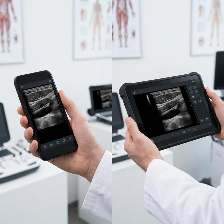
In modern dentistry, the transition from basic 2D screening X-rays to 3D Cone Beam Computed Tomography (CBCT) is fundamentally changing how clinicians diagnose, plan, and execute treatments. While 2D panoramic and periapical X-rays provide a general overview of dental structures, they flatten complex 3D anatomy into a single distorted plane—masking critical details that can mean the difference between clinical success and surgical failure.
For dental practice owners, specialists, and procurement directors, understanding the clinical applications of 3D CBCT imaging is essential. Investing in 3D diagnostic technology expands your in-house procedural scope, eliminates surgical surprises, lowers patient risk, and directly drives clinic revenue across every major dental specialty.
This guide details how 3D CBCT imaging is applied across implantology, endodontics, orthodontics, sleep dentistry, and oral surgery.
1. Dental Implant Planning & Guided Surgery
Placing dental implants without 3D imaging is like driving through an unfamiliar city without GPS. A 2D panoramic X-ray can only show bone height, leaving bone width, bone density, and ridge topography entirely unmapped.
Precise Bone Mapping & Grafting Decisions
3D CBCT cross-sections allow clinicians to evaluate exact bone dimensions before making the first incision:
-
Ridge Width Measurement: Instantly identify buccal or lingual bone concavities, undercut areas, and knife-edge ridges that require socket preservation or block bone grafting prior to implant placement.
-
Sinus Lift Planning: Measure sinus floor thickness, locate septa within the maxillary sinus, and assess mucosal membrane health to prevent sinus perforation during sinus elevation procedures.
Nerve Mapping & Critical Landmark Protection
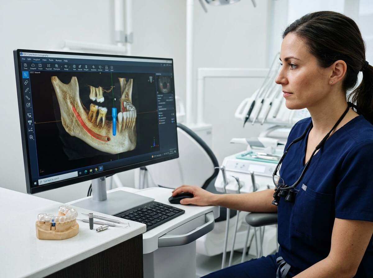
Surgical damage to the Inferior Alveolar Nerve (IAN) or Mental Foramen can cause permanent lip, chin, or tongue paresthesia.
-
CBCT software tools allow clinicians to highlight and draw the path of the nerve trunk directly onto the 3D volume in bright red or yellow.
-
Safety buffer margins (typically 2.0 mm) can be set around the nerve path to ensure drill depth settings never compromise neural tissue.
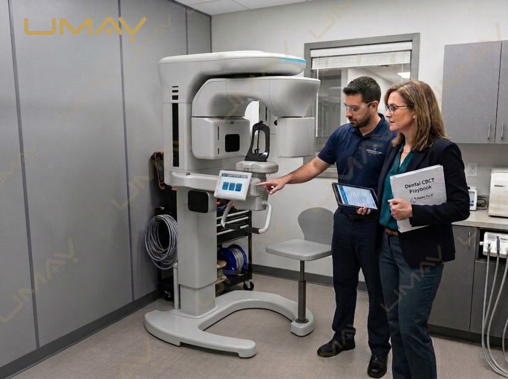
Technician explaining 3D CBCT scanner touchscreen interface to a clinic manager holding a CBCT playbook.
2. Orthodontics, Impacted Teeth & Airway Analysis
Orthodontists and pediatric dentists rely on 3D CBCT imaging to evaluate complex dental eruption paths, assess skeletal growth patterns, and diagnose sleep-disordered breathing.
Orthodontic & Airway CBCT Scope
———————————————————————–
| Clinical Need | 3D CBCT Clinical Benefit |
———————————————————————–
| Impacted Canines | Pinpoint exact 3D location & root resorption|
| Skeletal Asymmetry | Full 3D facial & craniofacial profiling |
| Airway / Sleep Apnea | Automated volumetric airway measurement |
| TAD Placement | Locate safe inter-radicular bone sites |
Managing Impacted Canines & Supernumerary Teeth
Impacted maxillary canines are among the most challenging teeth to exposure and align:
-
3D Localization: CBCT pinpoints whether the impacted tooth lies labially, palatably, or directly between adjacent incisor roots.
-
Resorption Checks: Determine if the crown of the impacted canine is actively resorbing the roots of lateral or central incisors—guiding the direction of orthodontic traction to avoid collateral damage.
Airway Volume & Sleep Dentistry Integration
Obstructive Sleep Apnea (OSA) and airway constrictions are increasingly treated in dental practices:
-
Large FOV CBCT scans (15×15 cm or larger) capture the entire pharyngeal airway passage from the nasal cavity to the epiglottis.
-
Diagnostic software automatically calculates total airway volume (in cubic centimeters) and highlights the narrowest cross-sectional area in red, helping clinicians design airway-expanding clear aligners, rapid palatal expanders (RPE), or mandibular advancement appliances.
3. Frequently Asked Questions (FAQ)
Is a 3D CBCT scan necessary for every routine dental patient?
No. CBCT scans should follow the ALADA principle (As Low As Diagnostically Acceptable). 3D scans should be ordered based on specific clinical needs—such as implant planning, endodontic re-treatment, impacted tooth evaluation, or unexplained jaw pain—rather than as a generic replacement for routine hygiene screenings.
What Field of View (FOV) is best for general dentists performing multiple specialties?
A Medium FOV unit (8*8 cm to 12*10 cm) is the ideal choice for general practices. It captures dual arches including the sinus floor, making it versatile enough for implants, full mouth restorative work, surgical extractions, and routine diagnostics.
Can 3D CBCT images detect dental caries (cavities)?
While CBCT can display large decay lesions, traditional 2D intraoral bitewing X-rays remain the primary standard for detecting early interproximal caries due to higher spatial resolution and zero streak artifacts from metal restorations.
How does Metal Artifact Reduction (MAR) work in clinical practice?
Metal fillings, crowns, and implants cause streak artifacts on 3D scans. Advanced MAR software algorithms filter out these scatter streaks during reconstruction, preserving tissue clarity around adjacent teeth and implants.
Conclusion: Elevating the Standard of Clinical Care
Integrating 3D CBCT imaging across your clinical workflows transforms your practice from a basic diagnostic clinic into a high-precision treatment center. Whether you are placing implants with digital surgical guides, locating hidden root canals, analyzing airway volume, or presenting treatment plans to patients, 3D CBCT technology provides the clarity, safety, and predictability needed to thrive in modern dentistry.
To be Continued…