The Ultimate Digital X-Ray Machine Buying Guide: How to Choose the Best DR System for Your Hospital or Clinic
Introduction
Investing in a digital X-ray machine is one of the most important decisions for hospitals, diagnostic imaging centers, orthopedic clinics, emergency departments, veterinary hospitals, and private medical practices. As healthcare providers continue to transition from analog and computed radiography (CR) systems to digital radiography (DR), selecting the right medical imaging equipment has become more complex than ever.
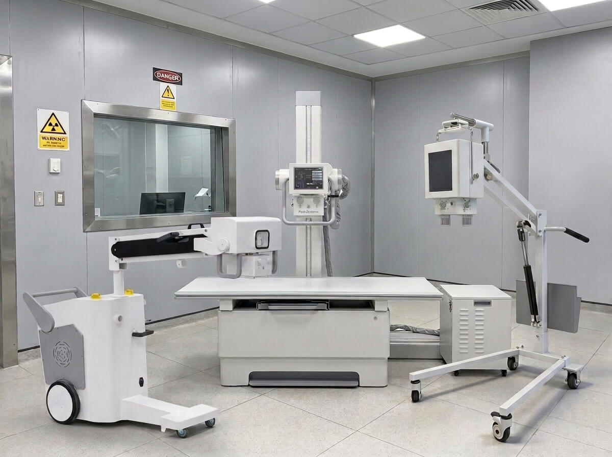
Today’s market offers hundreds of digital X-ray systems, including portable X-ray machines, mobile DR systems, floor-mounted X-ray units, ceiling-mounted radiography systems, and U-arm digital radiography equipment. Each system is designed for different clinical environments, patient volumes, budgets, and imaging requirements.
For medical equipment distributors, government procurement agencies, hospital project contractors, and healthcare investors, purchasing an X-ray machine is no longer simply about finding the lowest price. Buyers now evaluate image quality, detector technology, workflow efficiency, radiation dose management, artificial intelligence integration, software compatibility, after-sales service, warranty coverage, regulatory certifications, and long-term operating costs.
Whether you are purchasing your first digital radiography system, replacing an aging CR machine, expanding a diagnostic imaging department, or sourcing equipment from a reliable China X-ray machine manufacturer, this comprehensive buying guide will help you make an informed decision.
Throughout this guide, you will learn:
- How digital X-ray technology works
- Different types of DR systems
- Portable vs. mobile vs. fixed X-ray machines
- Flat panel detector technologies
- X-ray generator selection
- Image processing software
- Radiation safety considerations
- Hospital installation requirements
- Pricing factors
- Maintenance costs
- How to choose a reliable OEM or ODM manufacturer
By the end of this guide, you will understand exactly which digital X-ray solution best matches your hospital, clinic, imaging center, or distribution business.
Why More Hospitals Are Choosing Digital X-Ray Systems
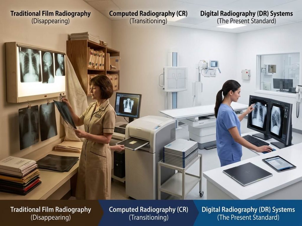
Medical imaging technology has evolved rapidly over the past decade. Traditional film-based radiography has largely disappeared, while computed radiography (CR) is gradually being replaced by fully digital DR systems.
Healthcare providers worldwide are upgrading because digital X-ray offers significant advantages, including:
- Faster image acquisition
- Higher diagnostic accuracy
- Lower radiation exposure
- Improved workflow efficiency
- Instant image sharing through PACS
- Better patient experience
- Reduced operating costs
- AI-assisted image analysis
- Environmentally friendly film-free workflow
As healthcare systems become increasingly digital, modern DR systems integrate seamlessly with Hospital Information Systems (HIS), Radiology Information Systems (RIS), Electronic Medical Records (EMR), and cloud-based PACS platforms.
These benefits make digital radiography an essential investment for both developed and emerging healthcare markets.
What Is a Digital X-Ray Machine?
A digital X-ray machine, also known as a Digital Radiography (DR) System, is a medical imaging device that uses X-ray radiation together with a digital detector to produce high-resolution diagnostic images almost instantly.
Unlike conventional film radiography, DR systems eliminate chemical film processing entirely.
Instead, X-rays passing through the patient’s body are captured by a Flat Panel Detector (FPD), converted into digital signals, processed by specialized imaging software, and displayed on a workstation within seconds.
This digital workflow enables physicians to diagnose patients much faster while significantly improving image quality and reducing examination time.
Digital X-ray systems are now widely used for:
- General radiography
- Orthopedic imaging
- Chest examinations
- Trauma diagnosis
- Emergency medicine
- Intensive care units (ICU)
- Surgical imaging support
- Outpatient clinics
- Community healthcare centers
- Veterinary hospitals
- Sports medicine
- Occupational health examinations
Because digital images can be stored electronically, hospitals also eliminate film storage costs while improving collaboration among radiologists and clinicians.
How Does a Digital Radiography System Work?
Understanding the imaging workflow helps buyers compare different systems more effectively.
A typical DR examination consists of several key components working together.
Step 1: X-Ray Generation
The X-ray generator supplies electrical power to the X-ray tube.
Inside the tube, high-energy electrons strike a tungsten target, producing X-ray photons that pass through the patient.
The generator controls several important parameters:
- Tube voltage (kVp)
- Tube current (mA)
- Exposure time
- Total radiation output
Modern high-frequency generators provide greater exposure consistency while reducing patient dose.
Step 2: X-Ray Beam Passes Through the Patient
Different tissues absorb X-rays differently.
For example:
- Bone absorbs more radiation.
- Soft tissue absorbs less.
- Air absorbs very little.
- Metal implants absorb the most.
This difference in absorption creates image contrast.
Step 3: Flat Panel Detector Captures the Image
Instead of using film or CR cassettes, digital systems use a Flat Panel Detector (FPD).
The detector immediately converts incoming X-rays into digital signals.
High-quality detectors offer:
- Better spatial resolution
- Higher detective quantum efficiency (DQE)
- Faster image acquisition
- Lower radiation dose
- Improved diagnostic confidence
Detector performance is one of the most important factors influencing overall image quality.
Step 4: Image Processing
Advanced software automatically enhances image quality using algorithms such as:
- Noise reduction
- Edge enhancement
- Dynamic contrast optimization
- Bone suppression
- AI-assisted positioning
- Automatic exposure optimization
These technologies help radiologists obtain consistent diagnostic images with minimal manual adjustment.
Step 5: Image Storage and Sharing
Images are stored digitally and transmitted through DICOM-compatible networks.
Hospitals can easily integrate the DR system with:
- PACS
- HIS
- RIS
- EMR
- Cloud imaging platforms
- Tele-radiology services
Doctors can review examinations from different departments or even remote locations, improving collaboration and reducing diagnosis time.