


Laptop Color Doppler Ultrasound System | Portable Shared Service Diagnostic Notebook Scanner | High-Sensitivity PW Doppler | CE & ISO 13485 Certified Factory OEM
Quick Info.
- SKU NO.: UMY-VEUS-030
- Device Classification: Class Ⅱ
- Warranty: 1 Year
- Power Source: Electric
- Transport Package: Carton or Wooden Box
- Origin: China
- Material: Metal, Plastic
- After-Sale Service: Online Technical Support
- Production Capacity: 3000 Sets/Year
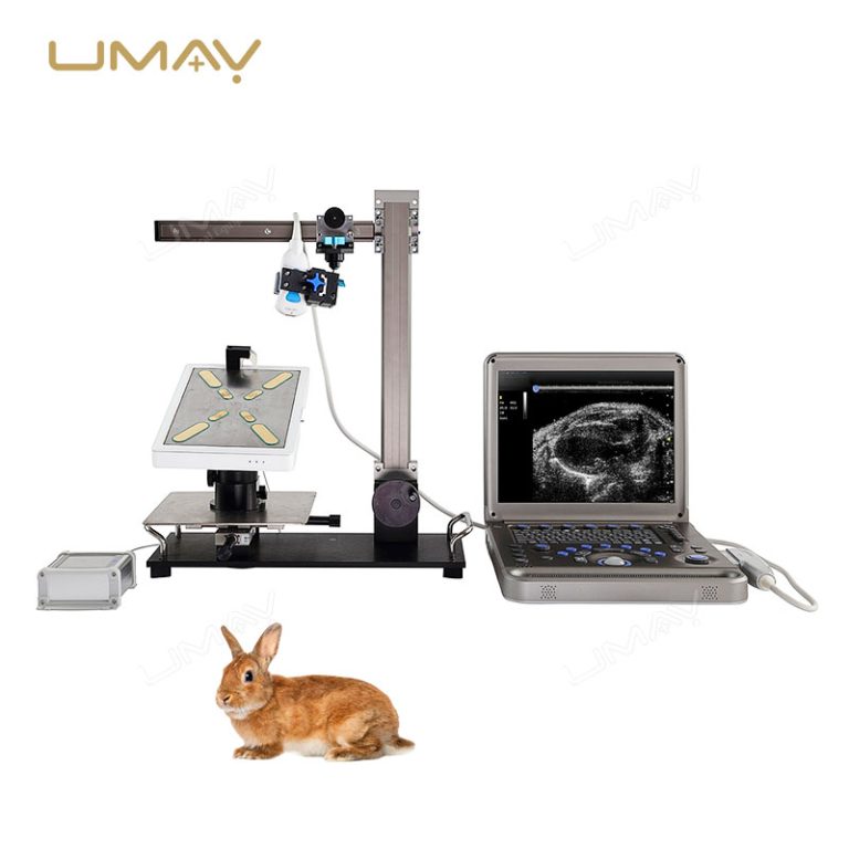
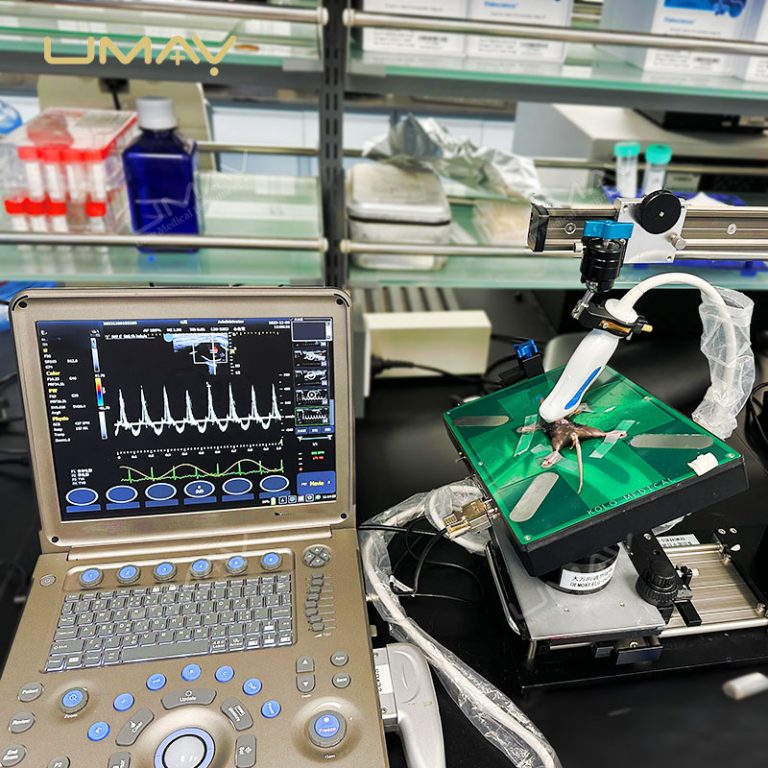
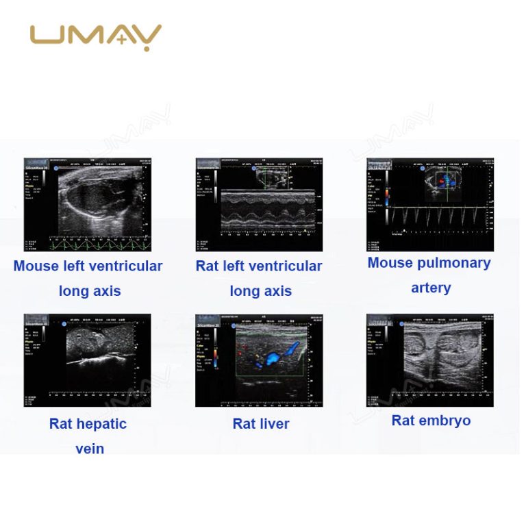
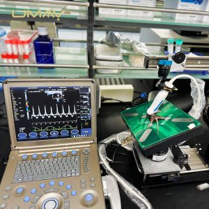
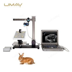
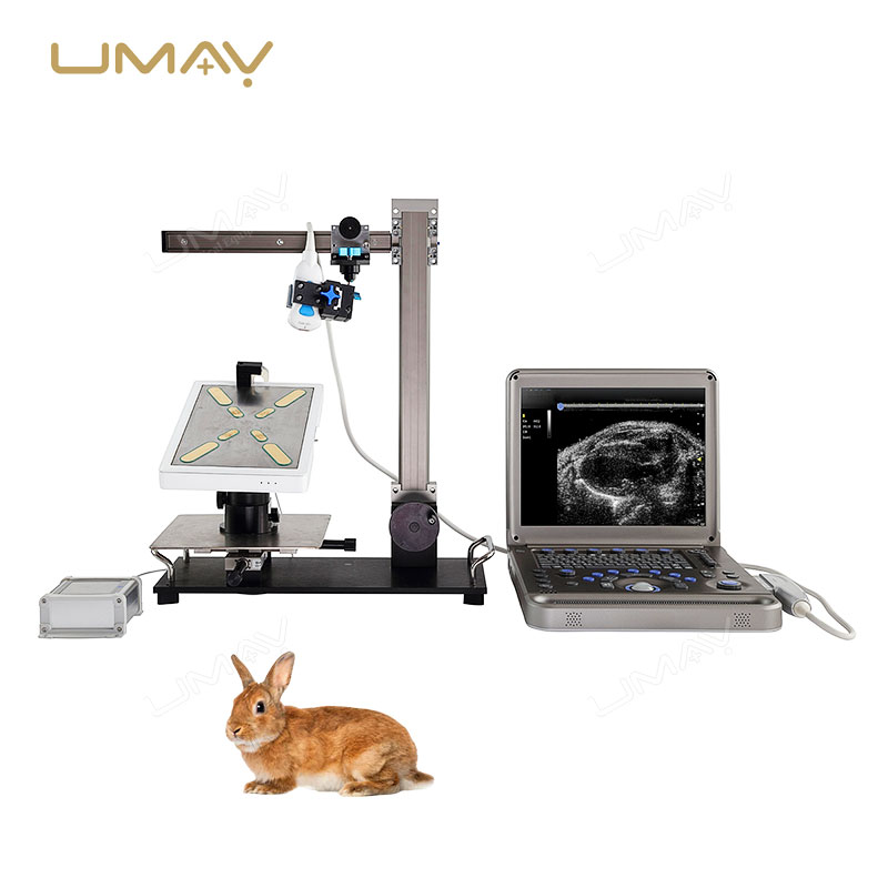
This ultra-high frequency ultrasound system is specifically designed for small animal research, featuring high-resolution imaging with a 30 MHz frequency for exceptional spatial clarity. The system integrates an advanced semiconductor CMUT probe, allowing for precise observation of blood vessels, abdominal organs, and embryos in small animals. The system supports multiple imaging modes, including B-mode, M-mode, C-mode, and Doppler imaging, enabling researchers to analyze dynamic cardiovascular conditions and tissue motion. Real-time imaging capabilities are ideal for repeated experiments and long-term monitoring. For a complementary diagnostic solution, explore our Portable Color Doppler Ultrasound Scanner for Obstetric Imaging.
The Specific Parameters
| ITEMS | PARAMETERS |
|---|---|
| System Host | |
| Main Console | Ultra-high frequency small animal ultrasound imaging system (UMYPUHF001) |
| Software | Skyline Ultrasound Imaging Software V1.0.5 |
| Imaging Modes | |
| B-Mode | 2D grayscale imaging for tissue structure, annular displacement, distance/thickness/area measurements |
| M-Mode | Motion mode for cardiac and vascular movement analysis; supports cardiac measurements (EF, FS, SV, CO, PV, Peak E/A, Peak E’/A’) |
| C-Mode | Color Doppler imaging for directional blood flow detection; adjustable velocity and color mapping |
| PW-Mode | Spectral Doppler imaging for blood flow velocity measurement |
| TVI/TVD Modes | Tissue Doppler imaging for quantitative analysis of local myocardial motion |
| AUTO Optimization | One-click image optimization |
| Probes | |
| Probe Model UM38-22K3 | Center Frequency: 30 MHz (suitable for mice and rats) |
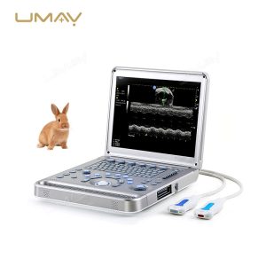
| Probe Model UM22-8K3 | Center Frequency: 15 MHz (suitable for large rats and rabbits) |
| Animal Applications | |
| Mice | Cardiovascular imaging, abdominal organs, lymph nodes, vascular imaging, tumor imaging (<13 mm), epidermal imaging, embryology |
| Rats | Cardiovascular imaging, embryology, abdominal imaging, vascular imaging, tumor imaging |
| Rabbits | Abdominal imaging, vascular imaging, ocular imaging |
| Measurement Packages | |
| Standard Package | Measurement package for small and large rodents, rabbits, and other experimental animals |
| Accessories | |
| Advanced Bracket Platform | Model H20; adjustable for mouse board and probe positioning |
| Mouse/Rat Board | Model P60; includes ECG, respiration, and heating modules |
| Mobile Trolley | Dedicated system trolley for mobility |
| Offline Workstation | WS01 with workstation software and Ukey-WSK01 |
| Power Cable | Included |
| Standard Documentation | Included |
Why Global POCUS Distributors & Private Clinics Partner With Us:
-
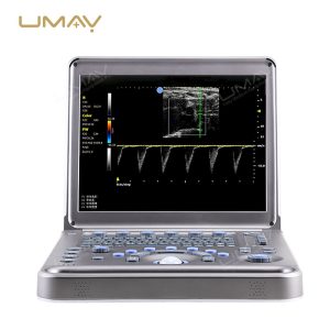
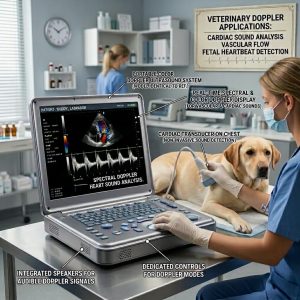
High-Sensitivity PW Doppler & Auto-Trace Intelligence: Capture tricky hemodynamic data with total confidence. Engineered with a premium system dynamic range, the platform flawlessly renders real-time pulsed-wave (PW) Doppler spectrum curves and color blood flow mapping, offering one-click auto-trace tracking for lightning-fast velocity metrics.
-
Anti-Glare Medical Display with Heavy-Duty Hinge: See clear data under any lighting condition. The high-contrast display terminal is layered with a specialized anti-reflective (AR) coating to eliminate glare from harsh operating room surgical lamps, supported by a reinforced structural hinge designed for thousands of seamless open-and-close cycles.
-
Rapid Cold-Boot Execution & High-Endurance Battery: Seconds save lives in emergency care. The optimized operating platform launches from cold power-on to active clinical scanning in under 25 seconds, backed by an integrated, premium lithium-ion battery pack that guarantees over 2 hours of continuous, uncompromised active scanning away from wall outlets.
-
Slim Native Transducer Hub with Modular Expansion: Maintain full application flexibility without the bulk. The sleek side-panel houses native, zero-force insertion probe sockets, and fully supports our external multi-port probe extenders and customized ergonomic docking carts to transition from a laptop to a full-station trolley in seconds.
-
Ruggedized, Impact-Resistant Ergo-Chassis: Built for relentless hospital transit. Constructed using a lightweight, military-grade anti-shock composite shell, the entire unit is physically optimized for comfortable hand-carrying between wards while shielding delicate internal beamformer boards from accidental vibrations or drops.
-
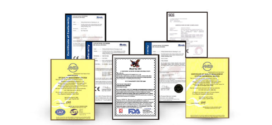
Tender-Ready OEM Customization Freedom: Dominate your local government and private hospital procurement bids. We fully empower our global distributor network by providing flexible white-label configurations, multi-language software localization packages, custom boot logos, and complete ISO 13485 technical dossiers.
💡 Core Specifications At a Glance (Customizable):
-
System Form Factor: Ergonomic Laptop/Notebook Compact Profile with Integrated Carrying Handle
-
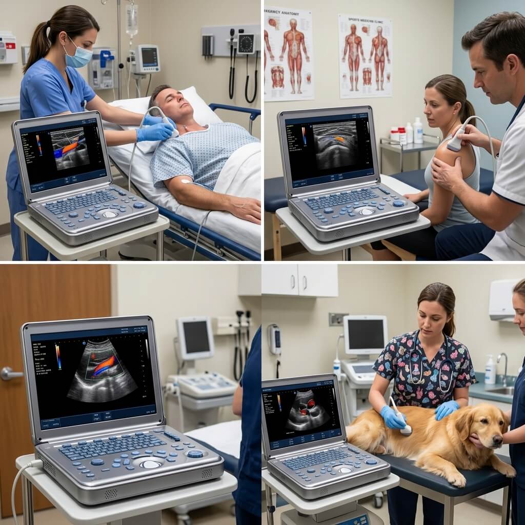
Imaging Capabilities: B, M, Color Doppler, Power Doppler, High-Definition PW Spectral Doppler
-
Display Terminal: Premium High-Definition Medical LCD with Anti-Glare Protective Layer
-
Workflow Efficiency: Full Tactile Physical Control Console with Backlit Trackball Navigation
-
Data Interoperability: Native DICOM 3.0 Compliance (Seamless integration with hospital PACS/RIS)
❓ Crucial B2B Procurement Q&A
Q1: How many native probe ports are built into the laptop, and can we expand them? A: The main notebook chassis features a sleek, space-saving native probe socket layout. For clinics requiring multiple transducers on standby, we provide an optional Modular Probe Extender Docking Station and a specialized ergonomic mobile trolley cart that expands the system to support 3 or more fully active, selectable transducer ports simultaneously.
Q2: Can the screen display handle intense ambient light and glare in emergency or surgical suites? A: Absolutely. The display retains crisp contrast for both grayscale tissue maps and color/spectral curves. This is achieved via a medical-grade anti-glare (AR) multi-layer coating applied to the display panel, ensuring excellent off-axis viewing clarity even directly underneath intense overhead surgical lights.
Q3: What is the exact battery performance during active, intensive Color and PW Doppler scanning? A: Many portable systems drain rapidly under heavy Doppler loads. Our system utilizes an intelligent, low-power architectural chipset that yields over 120 minutes of continuous, active scanning in demanding Color/PW Doppler modes on a single charge, plus an instant-on standby mode to conserve battery power during patient transitions.
Q4: Do you offer localized software and unbranded options for competitive regional medical tenders? A: Yes. We are an established OEM/ODM direct factory supplier. We can configure the system entirely without factory branding, laser-etch your custom distribution logo onto the housing, and completely localize the user interface software into languages such as Spanish, French, Portuguese, Russian, and Arabic to meet strict local ministry of health tender prerequisites.