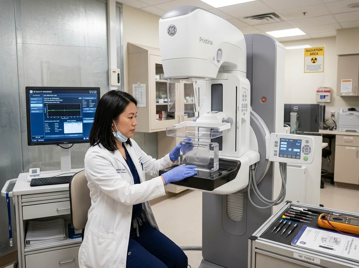
The Importance of Quality Control in Digital Mammography
In digital mammography, maintaining strict Quality Control (QC) is not merely a operational best practice—it is a strict regulatory requirement enforced by global health authorities (e.g., FDA MQSA in the US, EUREF in Europe, and IAEA protocols worldwide). Because early breast cancer screening relies on detecting subtle architectural distortions and microscopic calcifications (0.1 mm to 0.2 mm), even minor detector noise, beam quality shifts, or mechanical paddle misalignments can compromise diagnostic integrity or lead to unnecessary patient exposure.
A robust preventive maintenance and routine QC program ensures three critical outcomes:
-
Uncompromised Diagnostic Clarity: Maintains high Signal-to-Noise Ratio (SNR) and Contrast-to-Noise Ratio (CNR) across all patient exposures.
-
Radiation Safety Compliance: Guarantees that the Average Glandular Dose (AGD) stays well below strict statutory limits.
-
Equipment Longevity & ROI: Prevents premature failure of high-cost components such as flat-panel detectors and X-ray tubes.
This comprehensive operational guide details essential QC testing schedules, detector calibration protocols, X-ray tube care practices, and strategies for choosing the optimal service level agreement (SLA).
2. Structured Quality Control (QC) Schedule: Daily to Annual Testing
Quality control in mammography requires a layered testing routine executed by radiologic technologists, internal biomedical engineers, and certified medical physicists.
┌────────────────────────────────────────────────────────────────────────┐
│                      MAMMOGRAPHY QC TESTING TIMELINE                   │
├────────────────────────────────┬───────────────────────────────────────┤
│ DAILY                          │ • Detector Offset & Gain Auto-Calib   │
│ (Technologist)                 │ • ACR Accreditation Phantom Test      │
│                                │ • Visual Safety & Indicator Check     │
├────────────────────────────────┼───────────────────────────────────────┤
│ WEEKLY / MONTHLY               │ • Artifact Evaluation & Flat-Field    │
│ (Lead Technologist)            │ • Compression Force & Decompression   │
│                                │ • View Station Monitor Calibration    │
├────────────────────────────────┼───────────────────────────────────────┤
│ ANNUAL                         │ • Comprehensive Physics Audit         │
│ (Medical Physicist)            │ • Half-Value Layer (HVL) & AGD Test   │
│                                │ • Full Detector Pixel Map Recalib     │
└────────────────────────────────┴───────────────────────────────────────┘
Essential QC Tests Breakdown
| Test Frequency | QC Parameter | Testing Method & Equipment | Regulatory Acceptance Criteria |
| Daily | Phantom Image Quality | Scan standard ACR mammography phantom | Must resolve minimum 5 fibers, 4 speck groups, 4 masses without artifacts |
| Daily | System Warm-up & Zeroing | Automated software routine | Zero system errors; gantry motion within factory baseline |
| Weekly | Flat-Field Artifact Test | High-density acrylic block exposure | Zero grid lines, streak artifacts, or cluster bad pixels |
| Monthly | Compression Force | Calibrated bathroom scale or load cell | Automated force must hold between 111 N to 200 N (25–45 lbs) |
| Monthly | Diagnostic Monitor QC | TG18-QC or AAPM test pattern on 5MP/12MP display | 5% and 95% intensity patches clearly visible; zero geometric distortion |
| Annual | Beam Quality (HVL) & AGD | Aluminum filters & solid-state dosimeter | Average Glandular Dose < 3.0 mGy per image for standard 4.2 cm breast |
3. Flat-Panel Detector Calibration: Offset, Gain, and Bad Pixel Mapping
The flat-panel detector (FPD) is the single most delicate and expensive component inside a digital mammography unit. Over time, physical detector pixels undergo sensitivity changes, background electrical drift, and dark noise accumulation.
     STAGE 1: Dark Current / Offset Calibration (Measures internal electronic noise)
                                    │
                                    ▼
     STAGE 2: Gain Calibration (Normalizes pixel response across full field)
                                    │
                                    ▼
     STAGE 3: Bad Pixel Mapping (Identifies dead pixels & applies interpolation)
1. Dark Current (Offset) Calibration
Electronic noise is generated by readout circuits even when no X-rays strike the detector. Dark current calibration captures a zero-exposure frame to map baseline sensor noise. The acquisition software subtracts this offset map automatically from every clinical exposure to maintain crystal-clear image backgrounds.
2. Gain Calibration (Flat-Fielding)
Due to minor manufacturing variations, individual pixels on an Amorphous Selenium (a-Se) or Cesium Iodide (CsI) detector absorb radiation with slight sensitivity differences. Gain calibration subjects the entire detector surface to a uniform X-ray exposure through an attenuating phantom. The system calculates correction factors for every individual pixel to ensure perfectly uniform image brightness across the entire field.
3. Bad Pixel Mapping and Defect Masking
Over millions of exposures, individual pixels or entire readout lines may become unresponsive (“dead pixels”).
-
Defect Mapping Routine: Software scans identify unresponsive pixels during monthly QA runs.
-
Interpolation Algorithm: The acquisition workstation masks bad pixels by calculating weighted averages from adjacent healthy neighboring pixels.
-
Threshold Limit: If dead pixel clusters exceed manufacturer thresholds (e.g., >0.01% total area or adjacent defect clusters in critical diagnostic zones), detector replacement is required.
4. X-Ray Tube Maintenance & Thermal Management
Mammography X-ray tubes feature ultra-fine focal spots (0.1 mm for magnification, 0.3 mm for standard views) and heavy thermal loads. Preserving tube life requires vigilant thermal care.
               ┌─────────────────────────────────────────────────┐
               │    X-Ray Tube Heat Management Workflow          │
               └────────────────────────┬────────────────────────┘
                                        │
           ┌────────────────────────────┴────────────────────────────┐
           ▼                                                         ▼
┌──────────────────────────────────────┐  ┌──────────────────────────────────────┐
│       Daily Tube Warm-Up             │  │      Chiller & Liquid Cooling        │
├──────────────────────────────────────┤  ├──────────────────────────────────────┤
│ • Gradually elevates anode temp      │  │ • Maintains oil circulation cooling  │
│ • Prevents target thermal shock      │  │ • Monthly coolant level checks       │
│ • Mandatory after 4+ hours idle      │  │ • Prevents tube housing overheating  │
└──────────────────────────────────────┘  └──────────────────────────────────────┘
Best Practices for Tube Care:
-
Strict Warm-Up Protocols: Never execute high-mAs exposure on a cold tube target. Always perform a 3-step incremental low-mAs warm-up sequence every morning or whenever the gantry sits idle for more than 4 hours.
-
Monitor Accumulated Heat Units (HU): Modern acquisition consoles display remaining target heat capacity. During high-throughput 3D tomosynthesis screening sessions, allow brief cooling intervals between patients to avoid reaching thermal cut-off thresholds.
-
Cooling System Servicing: Inspect oil-to-air heat exchangers and liquid coolant lines every 6 months. Ensure fan radiators are free of accumulated dust, as elevated housing temperatures degrade high-voltage insulation oil rapidly.
5. Selecting the Right Service Level Agreement (SLA): OEM vs. ISO
When warranty coverage expires, hospital managers must select a ongoing service model. Choosing between Original Equipment Manufacturers (OEM) and certified Independent Service Organizations (ISO) involves balancing response speeds, technical depth, and overall budget.
┌────────────────────────────────────────────────────────────────────────┐
│                     SERVICE CONTRACT MODEL MATRIX                      │
├──────────────────────────┬───────────────────────┬─────────────────────┤
│ FULL SERVICE CONTRACT    │ SHARED RISK CONTRACT  │ PREVENTIVE MAINT.   │
│ Includes parts, labor,   │ Capped parts coverage │ PM visits only;     │
│ tube & detector coverage │ Lower annual fee      │ Parts & labor paid  │
│ Highest security/cost    │ Shared facility risk  │ Time & Materials    │
└──────────────────────────┴───────────────────────┴─────────────────────┘
OEM vs. ISO Comparison
| Service Factor | OEM (Original Equipment Manufacturer) | ISO (Independent Service Organization) |
| Annual Service Cost | High (Baseline Standard) | 20% to 40% Savings |
| Response Time (SLA) | Guaranteed 2–4 Hours | Flexible / Customized SLAs |
| X-Ray Tube & Detector Inclusion | Available in Premium Contracts | Optional or Capped Limits |
| Proprietary Software Access | Direct OEM License & Remote Diagnostics | Limited to Non-Proprietary Tools |
| Field Engineer Expertise | Factory-trained on specific model lines | Multi-vendor generalist experience |
Key SLA Clauses to Demand in Contracts:
-
Guaranteed Uptime Commitment: Demand a minimum 98% operational uptime guarantee (calculated on a quarterly basis), backed by financial penalty credits or contract extension days for non-compliance.
-
Glass Coverage (Tube & Detector): Verify whether the contract includes 100% full replacement coverage or pro-rated coverage for the X-ray tube and digital flat-panel detector.
-
Emergency Response Window: Ensure a maximum 4-hour on-site arrival guarantee for severe system downs, along with 24/7 phone and remote network diagnostic support.
6. Preventive Maintenance Checklist for Hospital Engineering Teams
-
[x] Daily Phantom Exposure Recorded: Pass/Fail scores logged in clinical QC tracking software.
-
[x] Mechanical Safety Interlocks Checked: Emergency stop buttons, gantry collision sensors, and manual compression overrides tested.
-
[x] Detector Offset & Gain Calibration: Executed and matched against baseline reference values.
-
[x] X-Ray Tube Chiller Inspection: Coolant level verified; heat dissipation radiator fins cleared of dust build-up.
-
[x] Compression Paddle Inspection: Checked for hair-line stress cracks, paddle latch looseness, or flex deformation.
-
[x] PACS & Workstation Audit: DICOM transmission speeds, diagnostic monitor calibration (DICOM GSDF), and hanging protocols verified.
7. Frequently Asked Questions (FAQ)
Q1: How often should an ACR phantom test be performed on a digital mammography machine?
A standard ACR phantom test must be performed daily (or before the first patient scan) by a radiologic technologist to ensure spatial resolution, contrast sensitivity, and lack of detector artifacts meet regulatory standards.
Q2: What is the average lifespan of a mammography digital flat-panel detector?
With proper daily calibration and climate-controlled room conditions, modern flat-panel detectors (both a-Se and CsI) typically last 6 to 8 years before pixel degradation or gain loss necessitates replacement.
Q3: What happens if the compression force test fails during monthly QC?
If the automated compression force drops below 111 N (25 lbs), images may suffer from motion blur and uneven tissue density. If it exceeds 200 N (45 lbs), it risks patient injury and discomfort. The unit must be recalibrated by an engineer before clinical use.
Q4: Is a shared-risk service contract recommended for a high-volume screening center?
For high-volume centers, a Full Service Contract with 100% tube and detector coverage is generally recommended over shared-risk models. High patient throughput increases component wear, making un-capped part replacement coverage financially safer.