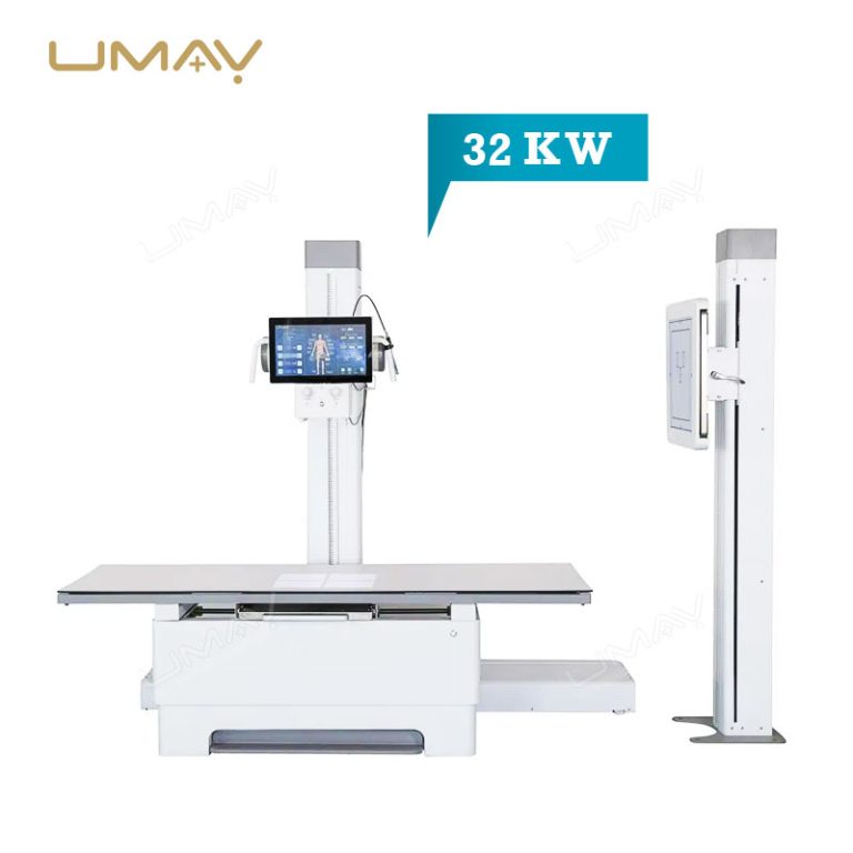


Floor-Mounted Fixed Digital Radiography System | High-Frequency 32kW Medical Diagnostic DR X-Ray Suite | Multi-Directional Floating Radiographic Table & Upright Bucky Wall Stand with Smart Touchscreen Tube Console
Quick Info.
- SKU NO.: UMY-XM-066
- Device Classification: Class Ⅱ
- Warranty: 1 Year
- Power Source: Electric
- Transport Package: Carton or Wooden Cases
- Origin: China
- Material: Aluminum & Magnesium Alloys and Plastic
- After-Sale Service: Online Technical Support
- Production Capacity: 1000 Sets/Year
This advanced 32KW Digital Radiography X ray Machine delivers exceptional diagnostic imaging quality with high frequency technology for superior contrast resolution. Designed specifically for hospital stationary installations, this system enables medical professionals to capture detailed anatomical structures while reducing radiation exposure for enhanced patient safety. The robust 32KW power capacity ensures consistent performance across a wide range of medical imaging applications from routine examinations to specialized diagnostic procedures.
The Specific Parameters
| ITEMS | PARAMETERS |
|---|---|
| X-ray Generator Specifications | |
| Power Output | 32KW / 400mA |
| Generator Frequency | 30kHz / 200kHz (optional) |
| Power Supply | 50Hz or 60Hz, wire diameter >4mm², internal resistance <0.50; supports 20/32kW (220V(SY)/380V(D)) |
| Exposure Control | |
| Tube Current (mA) | 10–400 |
| Tube Current-Time Product (mAs) | 0.4–320 |
| Tube Voltage (kV) | 40–125kV (1kV step) |
| Exposure Time | 1ms–10000ms |
| X-ray Tube and Heat Capacity | |
| Focus Size | Default 0.6*0.6mm / 1.2*1.2mm dual focus (Toshiba optional) |
| Anode Rotating Speed | 2800rpm |
| Anode Heat Capacity | 1300kHU |
| Radiography Table | |
| Table Dimensions | 73cm (W) × 200cm (L) |
| Table Movement | 74cm left/right, 20cm back/front |
| Flat Panel Detector | |
| Image Size | 17*17 inch (14*17 inch option available) |
| Pixel Matrix | 140μm |
| A/D Conversion | 16 bits |
| Spatial Resolution | 3.6 Lp/mm |
| Software | Professional Imaging Software |
| Computer Configuration | CPU: 15, 8G memory, 1T solid state drive, 2 pcs Gigabit network card |
-
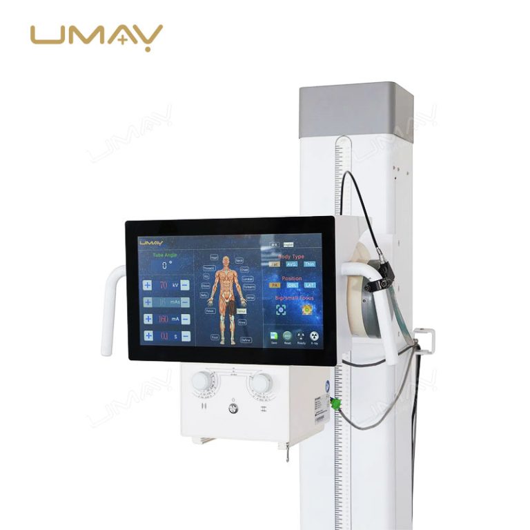
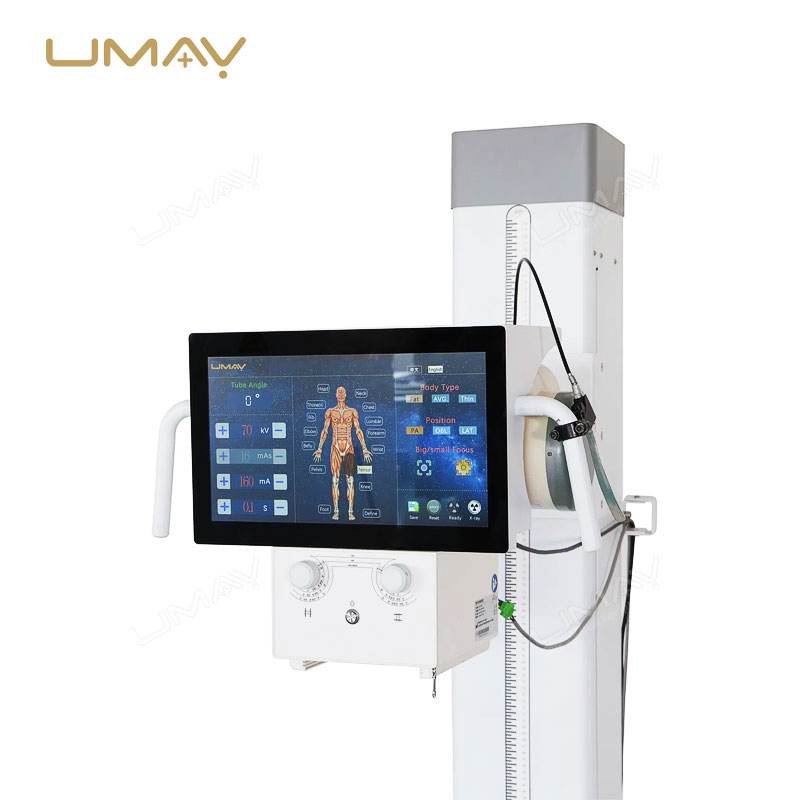
Intelligent Tube-Head Touchscreen Interface: Streamline bedside clinical operations. The vertical tube column is equipped with an advanced, large-format color touchscreen graphical user interface (GUI). Radiographers can manage Anatomical Programmed Radiography (APR) settings, verify patient metrics, and monitor system parameters directly in the examination room, maximizing patient face-time.
-
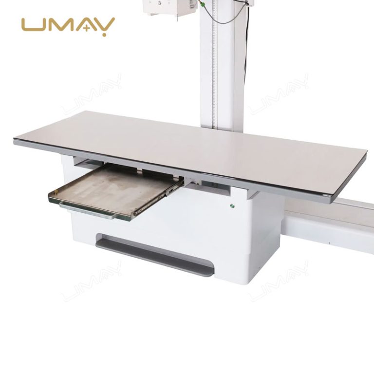
Heavy-Duty Multi-Directional Floating Table: Ensure effortless patient positioning. The spacious, low-absorption radiographic tabletop features smooth, multi-directional floating mechanics controlled by responsive electromagnetic foot locks. With an ultra-high weight bearing threshold, it easily accommodates bariatric patients while reducing physical strain on clinical staff.
-
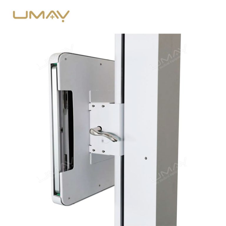
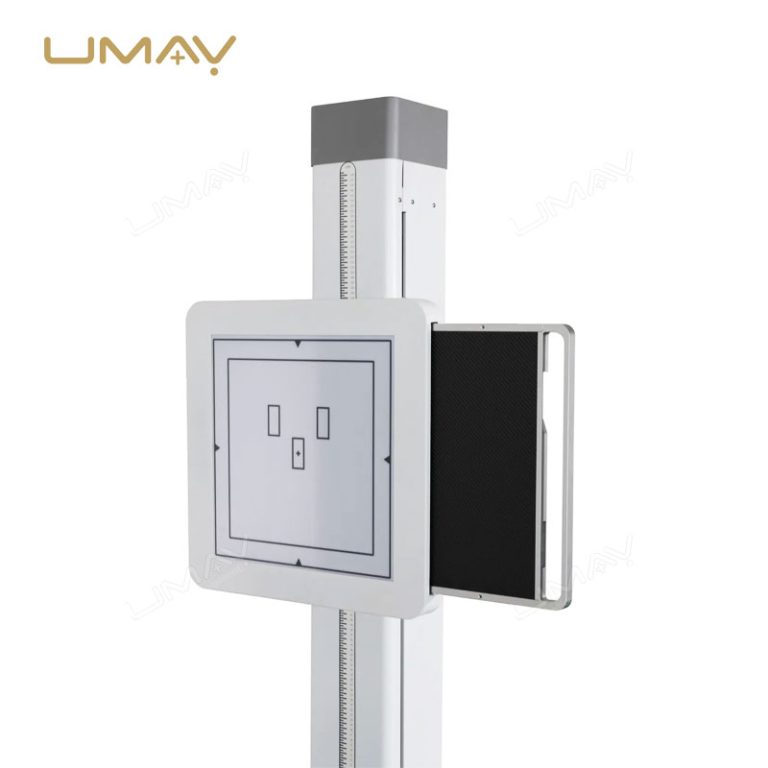
Independent Upright Bucky Wall Stand: Maximize chest and spinal examination throughput. The counterbalanced vertical stand moves fluidly to accommodate standing or seated wheel-chair patients. It houses a premium sub-surface bucky grid mechanism configured for seamless integration with high-resolution digital flat panel detectors.
-
High-Frequency Output Low-Dose Performance: Prioritize image quality and patient safety. Available in high-power output configurations up to 32kW, the internal high-frequency generator delivers ultra-short, highly stable exposure pulses. This minimizes motion artifacts and delivers crisp, high-contrast digital images at minimized radiation doses.
-
Robust Floor-Mounted Structural Footprint: Built for long-term clinical durability. The floor-mounted vertical column track provides rock-solid mechanical stability during rapid adjustments, eliminating image vibration and ensuring precise, repeatable geometric alignment over millions of exposure cycles.

-
System Layout: Complete Floor-Mounted Fixed Diagnostic X-Ray Suite
-
Generator Power Output: 32kW / 50kW / 65kW High-Frequency Options Available
-
User Interface: Integrated Smart Touchscreen Tube Console + Master PC Workstation
-
Patient Table Mechanics: Long-Travel Floating Tabletop with Electromagnetic Braking
-
Imaging Integration: Native DICOM 3.0 Protocol for Instant Hospital PACS/RIS Syncing
Q1: Can this fixed system be customized as a single shared-detector setup to meet tighter clinic budgets?
A: Yes, we offer flexible hardware configurations to accommodate different budgetary tiers. While we highly recommend our dual-detector configuration for high-volume hospitals, we can supply the system with a single shared wireless flat panel detector that can be manually swapped between the table tray and the wall stand bucky as needed.
Q2: How does the tube-head touchscreen console interact with our main operator workstation inside the control booth?
A: The system features full, bi-directional hardware and software synchronization. Any anatomical profile or exposure parameter (kV/mAs) adjusted on the tube-head touchscreen will update on the main control workstation instantly, and vice versa, preventing communication lag and eliminating manual entry errors.
Q3: What are the room layout and ceiling height requirements for installing this floor-mounted system?
A: Because this is a floor-mounted system rather than a ceiling-suspended layout, it has highly forgiving structural requirements. It can be installed in rooms with standard ceiling heights (typically down to $2.5\text{ meters}$), and does not require expensive structural steel ceiling reinforcement, drastically reducing your facility’s room renovation costs.
Q4: What types of post-processing software tools are included in the master workstation software package?
A: Our medical-grade image acquisition software comes pre-loaded with a comprehensive suite of advanced post-processing tools tailored for human anatomy. This includes automatic window leveling, tissue equalization, digital shuttering, electronic collimation, gridline suppression algorithms, and full annotation tools to guarantee diagnostic-ready images before sending them to the PACS array.