The Ergonomic Crisis and the Digital Shift in Microsurgery
For more than six decades, the traditional optical operating microscope has been the foundational centerpiece of surgical visualization. From the earliest binocular systems introduced in the 1950s to modern motorized apochromatic platforms, optical microscopes have allowed surgeons to operate on microscopic nerves, vessels, and tissues with extraordinary precision.
However, traditional microscopes come with an inherent ergonomic penalty: the surgeon must keep their eyes physically glued to binocular eyepieces for hours at a time.
Plaintext
                  THE MICROSURGEON'S PHYSICAL TOLL
+--------------------------------------------------------------------------+
| Fixed Neck Angle (20° to 45° Flexion) --> Cervical Spine Disc Strain     |
| Static Body Positioning (4 to 8 Hours) --> Chronic Lumbar & Shoulder Pain|
| Eye Fatigue & Facial Compression       --> Premature Surgical Retirement |
+--------------------------------------------------------------------------+
Studies consistently demonstrate that over 70% of microsurgeons—particularly in neurosurgery, spine surgery, and otolaryngology—suffer from occupational cervical spine disorders, lumbar pain, and musculoskeletal fatigue caused by prolonged, awkward neck flexion.
To address this ergonomic dilemma while taking advantage of high-speed digital imaging, medical device developers introduced the 4K 3D Digital Exoscope (extracorporeal telescope). By untethering the surgeon from the physical eyepieces and projecting a magnified, high-definition 3D image onto external monitors, the exoscope promises a new era of “heads-up” microsurgery.
This guide provides an in-depth comparative analysis of traditional optical operating microscopes versus next-generation digital exoscopes, examining optical physics, visual latency, ergonomic impact, clinical adoption patterns, and long-term procurement considerations.

2. Anatomy and Strengths of Traditional Optical Microscopes
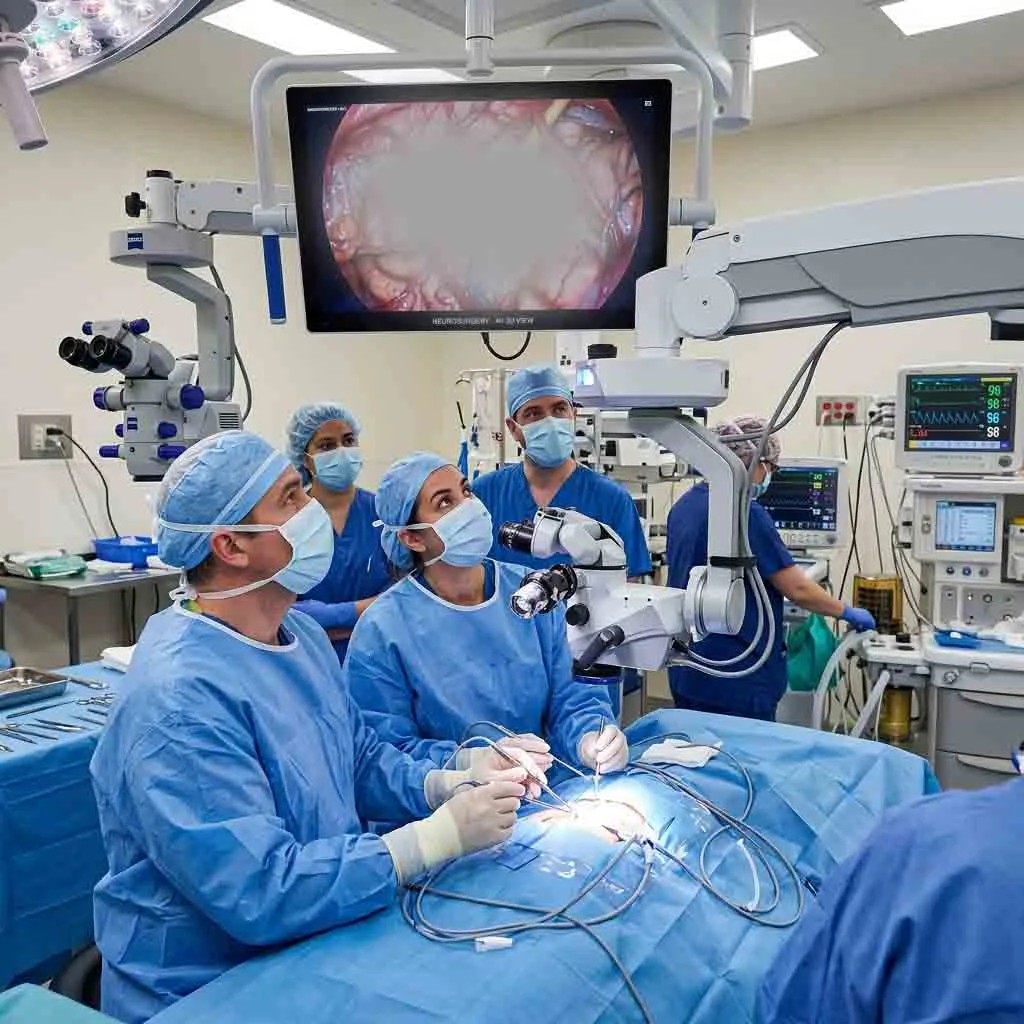
Traditional operating microscopes are pure optical instruments. Light travels from an internal illumination source (LED or Xenon), through the objective lens, bounces off the patient’s anatomical tissues, travels back up through the zoom drum and beam splitters, and enters the surgeon’s eyes through optical glass eyepieces.
Plaintext
               TRADITIONAL OPTICAL MICROSCOPE BEAM PATH
+-------------------------------------------------------------------------+
| Surgical Field  --> Objective Lens (Focal Point)                        |
| Optical Body    --> Apochromatic Glass Lens Array (Zoom Drum)           |
| Beam Splitter   --> Prisms split light to Binocular Eyepieces & Camera  |
| Visual Delivery --> Direct Photons into Surgeon's Retinas (Zero Lag)    |
+-------------------------------------------------------------------------+
Key Advantages of Traditional Optical Systems
1. Zero Latency (Speed-of-Light Visual Feedback)
Because light passes directly through optical glass lenses without electronic conversion, there is zero display lag. When a surgeon moves a pair of micro-forceps or ties an 10-0 nylon suture, the visual feedback is instantaneous. In high-stakes microvascular surgery, even a minor electronic delay can disrupt hand-eye coordination.
2. Natural Optical Stereopsis (True Depth Perception)
Optical microscopes provide true binocular vision. The slight physical separation between the two optical paths mimics the interpupillary distance of human eyes, creating natural stereoscopic depth perception. This allows surgeons to judge microscopic depth, tissue plane thickness, and instrument clearance instinctively without digital approximation.
3. Uncompressed High Dynamic Range (HDR)
High-end apochromatic glass lenses deliver pure, uncompressed optical contrast. The human eye can perceive subtle gradations in red and pink tissue hues, vascular pulsations, and translucent arachnoid membranes that digital sensors can sometimes blur, overexpose, or compress into digital noise.
Inherent Limitations of Optical Microscopes
-
Ergonomic Inflexibility: Even with 0–180 degree inclinable binocular tubes, the surgeon’s head must remain aligned with the oculars. When operating at extreme angles (e.g., steep lateral skull base approaches or high cervical spine cases), the surgeon must contort their neck and torso to maintain visual contact.
-
Restricted Assistant Viewing: While beam splitters allow an assistant to look through a secondary ocular tube, the light must be split (often 50/50), reducing the brightness available to both surgeons. Furthermore, the assistant is physically crowded against the primary surgeon around the patient’s head.
-
Bulky Footprint Over the Field: A conventional optical head housing multiple heavy glass elements, prism blocks, and motor drives is physically large. This bulky structure sits directly over the patient, occasionally restricting the trajectory of long surgical instruments like micro-drills, bayoneted forceps, and suction tubes.
3. What is a 4K 3D Digital Exoscope?
A digital exoscope is a specialized camera-based visualization system designed to replace traditional microscope eyepieces.
Rather than looking through ocular tubes, the surgical team positions a compact digital camera head—equipped with dual high-resolution 4K image sensors—between 200 mm and 500 mm above the surgical cavity. The camera captures two slightly offset video streams that are processed in real time and displayed on a 31-inch or 55-inch 4K 3D medical-grade monitor positioned directly in front of the surgeon.
Plaintext
                       DIGITAL EXOSCOPE ECOSYSTEM
+---------------------------------------------------------------------------+
| [ Compact 3D Camera Head ] ---> Positioned 200–500 mm above surgical site |
|              |                                                            |
|              v                                                            |
| [ Real-Time Image Processor ] -> Real-time edge enhancement & color filter|
|              |                                                            |
|              v                                                            |
| [ 55" 4K 3D Medical Display ] -> Viewed via passive polarized 3D glasses  |
|              |                                                            |
|              +----------------> Entire OR Team shares identical 3D view   |
+---------------------------------------------------------------------------+
The Concept of “Heads-Up Surgery”
The defining feature of exoscopic surgery is the “heads-up” posture. Wearing lightweight, passive polarized 3D glasses (identical to those used in modern 3D movie theaters), the surgeon sits or stands completely upright with their spine in a neutral posture.
The surgeon looks straight ahead at the large 3D monitor while their hands manipulate instruments within the surgical field below. This eliminates neck flexion, removes the physical weight of ocular pressure from the surgeon’s face, and dramatically reduces physical fatigue during procedures that exceed four hours.
Key Advantages of Digital Exoscopes
1. Unmatched Operating Ergonomics
The primary driver of exoscope adoption is physical health. By decoupling the surgeon’s eye line from the optical head, the camera can be tilted at extreme angles (e.g., looking upward under the skull base or horizontally through a tubular retractor) while the surgeon remains seated in an upright, neutral posture.
2. Unprecedented Surgical Workspace and Instrument Clearance
Because an exoscope camera head contains electronic image sensors rather than heavy glass prisms, it is significantly more compact than a traditional optical head.
-
Furthermore, exoscopes operate at extended working distances (typically 250 mm to 600 mm).
-
This leaves a large, unobstructed open space above the surgical cavity, preventing micro-instruments, drills, and robotic arms from colliding with the visualization unit.
3. Total Team Integration and Teaching Efficiency
In traditional microsurgery, only the primary surgeon and the direct assistant looking through the secondary ocular experience stereoscopic 3D depth. The rest of the team—scrub nurses, surgical technicians, residents, and anesthesiologists—watch a flat, low-contrast 2D monitor.
-
With an exoscope, everyone wearing 3D glasses sees the exact same high-definition 3D view.
-
Scrub nurses anticipate instrument exchanges because they can perceive surgical depth and cavity progress.
-
For teaching hospitals, medical residents and fellows learn anatomical spatial relationships far faster when viewing the procedure in true 3D on a large screen alongside the attending surgeon.
4. Digital Processing, Filters, and Augmented Reality
Digital video streams can be modified mathematically in real time:
-
Digital Color Enhancement: Software filters enhance vascular contrast without requiring physical glass filters.
-
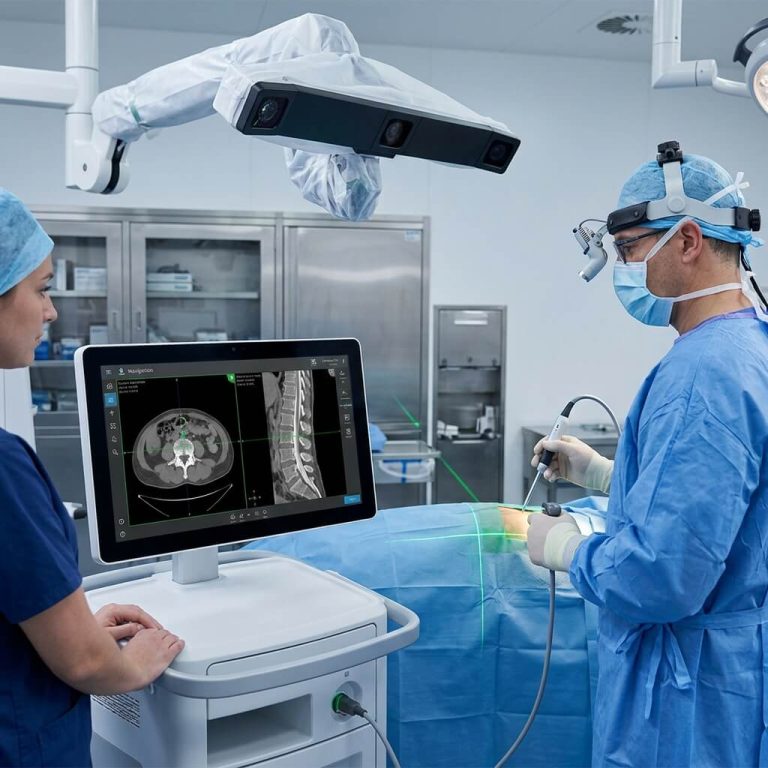
Integrated Picture-in-Picture (PiP): Preoperative MRI, CT scans, navigation tracking lines, and endoscope video feeds can be overlaid directly onto the primary 3D surgical monitor.
4. Head-to-Head Comparison: Optical Microscope vs. Digital Exoscope
The matrix below contrasts the physical, optical, and operational characteristics of traditional optical microscopes and modern 4K 3D digital exoscopes:
| Feature / Metric | Traditional Optical Operating Microscope | 4K 3D Digital Surgical Exoscope |
| Visual Delivery Method | Direct binocular eyepieces (Oculars) | Large 4K 3D flat-panel monitor with 3D glasses |
| Surgeon Working Posture | Fixed neck flexion; eyes tethered to oculars | Heads-up neutral posture; upright spine |
| Visual Latency (Lag) | Zero latency (Speed of light) | Minimal digital lag (typically 15 ms to 35 ms) |
| Depth Perception Mechanism | Pure binocular optical stereopsis | Dual-sensor digital stereopsis onto 3D monitor |
| Working Distance & Clearance | 200 mm to 400 mm; bulkier head over field | 250 mm to 600 mm; compact camera head |
| Team Visualization | Primary surgeon & 1 assistant (via beam splitter) | Entire operating room team shares identical 3D view |
| Illumination Source | High-intensity coaxial LED or Xenon | Coaxial LED with automated digital gain control |
| Digital Data Integration | Requires external add-on cameras & adapters | Native 4K video recording & PACS streaming |
| Hand-Eye Coordination | Direct alignment (Hands and eyes on same axis) | Dissociated axis (Hands down, eyes forward on monitor) |
| Average Capital Cost | Wide range ($35,000 – $180,000+ USD) | Premium tier ($120,000 – $300,000+ USD) |
5. The Critical Technical Battlegrounds: Latency, Resolution & Hand-Eye Coordination
While exoscopes offer clear ergonomic advantages, why haven’t they completely replaced traditional optical microscopes? The answer lies in the physiological limits of human hand-eye coordination.
Plaintext
                  THE EXOSCOPE LATENCY PIPELINE
+-------------------------------------------------------------------------+
| CMOS Sensor Exposure --> 3D Image Signal Processor --> Display Refresh  |
|      (~8 - 12 ms)                  (~5 - 10 ms)             (~5 - 10 ms)|
|                                                                         |
| TOTAL END-TO-END LATENCY: ~18 to 32 Milliseconds                        |
+-------------------------------------------------------------------------+
The Latency Threshold in Microsurgery
In consumer video, a latency (delay) of 50 to 100 milliseconds is unnoticeable. In microsurgery, however, where a surgeon is operating on a pulsatile 1-millimeter artery, latency becomes a critical performance factor:
-
Sub-30 Millisecond Standard: Modern medical-grade exoscopes must achieve an “end-to-end latency” (the time between physical movement and monitor display) of less than 30 milliseconds.
-
If latency exceeds 40 to 50 milliseconds, surgeons experience a subtle “rubber-band” sensation. When micro-suturing, the surgeon may over-correct needle trajectories because their eyes see the movement slightly after their hands have performed it. While experienced surgeons adapt to minor latency within a few hours, pure optical microscopes remain entirely immune to this issue.
Hand-Eye Axis Dissociation
With a traditional microscope, the surgeon’s eyes look downward along the same physical vector that their hands are working.
With an exoscope, the hands are working down in the patient’s wound, but the surgeon’s eyes are looking straight ahead at a monitor mounted 2 to 3 meters away on an equipment boom.
This hand-eye axis dissociation requires a cognitive adaptation curve. Surgeons must train their motor skills to manipulate instruments along one visual plane while looking at another—similar to the learning curve required when transitioning from open laparotomy to laparoscopic surgery.
6. Clinical Specialty Adoption: Where Are Exoscopes Winning?
The adoption rate of digital exoscopes varies considerably across surgical specialties, driven by anatomical depth and procedure duration.
Plaintext
                     SPECIALTY ADOPTION SPECTRUM
                                  |
     +----------------------------+----------------------------+
     |                                                         |
     v                                                         v
[ RAPID EXOSCOPE ADOPTION ]                   [ TRADITIONAL OPTICAL DOMINANCE ]
- Minimally Invasive Spine (MISS)             - Ophthalmology (Cataract, Cornea)
- Skull Base Tumor Resection                  - High-Magnification Microvascular
- Lateral Skull Base / ENT Neck Dissection    - Hand Surgery & Replantation
A. Spine Surgery: The Leading Edge of Exoscope Adoption
Spine surgery has adopted digital exoscopes faster than any other field.
-
Why it Works: Spine surgeons frequently work through long, narrow tubular retractors during microdiscectomies and laminectomies. The narrow corridor requires the visualization head to sit back, out of the way of long bone rongeurs, screwdrivers, and drills.
-
The exoscope’s long focal distance (300 to 500 mm) provides clear, deep lighting down the tube, while the heads-up posture allows the spine surgeon to maintain an upright, comfortable stance during long spinal fusion cases.
B. Neurosurgery (Cranial): A Balanced Coexistence
In cranial neurosurgery, exoscopes are heavily utilized for superficial tumor resections (meningiomas, convex gliomas) and complex skull base approaches where awkward angles make traditional eyepieces painful.
-
However, for deep vascular clipping of cerebral aneurysms and arteriovenous malformations, many senior neurosurgeons still prefer traditional optical microscopes for their uncompressed stereoscopic depth perception and zero-latency responsiveness.
C. Ophthalmology: Traditional Optics Still Dominate
Ophthalmology remains firmly anchored to traditional optical microscopes.
-
The Reason: Ophthalmic surgery relies heavily on the optical Red Reflex to visualize transparent lens capsules during cataract surgery. Digital image sensors, despite HDR advancements, can struggle to replicate the subtle photon reflection patterns produced by pure coaxial optical light bouncing off the vascular choroid.
-
While digital 3D heads-up systems (such as Alcon NGENUITY and Zeiss ARTEVO) exist, they are often paired with an optical microscope core rather than acting as standalone exoscopes.
D. Plastic & Reconstructive Surgery: Growing Curiosity
Reconstructive surgeons perform long free-flap operations (e.g., 6 to 10 hours for bilateral DIEP breast reconstruction) where surgeon fatigue is a primary factor in procedural complications.
-
The exoscope’s shared 3D monitor allows the plastic surgeon and the assistant to perform microvascular anastomosis without crowding around a small face-to-face ocular bridge.
-
However, some surgeons still find that suturing 10-0 and 11-0 nylon vessels (under 1.0 mm) is slightly faster and more predictable under pure optical magnification.
7. The Compromise: Hybrid Surgical Visualization Platforms
Recognizing that both optical and digital systems have distinct advantages, medical device engineering is increasingly moving toward Hybrid Operating Systems.
Plaintext
                     THE HYBRID VISUALIZATION MODEL
+---------------------------------------------------------------------------------+
|                  [ Traditional Apochromatic Optical Core ]                      |
|                                     |                                           |
|        +----------------------------+----------------------------+              |
|        |                                                         |              |
|        v                                                         v              |
| [ Physical Binocular Oculars ]                 [ Integrated 4K 3D Camera Head ] |
| - Used for critical micro-vascular ties       - Projects to 55" 3D Monitor      |
| - Instant zero-latency optical depth           - Used for heads-up dissection   |
+---------------------------------------------------------------------------------+
A hybrid surgical microscope incorporates a premium apochromatic optical head and traditional binocular oculars, but embeds dual 4K digital sensors directly inside the optical housing.
-
Clinical Workflow: The surgeon can perform initial bone drilling, craniotomy opening, and gross tumor debulking in the comfortable, heads-up position looking at the 55-inch 3D monitor.
-
When the operation reaches a critical microvascular phase—such as dissecting an adherent perforating artery off an aneurysm neck—the surgeon simply pulls the physical binocular eyepieces down and looks directly through the optical glass for zero-latency, maximum-fidelity control.
8. Procurement and Hospital Investment Analysis
For hospital purchasing directors, capital budget committees, and clinic owners, deciding between a traditional optical microscope, a digital exoscope, or a hybrid unit requires a balanced financial assessment.
Plaintext
               HOSPITAL PROCUREMENT DECISION FRAMEWORK
+---------------------------------------------------------------------------+
| Step 1: Analyze Specialty Mix (Spine-heavy favors Exoscopes;              |
|         Ophthalmology/Vascular favors Optical).                           |
| Step 2: Evaluate Operating Room Space (Exoscopes save ceiling/floor space |
|         but require high-end medical monitor boom arms).                  |
| Step 3: Assess Teaching Requirements (Academic centers benefit vastly     |
|         from shared 3D screen learning curves).                           |
| Step 4: Calculate Total Cost of Ownership (Optical systems have longer    |
|         10-15 year mechanical life; digital systems require software      |
|         and display upgrade cycles every 5-7 years).                      |
+---------------------------------------------------------------------------+
Capital Cost Considerations
-
Standard Optical Operating Microscopes: Range widely from $35,000 to $90,000 USD for high-quality standard units (ENT, Dental, General Microsurgery), rising to $150,000 to $250,000+ USD for premium neurosurgical models.
-
Standalone 4K 3D Digital Exoscopes: Typically command premium pricing, ranging from $150,000 to $350,000 USD, driven by multi-axis robotic camera arms, 4K 3D display monitors, and advanced image processing computer units.
Long-Term Operational Life (TCO)
-
Optical Systems: Consist of durable glass optics and robust mechanical counterbalances. With routine maintenance, optical microscopes often provide reliable clinical service for 12 to 15 years without structural obsolescence.
-
Digital Exoscope Systems: Depend on digital camera sensors, computer graphics processing units (GPUs), display panels, and operating software. Similar to enterprise IT hardware, digital systems are subject to faster technological obsolescence, typically requiring software patches and hardware display refresh cycles every 5 to 7 years.
Frequently Asked Questions (FAQ)
Q1: Can a digital exoscope completely replace a traditional surgical microscope today?
In specialized environments—such as dedicated minimally invasive spine centers or outpatient skull base surgery centers—yes, many surgical teams operate exclusively using digital exoscopes. However, across broad, multi-specialty hospital networks, traditional optical microscopes remain indispensable for complex microvascular repairs and ophthalmic procedures where zero latency and direct optical depth perception are non-negotiable.
Q2: Do surgeons experience headaches or eye strain from wearing 3D glasses during long operations?
Modern exoscopes utilize lightweight, passive polarized 3D glasses (weighing only a few grams) rather than heavy battery-powered active-shutter glasses. Because passive glasses do not flicker, most surgeons report significantly less eye strain and zero headaches compared to the physical pressure and orbital fatigue associated with leaning into hard rubber microscope oculars for hours.
Q3: Is 4K digital screen resolution actually sharper than an optical microscope?
No. High-quality optical glass operating microscopes have an effective visual resolution that exceeds even 8K digital sensors. Light entering the human eye through a multi-coated apochromatic lens provides an uncompressed, continuous photon stream. However, 4K digital exoscopes compensate for this by offering digital sharpening algorithms, digital magnification zoom, and localized contrast enhancement that make anatomical boundaries stand out clearly.
Q4: How long does it take for a microsurgeon to adapt to heads-up exoscopic surgery?
Most microsurgeons report an adaptation period of 5 to 10 surgical procedures to become comfortable with hand-eye axis dissociation and the remote monitor positioning. Once this spatial transition is mastered, surgeons frequently cite dramatic improvements in neck and upper back comfort.
Q5: Can an existing optical microscope be upgraded to a 3D digital heads-up display?
Yes. Many hospitals upgrade existing optical operating microscopes by attaching dual-sensor 4K 3D camera adapters to the existing optical beam splitter. This creates a functional hybrid system, allowing the surgical team to view the operation on a large 3D monitor while preserving the surgeon’s ability to look directly through the traditional binocular eyepieces at any time.
Summary
The choice between a traditional optical operating microscope and a 4K 3D digital exoscope is not a question of which technology is strictly superior, but rather which tool aligns with the surgical team’s clinical procedures, ergonomic demands, and institutional budget.
-
Choose a Traditional Optical Microscope when clinical priorities demand zero-latency visual responsiveness, natural stereoscopic depth perception, unmatched optical color fidelity, and a proven, 15-year mechanical lifespan.
-
Choose a 4K 3D Digital Exoscope when procedures are spine- or skull-base intensive, where surgeon ergonomics and physical longevity are critical concerns, where deep narrow corridors benefit from an open workspace, and where academic surgical teaching thrives on team-wide 3D visual immersion.