Executive Summary & Industry Overview
Selecting the right ultrasound machine for a medical facility, private clinic, or hospital department is one of the most critical capital expenditure decisions for healthcare administrators and procurement managers. Diagnostic ultrasound technology has advanced rapidly in recent years. Today’s commercial diagnostic market ranges from high-end cart-based color doppler ultrasound machines to ultra-lightweight portable laptop ultrasound scanners and handheld probe devices.
This comprehensive 2026 buying guide breaks down the essential factors involved in purchasing diagnostic ultrasound equipment. Whether you are searching for an affordable portable ultrasound machine price, comparing black and white ultrasound vs color doppler, or sourcing high-performance OB/GYN 4D ultrasound scanners directly from OEM medical equipment manufacturers, this article covers everything you need to make an informed, ROI-driven purchase.
1. Main Categories of Ultrasound Machines: Form Factors & Portability
Modern ultrasound diagnostic systems are classified primarily by their physical architecture, mobility, and computational performance. Selecting the correct form factor depends heavily on your facility’s daily workflow, space availability, and mobility requirements.
A. Cart-Based (Trolley) Ultrasound Systems
Cart-based or console ultrasound systems represent the traditional powerhouse in diagnostic imaging. Designed primarily for dedicated radiology departments, cardiology suites, and busy obstetric clinics, these units prioritize raw image processing power, large high-definition displays (typically 19 to 24 inches), and extensive probe connectivity.
-
Key Features: 3 to 5 active transducer probe ports, advanced cooling systems, motorized height adjustment, extended battery backup, and high-channel-count processing engines.
-
Best For: High-volume hospital departments, comprehensive vascular imaging, 3D/4D fetal diagnostics, and complex cardiac color doppler evaluations.
-
Pros & Cons: Unmatched diagnostic image resolution and probe versatility, but limited mobility outside the facility and higher price points.
B. Portable / Laptop Ultrasound Machines
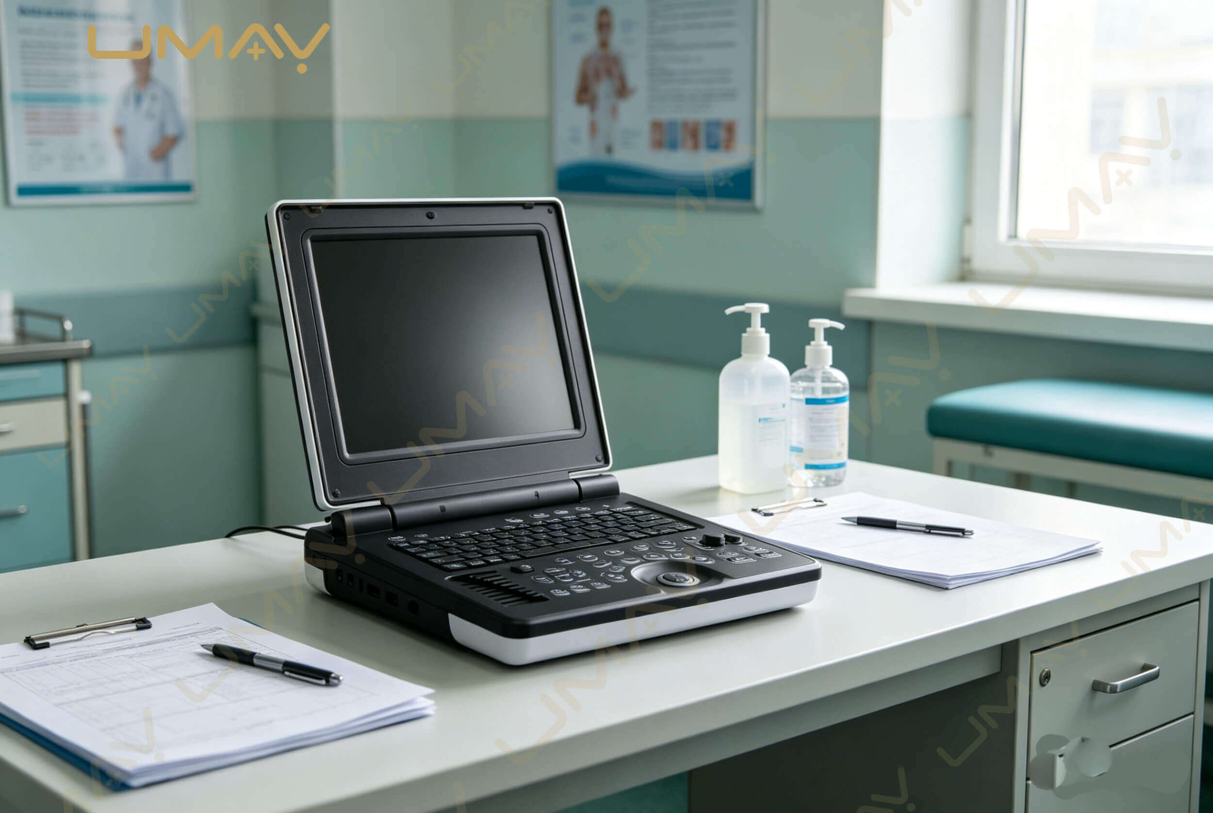
Portable laptop-style ultrasound scanners have revolutionized point-of-care diagnostics (POCUS). Combining the imaging capabilities of mid-tier cart systems with a compact clamshell design, portable color doppler machines offer flexibility for multi-room clinics, emergency rooms, and field medical applications.
-
Key Features: Fast boot-up times (under 30 seconds), integrated lithium-ion battery packs (2–4 hours continuous scanning), lightweight design (5 kg to 8 kg), and multi-probe connectors via quick-docking carts.
-
Best For: Emergency departments (ED), Intensive Care Units (ICU), mobile clinics, outpatient orthopedic centers, and general practice clinics.
-
Pros & Cons: Excellent mobility and space-saving footprint with near-cart-level image quality; slightly smaller screen size (15 to 17 inches).
C. Handheld & Wireless Ultrasound Scanners
Handheld ultrasound transducers connect directly to commercial smartphones or tablets via USB-C or high-speed Wi-Fi. Known as “pocket ultrasound” or “wireless transducer scanners,” these devices represent the newest tier of bedside diagnostic tools.
-
Key Features: All-in-one probe design (beamformer built into transducer handle), app-based user interface, cloud storage integration, and ultra-portable fit inside coat pockets.
-
Best For: Fast bedside triage, abdominal screening, vascular access guidance, quick bladder scans, and field emergency response.
| Form Factor Category | Mobility Level | Screen Size | Active Ports | Primary Clinical Use |
| Cart-Based Ultrasound | Stationary / Room-to-Room | 19″ – 24″ LED/OLED | 3 – 5 Ports | Radiology, Cardiology, OB/GYN, Vascular |
| Portable Laptop Ultrasound | High (Vehicle / Clinic) | 15″ – 17″ LCD | 1 – 3 Ports | POCUS, ICU, Anesthesia, MSK, Mobile Care |
| Handheld Probe Scanner | Ultra-High (Pocket) | Tablet / Phone | Integrated Probe | Bedside Triage, Vascular Access, Field Med |
2. Imaging Modes: B-Mode vs. Color Doppler vs. 3D/4D
When reviewing technical spec sheets from B2B medical ultrasound suppliers, understanding the available imaging software modes is essential for matching equipment capabilities with clinical requirements.
Black & White (B-Mode / 2D) Ultrasound
Brightness Mode (B-Mode) generates classic two-dimensional grayscale images of anatomical structures. Grayscale scanners remain widely used for routine abdominal screening, basic obstetric checks, and simple fluid identification. However, basic B-mode units lack blood flow velocity analysis.
Color Doppler & Spectral Imaging Modes
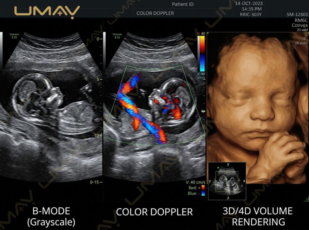
Color Doppler ultrasound overlays color-coded blood flow velocity data onto 2D anatomical images. Red and blue indicators represent the direction and velocity of hemodynamics relative to the transducer head.
-
Color Flow Mapping (CFM): Evaluates blood flow direction and velocity across cardiac chambers and major blood vessels.
-
Power Doppler Imaging (PDI): Offers superior sensitivity for low-velocity blood flow in small vessels (ideal for kidney perfusion and tumor microvascularity).
-
Pulsed Wave (PW) & Continuous Wave (CW) Doppler: Measures exact blood flow velocities, pressure gradients, and valve regurgitation in vascular and cardiac diagnostics.
Advanced 3D/4D & Elastography Imaging
Advanced commercial diagnostic scanners offer real-time three-dimensional rendering (3D) and continuous movement rendering (4D). 4D OB/GYN ultrasound machines allow prospective parents and clinicians to view fetal surface details in real time. Tissue Elastography measures tissue stiffness, assisting in early liver fibrosis assessment and breast mass differentiation.
3. Commercial Price Breakdown & Budgeting Guide (2026 Market Analysis)
Ultrasound machine pricing varies widely based on technology tier, transducer channel density, brand recognition, and included software packages. Below is a breakdown of global market pricing for new diagnostic ultrasound machines in 2026.
| Market Tier & Type | Price Range (USD) | Included Features & Specifications | Target Buyer Profile |
| Entry-Level / B&W Portable | $2,000 – $6,000 | 2D Grayscale, 1-2 Basic Probes, 12-15″ Screen, Basic Measurement Software | Small Clinics, Veterinary Practices, Basic Screening |
| Mid-Range Portable Color Doppler | $6,000 – $16,000 | Color Doppler, PW/Power Doppler, 2 Active Ports, THI, Cardiac/OB Software | Community Hospitals, Urgent Care, MSK Clinics, POCUS |
| High-End Cart-Based Color Doppler | $16,000 – $38,000 | 3D/4D Imaging, Auto-EF, Strain Rate, 4 Active Ports, Touchscreen Control, DICOM 3.0 | Specialist Clinics, Regional Hospitals, OB/GYN Centers |
| Premium Clinical & Research Systems | $38,000 – $80,000+ | Single Crystal Probes, AI-assisted Diagnostics, Contrast Imaging, Shear Wave Elastography | Tertiary Hospitals, Academic Research, Cardiac Centers |
💡 Pro Buyer Tip on Total Cost of Ownership (TCO): Never evaluate an ultrasound machine based solely on the upfront sticker price. Always account for additional expenses including extra probe transducers ($1,000–$4,000 per probe), shipping/import duties, extended warranty packages, DICOM connectivity software licenses, and ongoing maintenance service contracts.
4. Key Technical Specifications to Evaluate Before Buying
To ensure maximum ROI and clinical utility, review these crucial hardware and software specifications when communicating with B2B medical equipment vendors:
-
Transducer Probe Frequency & Channels: Higher channel counts (e.g., 64, 128, or 256 channels) deliver superior signal processing, finer spatial resolution, and clearer tissue penetration. Ensure frequency ranges match target anatomy (e.g., 2.0–5.0 MHz for deep abdominal scans; 6.0–15.0 MHz for superficial MSK/vascular scans).
-
Tissue Harmonic Imaging (THI): THI technology uses harmonic sound frequencies generated as ultrasound pulses travel through tissue. This significantly reduces clutter, improves contrast resolution, and enhances border visualization in difficult-to-image patients.
-
DICOM 3.0 Integration & Cloud Connectivity: Ensure the unit supports DICOM Worklist, Store, Print, and Structured Reporting. Seamless integration with your hospital’s PACS (Picture Archiving and Communication System) or clinic EMR is essential for efficient patient workflow.
-
Battery Life & Boot Speed: For mobile emergency and point-of-care environments, verify that portable units offer hot-swappable batteries and boot times of under 30 seconds from standby mode.
5. How to Choose the Right Ultrasound Machine by Clinical Application
Different clinical specialties demand specific hardware configurations, software measurement modules, and probe combinations:
OB/GYN & Women’s Health
Requires high-resolution 2D imaging alongside 3D/4D volumetric rendering. Essential probes include a convex abdominal probe (for routine obstetric scanning) and a transvaginal probe (EBL/TV) for early pregnancy and gynecological diagnostic evaluations. Automatic fetal biometry calculation tools speed up routine patient visits.
Cardiology & Vascular Diagnostics
Demands high frame rate imaging and continuous wave (CW) Doppler. Essential probes include a phased array cardiac probe and high-frequency linear vascular transducer. Dedicated cardiology software packages must include Auto-EF (Ejection Fraction), TDI (Tissue Doppler Imaging), and Stress Echo capabilities.
Musculoskeletal (MSK) & Pain Management
Requires ultra-high frequency linear probes (up to 18 MHz) to visualize superficial nerves, tendons, and joint spaces clearly. High spatial resolution and needle enhancement visualization software are crucial for guiding injections accurately.
General Practice & Bedside POCUS
Favors versatile, lightweight laptop portable ultrasound machines equipped with a dual-head probe or a combination of convex and linear transducers. Prioritize quick boot time, intuitive touch interface, and durable chassis design.
6. Navigating B2B Procurement, Import Logistics & Supplier Verification
When purchasing diagnostic medical equipment internationally from manufacturers or wholesale exporters, risk mitigation is critical. Follow this structured checklist before executing a purchase order:
-
International Regulatory Certifications: Confirm the manufacturer holds valid ISO 13485 quality management certification along with relevant destination market approvals, such as CE Mark (MDR) for Europe or FDA 510(k) clearance for the US.
-
OEM/ODM Customization Capabilities: For wholesale medical distributors, verify if the supplier provides OEM custom branding, language localization, and software UI modification.
-
Warranty & Spare Parts Availability: Insist on a minimum 18-to-24-month comprehensive warranty covering main board components and probes. Verify that the supplier maintains a supply of spare parts (power boards, trackballs, display cables) for fast dispatch.
-
After-Sales Technical Support: Ensure the factory provides remote technical support via video call or online diagnostic software to troubleshoot operational issues without delay.
7. Frequently Asked Questions (FAQ) for Ultrasound Buyers
Q1: What is the difference between a 3D and 4D ultrasound machine?
A 3D ultrasound scanner captures static three-dimensional volume images of internal organs or fetuses. A 4D ultrasound scanner captures 3D images continuously in real-time, creating a live moving video of fetal motion or cardiac structure dynamics.
Q2: Can I use a portable ultrasound machine for full cardiac diagnostic examinations?
Yes. Premium portable color doppler ultrasound machines equipped with phased array transducers, Continuous Wave (CW) Doppler, and cardiac measurement packages offer diagnostic capabilities comparable to mid-range cart units.
Q3: How long does a commercial ultrasound machine typically last?
With regular preventative maintenance, transducer care, and software updates, a quality commercial ultrasound machine has a typical operational lifespan of 7 to 10 years.
Q4: Why are linear probes used for vascular scans while convex probes are used for abdominal scans?
Linear probes utilize high frequencies (6–15 MHz) providing crisp resolution at shallow depths, ideal for superficial blood vessels and small parts. Convex probes utilize lower frequencies (2–5 MHz) with a curved beam structure, allowing deep acoustic penetration for abdominal and pelvic organs.
Conclusion: Maximizing Value on Your Ultrasound Investment
Investing in the right ultrasound machine requires balancing diagnostic performance, mobility requirements, probe versatility, and long-term operating costs. By understanding the distinction between cart-based and portable color doppler systems, selecting application-appropriate probes, and verifying international B2B suppliers, healthcare providers can secure reliable diagnostic imaging solutions that enhance patient care and deliver maximum financial ROI.