Introduction: Conquering the “Dark Continent” of the GI Tract
For gastroenterologists, the human gastrointestinal (GI) tract consists of three main segments: the upper GI tract (esophagus and stomach), the lower GI tract (colon and rectum), and the middle segment known as the small intestine (small bowel).
While traditional push gastroscopy easily views the stomach, and colonoscopy views the large intestine, the small bowel—measuring roughly 6 meters (20 feet) in length with complex, winding loops—was historically considered the “dark continent” of the digestive system. Reaching deep into the small bowel previously required highly invasive, uncomfortable, and sometimes risky procedures like balloon-assisted enteroscopy or intraoperative enteroscopy.
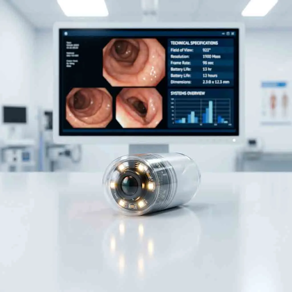
The advent of Wireless Capsule Endoscopy (WCE) changed everything. By simply swallowing a vitamin-sized camera, patients allow doctors to capture tens of thousands of high-resolution images of the entire small bowel mucosa non-invasively.
This guide breaks down the specific medical conditions diagnosed via capsule endoscopy, the step-by-step patient workflow, and the clinical safety protocols used to prevent complications.
2. Primary Diagnostic Indications: When is Capsule Endoscopy Used?
Capsule endoscopy is not a replacement for traditional endoscopy in every scenario. It is a highly specialized tool deployed for specific clinical indications, primarily focused on small bowel pathology.
A. Obscure Gastrointestinal Bleeding (OGIB)
The most common indication for capsule endoscopy is Obscure GI Bleeding. This occurs when a patient has ongoing internal bleeding—manifesting as unexplained iron-deficiency anemia, positive fecal occult blood tests, or visible dark stools (melena)—but both a standard upper endoscopy and a colonoscopy have failed to find the bleeding source.
-
What the Capsule Finds: The capsule is highly effective at identifying small bowel angioectasias (abnormal, fragile blood vessels), actively bleeding erosions, and Cameron lesions that standard scopes miss.
B. Crohn’s Disease (Inflammatory Bowel Disease)
Crohn’s disease can affect any part of the GI tract, but it frequently targets the terminal ileum and deep small bowel.
-
What the Capsule Finds: Capsule endoscopy is the gold standard for detecting early, subtle mucosal changes associated with Crohn’s disease, such as aphthous ulcers (small mucosal breaks), erythema (redness), mucosal edema (swelling), and cobble-stoning. It is also used to evaluate mucosal healing in patients undergoing biologic therapies.
C. Small Bowel Tumors and Polyps
While primary small bowel cancer is rare, benign and malignant tumors do occur.
-
What the Capsule Finds: The pill camera helps detect Gastrointestinal Stromal Tumors (GIST), neuroendocrine tumors (carcinoids), lymphomas, and adenocarcinomas. It is also a vital surveillance tool for patients with hereditary polyposis syndromes, such as Peutz-Jeghers syndrome and Familial Adenomatous Polyposis (FAP), who need regular monitoring for small bowel polyps.
D. Celiac Disease (Gluten-Sensitive Enteropathy)
Celiac disease is traditionally diagnosed via blood tests and a duodenal biopsy. However, capsule endoscopy plays a critical role in patients who cannot tolerate traditional endoscopy or those who have “refractory Celiac disease” (symptoms persist despite a strict gluten-free diet).
-
What the Capsule Finds: The camera captures high-definition images of villous atrophy (flattening of the finger-like projections in the intestine), mucosal scalloping, and mucosal fissures characteristic of severe gluten intolerance.
3. The Patient Workflow: Step-by-Step Preparation & Procedure
One of the greatest advantages of capsule endoscopy is patient comfort. The procedure requires no sedation, no anesthesia, and no recovery room time. However, achieving clear diagnostic images requires strict patient compliance regarding bowel preparation.
Plaintext
[ Capsule Endoscopy Workflow Timeline ]
------------------------------------------------
Day -1 (Prior to Exam):
- 12:00 PM: Switch to a strict clear liquid diet.
- 06:00 PM: Take prescribed bowel preparation (e.g., PEG laxative).
- 10:00 PM: Begin complete fasting (NPO) – no food or water.

Day 0 (Day of Exam):
- 08:00 AM: Patient arrives at clinic; sensor belt is attached.
- 08:15 AM: Patient swallows the capsule with a small sip of water containing simethicone.
- 10:15 AM (2 Hrs Post-Swallow): Patient may drink clear liquids.
- 12:15 PM (4 Hrs Post-Swallow): Patient may eat a light snack.
- 04:00 PM (8 Hrs Post-Swallow): Patient returns to clinic; equipment is removed.
- Next 24-48 Hrs: Patient confirms capsule excretion in stool.
------------------------------------------------
1. Bowel Preparation (Clearing the View)
The small intestine must be completely empty of food debris, bile, and opaque fluids. Patients typically consume a clear liquid diet the day before the procedure. Gastroenterologists often prescribe a mild polyethylene glycol (PEG) laxative to flush the bowel, alongside simethicone drops just before swallowing the capsule to eliminate intestinal gas bubbles that could block the camera lens.
2. Swallowing the Capsule & Sensor Placement
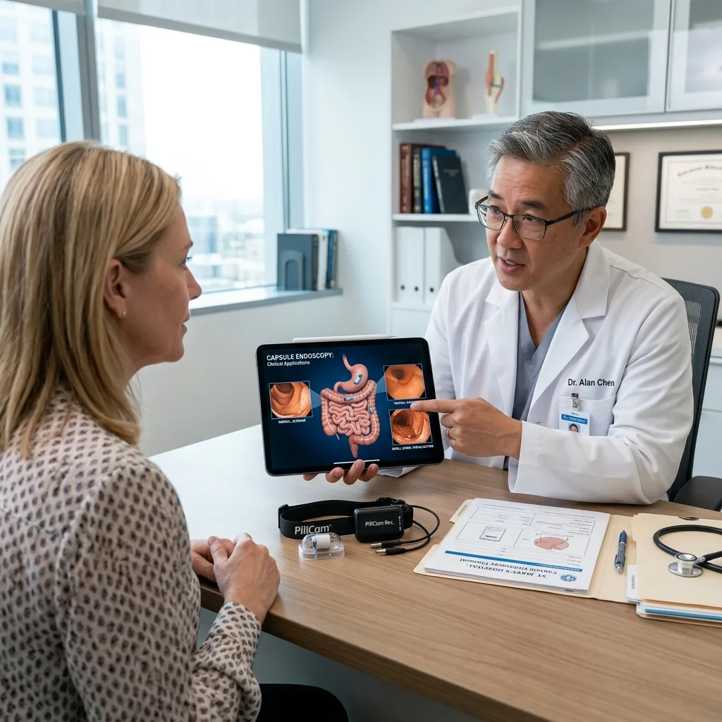
On the morning of the exam, the nurse secures a sensor array belt around the patient’s abdomen. The data recorder is connected to the belt and slung over the shoulder. The patient then swallows the flashing, pill-sized capsule with a small cup of water.
3. The Ambulatory Phase
Once swallowed, the patient leaves the clinic and goes about their normal daily activities. Walking and light movement are actually encouraged, as gravity and physical activity help stimulate normal gastrointestinal peristalsis, moving the capsule smoothly through the digestive tract.
4. Excretion and Equipment Return
After 8 to 12 hours (depending on the battery life of the specific capsule model), the recording is complete. The patient returns the sensor belt and data recorder to the clinic. The single-use capsule will pass naturally in a bowel movement within 1 to 3 days. It is safely flushed down the toilet.
4. Understanding and Preventing “Capsule Retention”
The primary clinical risk associated with capsule endoscopy is capsule retention. Retention occurs when the capsule gets stuck in the digestive tract for more than two weeks, preventing it from passing naturally.
Why Does Retention Occur?
Retention is almost never an issue in a healthy GI tract. It happens when the patient has an undiagnosed stricture (a severe narrowing of the intestine). Strictures are most commonly caused by:
-
Severe, long-standing Crohn’s disease resulting in scar tissue.
-
Previous abdominal surgeries causing intestinal adhesions.
-
Radiation enteritis (damage from previous cancer radiation therapies).
-
Large, obstructive small bowel tumors.
-
Chronic use of NSAIDs (Non-Steroidal Anti-Inflammatory Drugs) leading to NSAID-induced enteropathy and diaphragm-like strictures.
The Solution: The Patency Capsule
If a gastroenterologist suspects a patient might have a stricture, they will perform a safety test using an Agile Patency Capsule before administering the actual video capsule.
A patency capsule is a dissolvable “dummy” pill that is the exact same size and weight as the real camera capsule. It contains a tiny RFID scanner inside a body of compressed barium and lactose.
-
If the GI tract is clear: The patient excretes the patency capsule intact within 30 hours, giving the doctor the “green light” to use the real video capsule safely.
-
If the GI tract has a stricture: The patency capsule will get temporarily stuck. However, because it is designed to dissolve, intestinal fluids will break down the capsule within 40 to 80 hours, allowing it to collapse and pass harmlessly, thereby preventing a medical emergency.
5. Contraindications: Who Should NOT Have a Capsule Endoscopy?
While highly safe, capsule endoscopy is contraindicated (not recommended) for certain patient populations:
-
Patients with Known GI Obstructions: Anyone with suspected bowel obstruction, strictures, or fistulas should not swallow a permanent capsule (without a successful patency test).
-
Swallowing Disorders (Dysphagia): Patients with severe dysphagia or Zenker’s diverticulum risk aspirating the capsule into the airway. In these cases, doctors can use a traditional endoscope to manually place the capsule directly into the duodenum.
-
Severe Gastroparesis: Patients with delayed gastric emptying may have the capsule sit in the stomach for 12 hours until the battery dies, failing to image the small bowel. Prokinetic drugs (like metoclopramide) may be administered to force the capsule into the small intestine.
-
Pregnancy: As a standard medical precaution, capsule endoscopy is generally avoided during pregnancy unless absolutely clinically necessary.
-
Cardiac Pacemakers (Relative Contraindication): Older guidelines prohibited capsule use in patients with pacemakers due to fear of radiofrequency interference. However, modern clinical studies have shown that capsule endoscopy is overwhelmingly safe for patients with modern pacemakers and implantable cardioverter-defibrillators (ICDs), though it is still managed with caution.
6. Image Interpretation: How Doctors Read the Results
Once the data recorder is returned to the clinic, the data is downloaded to a proprietary software workstation. The 12-hour journey produces between 50,000 and 60,000 individual photographs.
The clinical reading software compiles these images into a continuous video. Using a specialized medical monitor, the gastroenterologist reviews the video at variable speeds (e.g., 10 to 25 frames per second). The doctor looks for specific landmarks to assess transit time:
-
First Gastric Image: The moment the capsule enters the stomach.
-
First Duodenal Image: The moment it passes through the pyloric sphincter into the small bowel.
-
First Cecal Image: The moment it passes the ileocecal valve into the colon.
The reading process traditionally takes 45 to 90 minutes. The doctor identifies and bookmarks pathological images, such as bleeding, polyps, or ulcers, and compiles them into a final diagnostic report. (In modern clinics, Artificial Intelligence is now being used to drastically reduce this reading time—a topic we will explore in detail in Post 4).
Frequently Asked Questions (FAQ)
Q1: Does swallowing the capsule hurt?
No. The capsule is completely smooth, made from biocompatible plastic, and is easily swallowed with a sip of water. Patients do not feel the capsule as it moves through the stomach and intestines.
Q2: Can I have an MRI test if I have swallowed an endoscopy capsule?
No. This is a critical safety rule. Patients must absolutely not undergo a Magnetic Resonance Imaging (MRI) scan until they have positively confirmed that the capsule has been excreted in their stool. The powerful magnetic field of an MRI machine can interact with the metal and battery inside the capsule, causing severe internal tissue tearing.
Q3: What happens if the capsule battery dies before it finishes the journey?
If the patient has slow bowel motility, the battery (which lasts 12 to 16 hours) may die before the capsule reaches the large intestine. This is called an “incomplete study.” If the suspected pathology was in the distal (lower) small bowel, the doctor may need to repeat the test, sometimes prescribing a prokinetic medication to speed up intestinal transit.
Q4: Can the capsule take biopsies or stop bleeding?
No. Standard wireless capsule endoscopes are purely diagnostic imaging tools. They cannot take tissue samples (biopsies), remove polyps, or cauterize bleeding blood vessels. If a severe lesion is found, a traditional device-assisted enteroscopy or surgery will be scheduled for treatment.
Q5: How do I know when the capsule has passed?
Patients are instructed to check their stool for 1 to 3 days following the procedure. The capsule flashes a bright light and is relatively easy to spot. If a patient is unsure whether the capsule has passed after two weeks, a simple abdominal X-ray will be taken to confirm it is no longer in the body.
Summary
The clinical application of medical endoscopy capsules has profoundly improved the diagnosis of small bowel diseases. By following proper patient preparation protocols and utilizing safety measures like patency capsules, healthcare providers can safely and accurately diagnose Obscure GI Bleeding, Crohn’s disease, and small bowel tumors without subjecting patients to invasive surgical procedures.