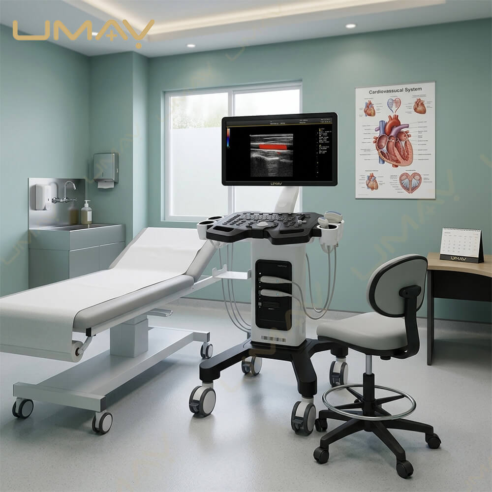
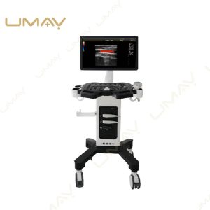
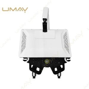
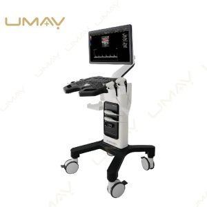



High-Frequency Trolley Color Doppler Ultrasound Machine | Institutional Cart-Based Diagnostic Ultrasound System | Full Digital Shared-Service Human Body Ultrasound Scanner with Multiple Active Probe Ports, CFM, PW, & Continuous Wave Doppler
Quick Info.
- SKU NO.: UMUS074
- Device Classification: Class Ⅱ
- Warranty: 1 Year
- Power Source: Electric
- Transport Package: Carton
- Origin: China
- Material: Metal, Plastic
- After-Sale Service: Online Technical Support
- Production Capacity: 2600 Sets/Year
Trapezoidal Imaging
Trapezoid imaging is a kind of expanded imaging, which is transformed
into a trapezoid based on the original rectangle, and the left and right sidesare expanded to a certain extent, achieving a wider field of view. The princi-ple of ultrasound imaging is to scan the human body with ultrasonic soundbeams, and obtain images of internal organs by receiving and processing the reflected signals.
Directional Power Doppler Imaging (DPDI)
Directional Power Doppler employs a small sample volume with highresolution to produce images with two-color directional information andless ‘blooming’ of color for more realistic representation of defect size. Itcan make up for the lack of power Doppler that can not display thedirection of blood flow, and increase the direction information.
Tissue Harmonic Imaging (THI)
It improves image clarity by improving tissue contrast resolution, and
spatial resolution, and eliminating near-field artifacts. It is mainly used forthe diagnosis of cardiovascular and abdominal diseases. It plays an import-ant role in evaluating the lesion area and boundary division of patients
with imaging difficulties.
The Specific Parameters
| ITEMS | PARAMETERS |
|---|---|
| Application Scene | |
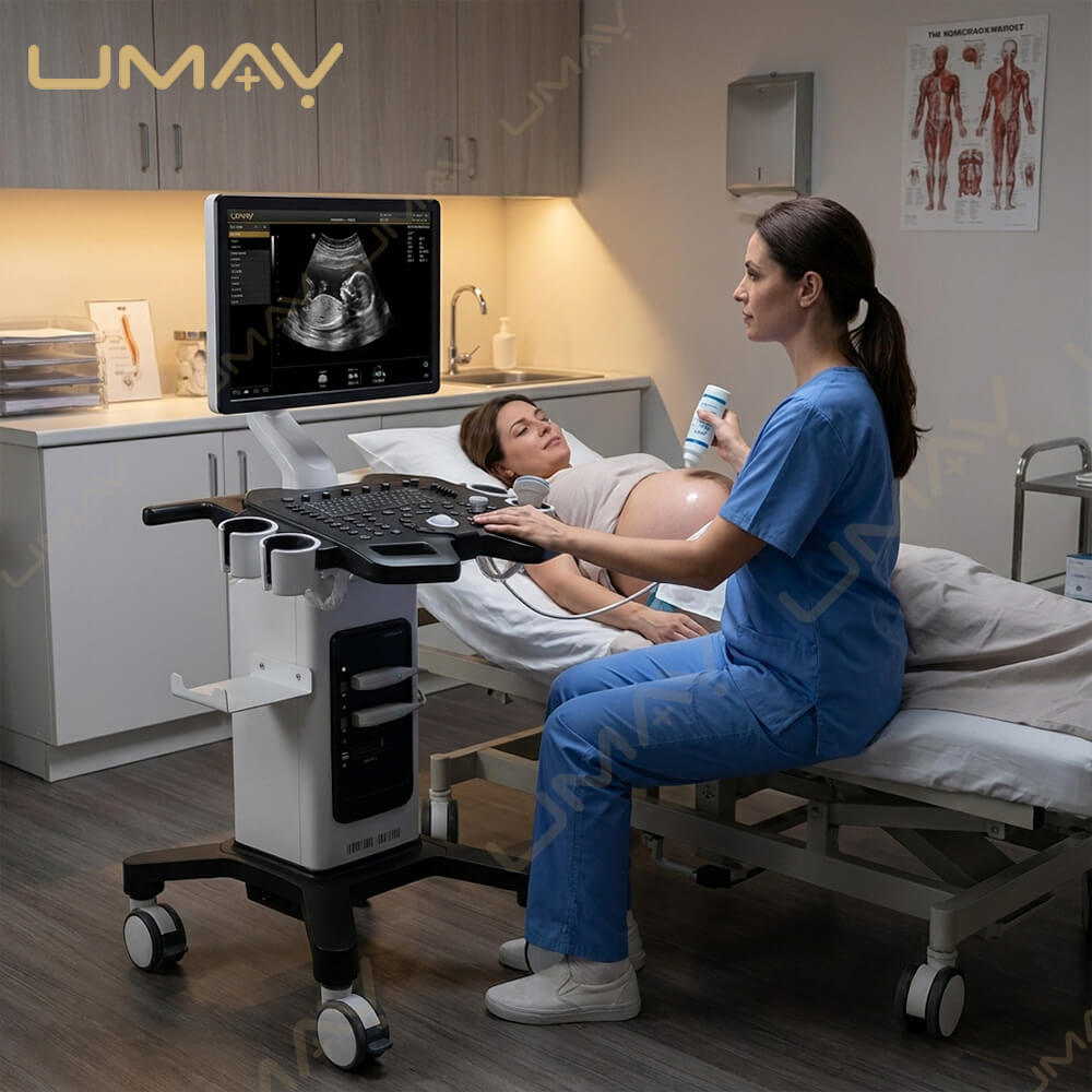
| Target Clinical Areas | Abdomen, gynecology, obstetrics, pediatrics, urology |
| Target Organs & Systems | Transvaginal, superficial, blood vessels, small organs |
| Display | |
| Display Type | 15 inch high-resolution color LCD |
| Color Modes | 8 groups fundamental + 8 harmonic frequencies |
| System Configuration | |
| Probe Interfaces | 4 interfaces, non-expandable |
| Rotating Arm | 360° rotation (up, down, left, right) |
| Operating System Languages | Chinese, English, Spanish (extendable) |
| Image Processing | |
| False Colors | 7 types |
| Gain Adjustment | B / M independently adjustable |
| Image Magnification | Up to 10× |
| Image Depth | ≥300mm |
| Sector Angle Adjustment | 10° to -50° range |
| Clipboard | Real-time image storage |
| Color Doppler Imaging | |
| Doppler Gain | 100% dB, continuously adjustable |
| PRF | ≥16 gears, 0.7kHz–22kHz |
| Angle Correction | 85° ~ -85°, 5° step size |
| Spectrum Reversal | Yes |
| Wall Filter | ≥3 gears adjustable |
| Spectrum Doppler | |
| PWD Modes | Linear, Convex, Sector |
| Pulse Frequency | 1.82–5.54kHz, adjustable |
| Spectrum Flip | Yes |
| Envelope Modes | Multiple types, real-time |
| Doppler Frequencies | 5 groups (linear/convex) |
| Display Control | Reverse display: left, right, up, down |
| Image Storage | |
| Storage Methods | Hard disk & USB disk |
| Playback Capacity | Grayscale image playback ≥1000 |
| System Functions | |
| Preset Modes | Obstetrics, vascular, cardiac, pediatrics, etc. |
| Image Management | Annotation, archiving, body mark, puncture |
| One-Key Recovery | Yes |
| Reset Function | Yes |
| Measurement & Analysis | |
| General Measurements | Distance, area, perimeter, angle, time, flow rate |
| Obstetric Measurements | Weight, gestational age, fetal index |
| Cardiac Measurements | Ejection index, end-diastole, acceleration |
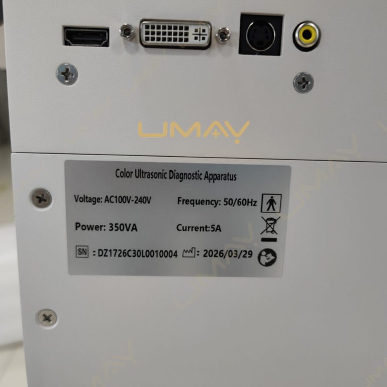
| Input / Output | S-Video, USB, LAN |
Core Systems Engineering Advantages for Medical Sourcing Networks:
-
Intelligent Software-Driven Beamforming Engine: Expand your diagnostic limitations. The core computing architecture integrates state-of-the-art Spatial Compound Imaging (SCI) and Speckle Reduction Technology (SRI). This drastically minimizes acoustic artifacts, enhances fine border delineation, and sharpens edge contrast for structural evaluations.
-
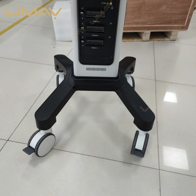
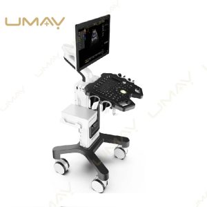
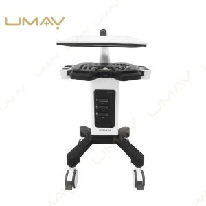
Simultaneous Multi-Probe Port Integration: Eliminate mechanical wear and clinical downtime. The low-profile cart body houses a side-mounted multi-port transducer bay, keeping all necessary probes safely docked, actively powered, and ready to swap with a single keystroke.
-
Intuitive Control Console with Multi-Zone TGC: Take total command of acoustic uniformity. The spacious control panel blends tactile alphanumeric shortcut keys with a sliding multi-segment Time Gain Compensation (TGC) matrix, giving the operator real-time amplification authority from shallow vascular pathways down to deep pelvic examinations.
-
Agile Trolley Footprint with Dual Braking Wheel Systems: Designed for intense daily hospital workflows. The sleek outer structural shield protects sensitive high-voltage processing components, while the low-vibration medical casters and secure locking mechanisms ensure effortless transit through tight intensive care units (ICU) and emergency bays.
📊 Comprehensive Technical Specifications:
| Technical Parameter Component | Standard System Configuration Attribute |
| System Architectural Layout | Ergonomic Mobile Trolley / Wheeled Cart Diagnostic Suite |
| Primary Scanning Configurations | B, 2B, 4B, M, Color Doppler (CFM), PDI, DPDI, PW Doppler, THI |
| Transducer Port Bay Capacity | Multiple Fully Active Ports with Automated Software Switching |
| Display Monitor Architecture | High-Definition Wide LED Panel with Multi-Angle Tilt & Swivel |
| Workflow Network Protocols | Native DICOM 3.0 compliance for PACS / RIS Connectivity |
❓ Crucial B2B Component Sourcing Q&A
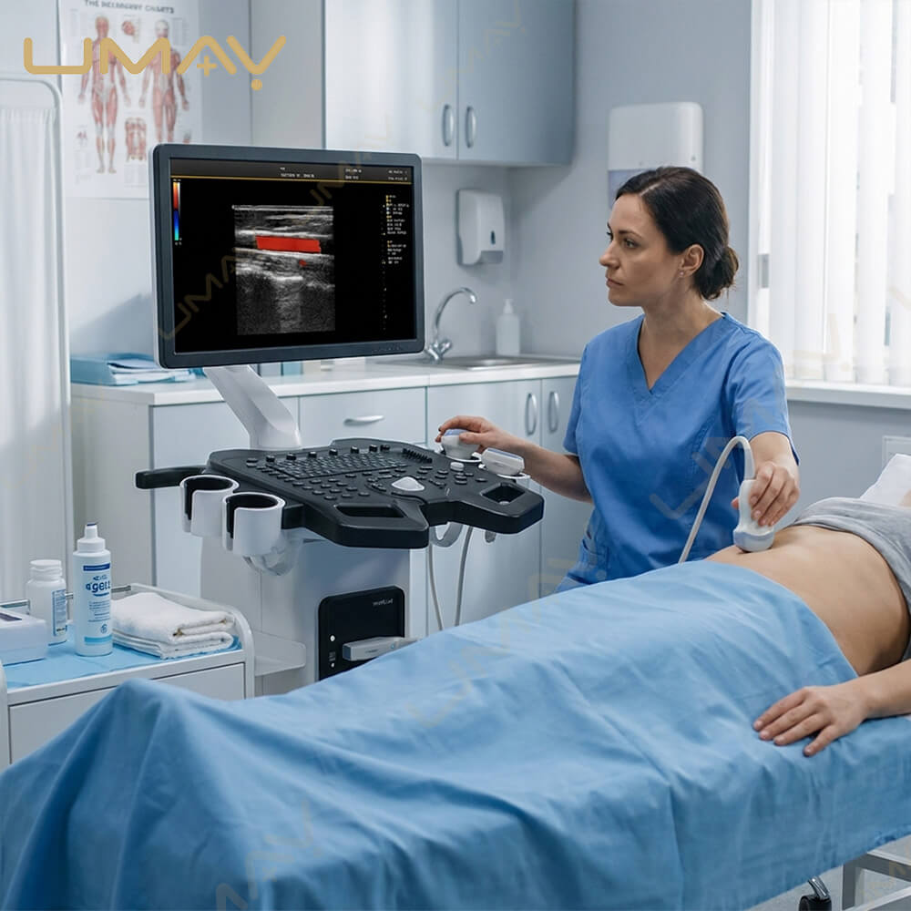
Q1: What specific transducer arrays does your factory manufacture to support the vascular mode shown on screen?
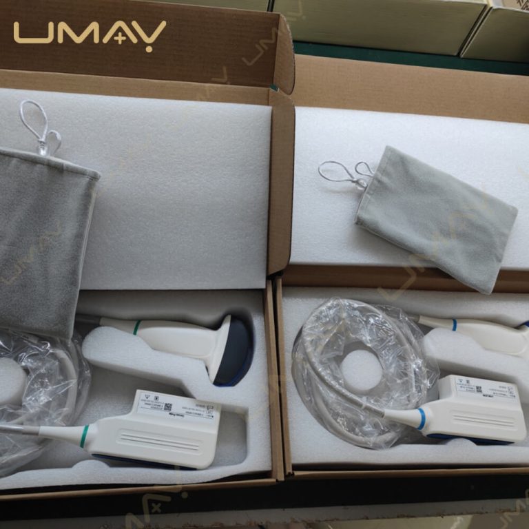
A: We engineer a full catalog of high-density multi-frequency transducers. For the superficial vascular and small parts studies shown on the display, we recommend our premium high-frequency linear array probe (ranging from $5.0\text{ MHz}$ to $12.0\text{ MHz}$). Additionally, our factory supplies wide-band convex arrays for abdominal exams, phased arrays for adult/pediatric cardiology, and micro-convex assemblies for specialized pediatrics.
Q2: Does this cart system feature an automated image optimization feature for rapid clinical triage?
A: Yes, absolutely. To speed up high-volume patient workflows, the console features a dedicated one-key automatic optimization macro button. When pressed, the internal software instantly analyzes the current gray-scale or Doppler mapping returns and self-adjusts overall gain, dynamic range, and wall filters to deliver the cleanest possible image balance without manual slider repositioning.
Q3: How does your factory handle shipping protection and custom OEM branding configurations for global medical device distributors?
A: Every trolley ultrasound system is packed inside a heavy-duty, multi-layered anti-shock wooden crate to ensure perfect protection during international ocean or air freight transport. For qualified distribution networks, we provide extensive OEM/ODM private label options, including custom startup splash screens, personalized housing logo badge placements, and custom-tailored local language software updates.