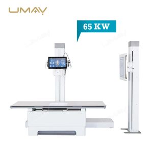


Stationary Digital Radiography Room Equipment | Complete General Radiography X-Ray Suite | High Frequency Diagnostic X-Ray Apparatus 65KW with Floating Bucky Table and Upright Chest Stand
Quick Info.
- SKU NO.: UMY-XM-068
- Device Classification: Class Ⅱ
- Warranty: 1 Year
- Power Source: Electric
- Transport Package: Carton or Wooden Cases
- Origin: China
- Material: Aluminum & Magnesium Alloys and Plastic
- After-Sale Service: Online Technical Support
- Production Capacity: 1000 Sets/Year
This professional grade 65KW Digital Radiography X ray Machine delivers unmatched diagnostic imaging capabilities with cutting edge high frequency technology for maximum resolution and contrast. Engineered for permanent hospital installation, this powerful system enables healthcare professionals to capture highly detailed anatomical images while significantly reducing radiation exposure for enhanced patient safety. The exceptional 65KW power capacity supports continuous high volume operation across all medical imaging scenarios from routine examinations to complex specialty diagnostics.
The Specific Parameters
| ITEMS | PARAMETERS |
|---|---|
| X-ray Generator Specifications | |
| Power Output | 65KW / 800mA |
| Generator Frequency | 30kHz / 200kHz (optional) |
| Power Supply | 50Hz or 60Hz, wire diameter >4mm², internal resistance <0.50; supports 65kW (only 380V) |
| Exposure Control | |
| Tube Current (mA) | 10–800 |
| Tube Current-Time Product (mAs) | 0.4–1000 |
| Tube Voltage (kV) | 40–150kV (1kV step) |
| Exposure Time | 1ms–10000ms |
| X-ray Tube and Heat Capacity | |
| Focus Size | Default 0.6×0.6mm / 1.2×1.2mm dual focus (Toshiba option) |
| Anode Rotating Speed | 8400rpm |
| Anode Heat Capacity | 1300kHU |
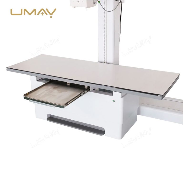
| Radiography Table | |
| Table Dimensions | 73cm (W) × 200cm (L) |
| Table Movement | 74cm left/right, 20cm back/front |
| Flat Panel Detector | |
| Image Size | 17×17 inch (14×17 inch option available) |
| Pixel Matrix | 140μm |
| A/D Conversion | 16 bits |
| Spatial Resolution | 3.6 Lp/mm |
| Software | Professional Imaging Software |
| Computer Configuration | CPU: 15, 8G memory, 1T solid state drive, 2 pcs Gigabit network card |
-
High-Frequency 630mA Diagnostic Generator: Experience uncompromising image contrast across all human tissue densities. The advanced high-voltage generator delivers rapid, crisp exposures, effectively cutting down motion artifacts during thoracic or complex abdominal studies while keeping the cumulative radiation entry dose minimal.
-
4-Way Floating Mechanical Bucky Table: Maximize patient positioning comfort and save valuable time. The heavy-duty tabletop glides effortlessly across a wide horizontal and lateral range, supported by ultra-quiet electromagnetic brakes. This allows technologists to align patients precisely beneath the collimator with minimal physical repositioning.
-
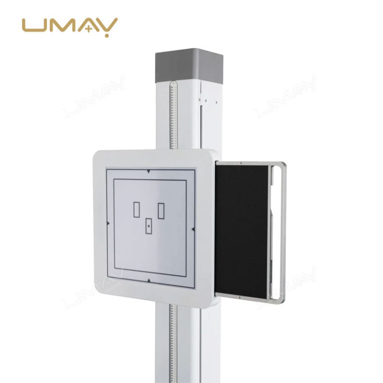
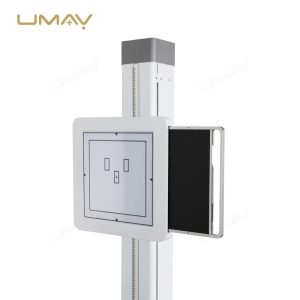
Counterbalanced Upright Chest Stand Suite: Streamline standing and seated examinations. The vertical Bucky chest stand features a precision-calibrated counterbalance mechanism, enabling clinicians to smoothly adjust the height of the digital panel housing with a single hand to accommodate various patient statures, from pediatrics to adults.
-
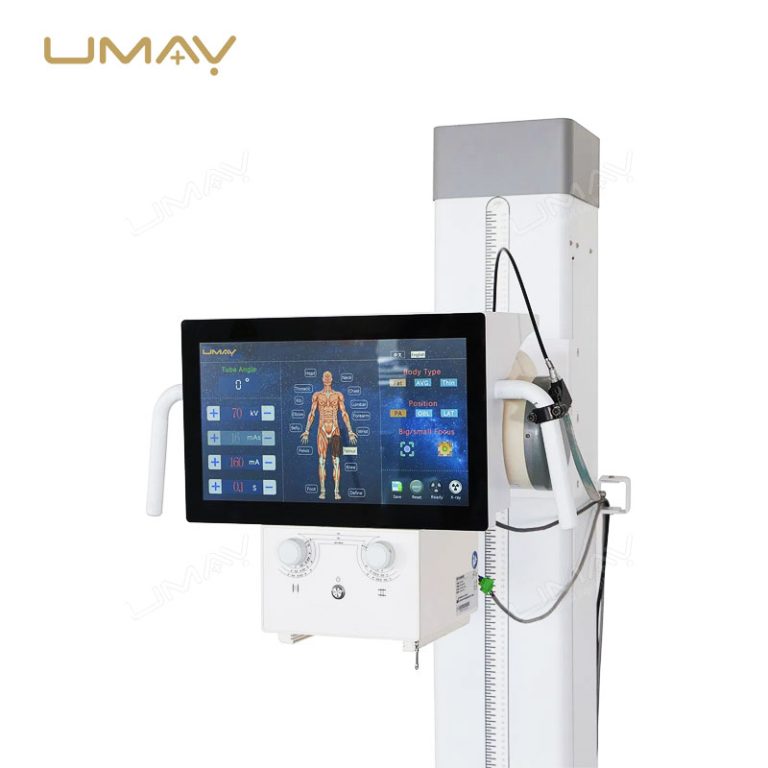
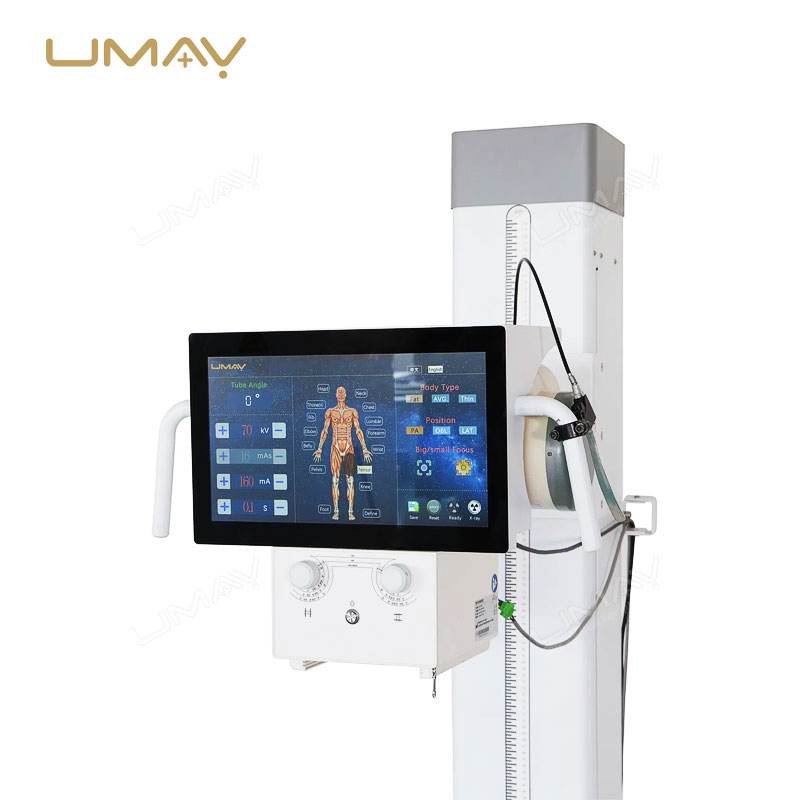
Smart Interactive Tube Head Touch Console: Improve operational efficiency and accuracy inside the examination room. Technologists can easily adjust milliamperage ($\text{mA}$), kilovoltage ($\text{kV}$), and localized Anatomical Programmed Radiography (APR) protocols right next to the patient, eliminating repetitive steps back to the control booth.
-
Seamless DICOM 3.0 Hospital Network Sync: Protect your digital data workflow. The bundled acquisition workstation software integrates natively with local PACS networks, RIS platforms, and cloud storage servers, allowing instant file transfers, easy multi-frequency image post-processing, and swift diagnostic readouts.

💡 Core Specifications At a Glance:
-
System Layout: Full-Scale Stationary General Radiography X-Ray Room Suite
-
Power Capability: High-Frequency Output Option Rated at $500\text{mA}$ / $630\text{mA}$ Configurations
-
Table Architecture: 4-Way Smooth Floating Radiographic Table with Electromagnetic Locks
-
Chest Stand Mechanics: Vertically Gliding Counterbalanced Upright Bucky Stand
-
Software Connectivity: Open-Architecture Digital Acquisition Suite with Full DICOM Support
❓ Crucial B2B Procurement Q&A
Q1: What is the maximum weight capacity of the floating Bucky table included in this stationary digital radiography room equipment?
A: The heavy-duty radiographic table is engineered with a reinforced steel sub-frame and a high-tensile, low-absorption composite tabletop. It safely supports a maximum patient weight capacity of up to 200kg without experiencing mechanical deflection or compromising the smooth movement of the 4-way floating tabletop.
Q2: Can the upright chest stand be outfitted with a high-ratio anti-scatter grid for advanced lung or spinal imaging?
A: Yes. Both the upright chest stand and the floating table Bucky assembly come pre-configured to accept standardized high-ratio stationary or moving anti-scatter grids (such as 10:1 or 12:1 grid ratios). This ensures maximum scatter-radiation filtering, resulting in exceptional contrast resolution during human chest and orthopedic assessments.
Q3: Does the 65Kw high frequency diagnostic X-ray apparatus require specific specialized environmental cooling in the x-ray room?
A: The X-ray tube unit features high thermal storage capacity anodes paired with advanced oil-cooled housing insulation. While standard medical climate control is recommended for optimal electronics lifespan inside the generator and workstation cabinet, the tube assembly itself requires no external water cooling lines or dedicated chillers.
Q4: Is the system capable of supporting automatic stitching for full-spine or long-leg orthopedic imaging?
A: Our digital imaging workstation software features an optional advanced image stitching module. When capturing sequential exposures of long bone structures or full spinal columns on the upright chest stand, the algorithm intelligently aligns and fuses the frames using anatomical landmarks, providing perfect geometric accuracy for orthopedic evaluations.