Executive Overview: The Acoustic Heart of Diagnostic Imaging
The ultrasound transducer (probe) is the core hardware component in any diagnostic ultrasound system. Functioning as both transmitter and receiver, the probe determines spatial resolution, acoustic penetration depth, field of view (FOV), and clinical versatility. For clinical directors and equipment buyers, selecting the correct configuration of probes is as critical as choosing the main console.
Matching the right transducer architecture—whether linear, convex, phased array, endocavity, or 4D volume—to specific clinical specialties prevents premature diagnostic limitations, optimizes capital expenditure, and maximizes patient throughput in busy radiology and cardiology departments.
1. Piezoelectric Physics & Acoustic Frequency Dynamics
Ultrasound transducers operate via the Piezoelectric Effect, converting electric pulses into mechanical sound waves (and vice versa) using synthetic ceramic elements (such as Lead Zirconate Titanate, or PZT) or advanced Single Crystal materials (PMN-PT).
Frequency vs. Spatial Resolution & Penetration Trade-Off
💡 Material Tech Innovation — Single Crystal Transducers: Advanced Single Crystal (PMN-PT) technology replaces traditional polycrystalline PZT ceramics with a uniform single-crystal structure. This yields up to 90% piezoelectric conversion efficiency, providing broader bandwidth, cleaner Doppler signals, and up to 20% deeper penetration at higher frequencies.
2. Core Ultrasound Transducer Categories
A. Linear Array Transducers
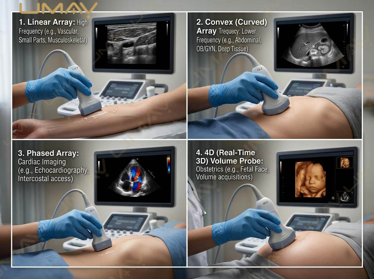
Linear probes feature piezo elements arranged in a straight line, generating a rectangular field of view with parallel acoustic beams.
-
Frequency Range: High (5.0 – 18.0 MHz)
-
Primary Applications: Vascular imaging (carotids, DVT), Thyroid, Breast, Small Parts, Musculoskeletal (MSK), and Nerve Blocks.
B. Convex Array (Curved Linear) Transducers
Convex transducers feature a curved crystal face, producing a wide fan-shaped sector FOV with superior deep-tissue penetration.
-
Frequency Range: Low to Mid (2.0 – 6.0 MHz)
-
Primary Applications: General Abdomen (Liver, Kidneys, Gallbladder), Obstetrics & Gynecology (OB/GYN), Renal vascularity.
C. Phased Array Transducers
Phased array probes utilize a small footprint with tightly packed elements that are fired sequentially with precise time delays, steering the acoustic beam electronically without moving parts.
-
Frequency Range: Low (1.5 – 5.0 MHz)
-
Primary Applications: Adult & Pediatric Cardiology, Transcranial Doppler (TCD), Emergency Medicine (FAST exam through intercostal spaces).
D. Endocavity Transducers (Transvaginal / Transrectal)
Endocavity probes possess a long, ergonomic shaft and a tightly curved tip designed for internal cavity imaging, offering ultra-wide viewing angles ($> 150° – 180°).
-
Frequency Range: Mid to High (4.0 – 10.0 MHz)
-
Primary Applications: Early OB/GYN, Pelvic Floor, Prostate Biopsy, Endometrial evaluation.
E. 3D/4D Volume Transducers
4D transducers incorporate internal motorized sweeping mechanisms or 2D matrix arrays that sweep acoustic planes continuously in real time to generate volumetric renderings.
-
Frequency Range: Mid (2.0 – 8.0 MHz)
-
Primary Applications: 4D Fetal Surface Rendering, Complex Congenital Heart Disease (3D Echo), Gynecological Structural Anomalies.
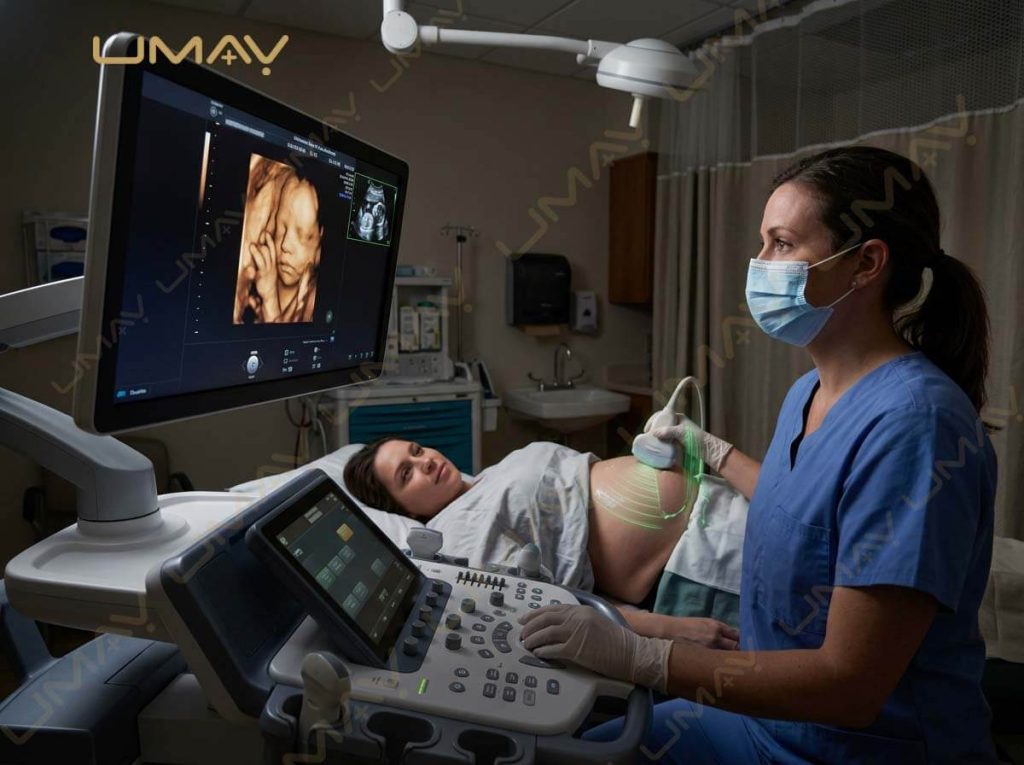
Sonographer performing an obstetric 4D ultrasound scan on an expectant mother, displaying real-time volumetric fetal features on a large monitor.
3. Side-by-Side Probe Comparison Matrix
| Transducer Type | Frequency Range | Footprint & FOV Shape | Penetration Depth | Primary Clinical Target |
| Linear Array | 5.0 – 18.0 MHz | Flat / Rectangular FOV | Shallow (2 – 7 cm) | Vascular, Thyroid, MSK, Small Parts |
| Convex (Curved) | 2.0 – 6.0 MHz | Curved Sector / Fan FOV | Deep (12 – 28 cm) | Abdominal, OB/GYN, Renal |
| Phased Array | 1.5 – 5.0 MHz | Small Point / Wide Sector | Deep (15 – 25 cm) | Echocardiography, FAST, Intercostal |
| Endocavity | 4.0 – 10.0 MHz | Curved Tip / Wide FOV (>180°) | Medium (4 – 12 cm) | Transvaginal OB/GYN, Prostate Biopsy |
| 4D Volume (OB/GYN) | 2.0 – 8.0 MHz | Bulky Motorized Curved Face | Deep (10 – 22 cm) | Real-Time 3D/4D Fetal & Pelvic Imaging |
4. Key B2B Procurement & Hardware Specifications
When reviewing technical datasheets and purchasing proposals from OEM manufacturers, procurement committees should check these critical indicators:
-
Probe Connector Architecture: Pinless high-density connectors (e.g., zero-insertion-force) minimize signal noise, reduce bent pin damage, and extend probe socket life compared to traditional pin connectors.
-
IPX7 Waterproof Rating: Endocavity and intraoperative transducers must feature IPX7 fluid immersion protection to withstand rigorous high-level chemical disinfection protocols (e.g., Cidex, OPA).
-
Multi-Frequency Harmonics Support: Verify that probes support Dynamic Frequency Tuning (DFT) and Tissue Harmonic Imaging (THI) across multiple central frequencies on a single crystal head.
-
Strain Relief & Cable Shielding: Reinforced cable-to-housing strain relief prevents signal dropouts caused by frequent twisting and bending in high-volume clinics.
5. Frequently Asked Questions (FAQ)
Q1: What is the main advantage of Single Crystal probes over standard PZT probes?
Single Crystal transducers offer a significantly broader frequency bandwidth and higher energy conversion efficiency. This allows a single probe to cover the frequency range of two traditional probes while delivering superior acoustic penetration and contrast resolution in hard-to-image patients.
Q2: Why is a Phased Array probe mandatory for cardiac imaging instead of a Convex probe?
Cardiac imaging requires negotiating narrow intercostal windows between human ribs. A Phased Array probe has a tiny footprint , electronically steering acoustic beams outwards into a wide sector to visualize the entire heart without rib bone shadows.
Q3: What is the difference between Mechanical 4D and Matrix 4D volume probes?
Mechanical 4D probes use an internal motor to physically sweep a curved array back and forth inside an oil-filled housing. Matrix 4D probes utilize thousands of active elements in a stationary grid, steering the beam electronically for instantaneous 4D acquisition without mechanical wear or acoustic lag.
Conclusion: Optimizing Transducer Selection for Institutional ROI
Investing in the correct mix of high-durability, high-sensitivity ultrasound probes directly impacts clinical workflow, diagnostic throughput, and total cost of ownership. By balancing core clinical needs with advanced technology like Single Crystal elements and pinless connectors, healthcare facilities ensure long-term diagnostic success.