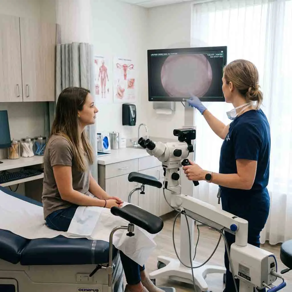
1. Optical Architecture: Lenses, Zoom Drums & Image Formation
The optical head is the heart of the colposcope. It captures light reflected from the cervical epithelium, magnifies it without distortion, and delivers it to the clinician’s eyes or a digital sensor.
Plaintext
                      OPTICAL RESOLUTION & COLOR ALIGNMENT
+-------------------------------------------------------------------------+
| STANDARD ACHROMATIC LENS:                                               |
| Red & Blue wavelengths aligned; Green slightly offset                   |
| Result: Mild color fringing under intense illumination                  |
|                                                                         |
| ADVANCED APOCHROMATIC LENS:                                             |
| Red, Green, and Blue wavelengths focused on the exact same focal plane  |
| Result: True-to-life tissue colors, razor-sharp lesion boundaries       |
+-------------------------------------------------------------------------+
A. Apochromatic vs. Standard Achromatic Lenses
Standard glass lenses bend different colors of light at varying angles, a physical phenomenon known as chromatic aberration:
-
Standard Achromatic Optics: Correct for two primary wavelengths (typically red and blue). While adequate for basic general examinations, they can create faint purplish or yellowish halos around sharp, high-contrast edges when subjected to intense light.
-
Apochromatic Optics: Correct for three color wavelengths (red, green, and blue) simultaneously on the exact same focal plane. Apochromatic systems provide superior chromatic fidelity, flat-field distortion correction, and razor-sharp border definition. This level of clarity is vital when attempting to differentiate between faint, benign metaplastic acetowhitening and high-grade cervical intraepithelial neoplasia (CIN 2/3).
B. Magnification Mechanics: Step-Zoom vs. Continuous Optical Zoom
Colposcopes utilize two distinct mechanical methods to shift magnification:
Plaintext
                  MAGNIFICATION MECHANISM COMPARISON
+-------------------------------------------------------------------------+
| 3-STEP / 5-STEP CHANGER DRUM            CONTINUOUS OPTICAL ZOOM         |
| - Rotating internal turret              - Sliding internal lens groups  |
| - Fixed presets (e.g., 4x, 8x, 15x)     - Seamless fluid range (4x-20x) |
| - High durability, zero motor failure   - Precision motorized or manual |
| - Rapid, standardized clinical workflow - No blackout or click between steps|
+-------------------------------------------------------------------------+
1. Step-Magnification Changers (3-Step or 5-Step)
A rotating mechanical barrel inside the optical head holds pairs of lenses of varying power. As the clinician turns an external dial, the barrel clicks into place:
-
Typical 3-Step Setup: 3.75x, 7.5x, 15x
-
Typical 5-Step Setup: 3x, 5x, 8x, 13x, 21x
-
Advantages: Extremely reliable, cost-effective, and mechanically robust with no motors or electronic potentiometers to fail. Standardized magnification presets also simplify clinical research and follow-up documentation because image scales remain consistent across visits.
2. Continuous Optical Zoom (Stepless)
The internal lens groups move smoothly along a machined track, increasing and decreasing magnification fluidly without interruptions:
-
Advantages: Allows the clinician to dial in the exact framing desired, zooming in on an atypical vessel loop without losing visual contact or experiencing mechanical clicks.
-
Parfocality Requirement: If selecting a continuous zoom colposcope, buyers must confirm that the optics are strictly parfocal—meaning the image remains in crisp focus as the clinician zooms from the lowest magnification to the highest without requiring refocusing.
2. The Critical Working Distance: Instrument Clearance vs. Ergonomics
The working distance (often corresponding to the focal length of the objective lens) is the physical distance between the front glass surface of the colposcope’s objective lens and the patient’s cervical os.
Plaintext
                     THE WORKING DISTANCE CLEARANCE ZONE
+-------------------------------------------------------------------------+
| [ Colposcope Lens Head ]                                                |
|            |                                                            |
|            |<--- WORKING DISTANCE (Optimal: 250 mm to 300 mm) --->|     |
|            |                                                      |     |
|            +--- [ Biopsy Forceps & Endocervical Speculums ] ------+     |
|            |    (Requires 200 mm to 240 mm of maneuvering room)   |     |
|            v                                                      v     |
|     Vaginal Opening                                            Cervix   |
+-------------------------------------------------------------------------+
Why Working Distance Dictates Clinical Success
If the working distance is too short, the procedure becomes physically awkward or impossible:
-
Too Short (< 200 mm): The colposcope head must sit dangerously close to the patient’s perineum. Long cervical biopsy forceps (such as Tischler or Kevorkian instruments, which measure 200 mm to 250 mm in shaft length), endocervical speculums, or smoke evacuators will physically strike the front of the microscope lens, contaminating sterile tools or scratching expensive optical glass.
-
Too Long (> 350 mm): The colposcope sits far away from the patient, requiring the clinician to lean forward excessively with outstretched arms to manipulate instruments inside the vaginal canal, resulting in rapid shoulder and lumbar muscle strain.
The Clinical Gold Standard: 250 mm to 300 mm
A focal length of 280 mm to 300 mm is recognized globally as the optimal sweet spot for gynecology. It provides ample physical clearance for instrument articulation, allows direct line-of-sight lighting, and maintains comfortable, natural arm posture for the examining physician.
Fine-Focus Mechanisms: Coaxial Micro-Drives
Gross positioning of the colposcope is achieved by rolling the floor stand or moving the articulated arm. However, landing on the exact microscopic focal plane requires a fine-focus mechanism:
-
Look for an optical head equipped with a smooth, coaxial fine-focus control knob (offering 40 mm to 50 mm of travel).
-
The drive must have fluid resistance—loose gears will slip out of focus when the clinician releases the knob, while overly stiff gears cause jerky, jarring visual shifts.
3. Illumination Engineering: Why Medical Cold LED Is Mandatory
Lighting can make or break a colposcopic evaluation. The cervix resides at the deep end of a narrow, shadowed canal. To illuminate this cavity, all modern colposcopes utilize coaxial illumination—the light beam travels along the exact same central optical axis as the viewing lenses, completely eliminating shadows inside the vagina.
However, the nature of the light source itself has undergone a massive technological shift over the past decade.
Plaintext
                  LIGHTING TECHNOLOGY IN MODERN COLPOSCOPY
+-------------------------------------------------------------------------+
| PARAMETER             | OLD HALOGEN SYSTEMS    | MODERN MEDICAL COLD LED|
+-----------------------+------------------------+------------------------+
| Color Temperature     | 3000K - 3200K (Yellow) | 5500K - 6000K (Daylight|
| Color Rendering (CRI) | Average (Distorts red) | Exceptional (CRI >= 90)|
| Tissue Heat Emission  | High (Causes drying)   | Near Zero (Cold Light) |
| Lamp Lifespan         | ~50 to 100 Hours       | 50,000+ Hours          |
| Maintenance Overhead  | Costly bulb inventory  | Virtually Maintenance-Free|
+-------------------------------------------------------------------------+
The Three Critical Illumination Metrics
1. Color Rendering Index (CRI)
The Color Rendering Index measures how accurately a light source reveals the true colors of physical objects compared to natural sunlight (rated at 100 CRI).
-
In colposcopy, differentiating between pink squamous epithelium, red inflamed stroma, translucent glandular cells, and white acetowhite plaques requires exceptional color fidelity.
-
Procurement Rule: Never purchase a colposcope with a CRI below 85. High-precision clinical units demand a CRI of 90 or greater to prevent diagnostic color distortion.
2. Color Temperature: The 5500K Daylight Target
Halogen bulbs burn with a warm, yellowish cast (around 3000K). This amber tint artificially masks subtle acetowhite changes and makes vascular capillaries harder to spot.
-
Modern Medical LEDs emit crisp, daylight-balanced white light (between 5500K and 6000K).
-
This neutral white illumination provides pure background contrast, allowing chemical staining reactions to stand out instantly.
3. Tissue Safety & Cold Light Mechanics
Halogen and Xenon lamps generate intense infrared thermal radiation. If focused into the vaginal canal during a prolonged 20-minute examination or minor outpatient procedure, this heat can dry out cervical mucus, cause thermal tissue irritation, and produce uncomfortable warmth for the patient.
-
High-intensity LEDs generate light using semiconductors rather than heated filaments.
-
The light emitted from the front objective is true cold light, preserving tissue hydration and patient comfort while directing internal heat safely away through rear aluminum heat sinks.
4. The Green Filter: Optical Glass vs. Digital Image Processing
The green filter is an indispensable diagnostic feature on any colposcope, serving as the primary optical tool for evaluating abnormal cervical micro-vascular architecture.
Plaintext
                     THE GREEN FILTER ABSORPTION PHENOMENON
+-------------------------------------------------------------------------+
| Optical Emission: Green Light Wavelength (~540 nm)                      |
|                                                                         |
| Cervical Tissue Interaction:                                            |
| - Normal Epithelial Tissue  --> Reflects green light (Appears pale grey)|
| - Hemoglobin in Blood Vessels--> AVIDLY ABSORBS GREEN LIGHT             |
|                                                                         |
| Visual Result: Blood vessels turn deep black against a high-contrast bed|
+-------------------------------------------------------------------------+
Why Green Light Highlights Capillaries
Human blood appears red because oxygenated hemoglobin reflects red wavelengths while absorbing green wavelengths.
-
When the clinician shines green light onto the cervix, the red capillaries absorb the photons completely.
-
Instead of appearing as faint red lines against a pink tissue background, the blood vessels stand out as sharp, high-contrast, black or dark purple patterns.
Optical Glass Filter vs. Digital Software Filter
-
Mechanical Optical Green Filter: A physical, precision-polished green glass disc housed inside the optical head. The clinician flips a manual lever or switch, sliding the glass directly into the optical path.
-
Advantage: Delivers pure, analog optical filtering with zero digital noise, zero pixelation, and maximum optical dynamic range. This is the preferred choice of seasoned colposcopists.
-
-
Electronic / Digital Green Filter: In digital video colposcopes, the image sensor captures the standard color feed, and onboard software algorithmically isolates and enhances the green channel while subtracting red components.
-
Advantage: Convenient, requires no moving physical parts, and allows for variable levels of digital contrast enhancement on an external display.
-
5. Mechanical Architecture: Stand Selection & Spatial Ergonomics
A colposcope’s optical head must remain rock-steady during high-magnification examinations and fine-needle biopsies. The physical stand dictates how easily the unit moves around the examination table and how much floor space it consumes.
Plaintext
                       STAND CONFIGURATION SPECTRUM
                                     |
     +-------------------------------+-------------------------------+
     |                               |                               |
     v                               v                               v
[ VERTICAL ROLLING STAND ]    [ ARTICULATED SWING-ARM ]       [ CHAIR-INTEGRATED MOUNT ]
- Single vertical post        - Counterbalanced spring arm    - Bolted directly to exam table
- Compact, budget-friendly    - Extreme multi-angle reach     - Zero footprint on OR floor
- Limited vertical travel     - Smooth "one-finger" positioning- Dedicated to single chair
A. The Upright Vertical Rolling Stand
-
Structure: A heavy, star-shaped caster base holding a fixed vertical metal column. The optical head mounts to a horizontal cross-arm and moves up and down on a vertical friction track.
-
Pros: Minimal footprint, low cost, and very easy to wheel into a corner or storage closet when not in use.
-
Cons: Limited range of articulation. Positioning the optical head requires loosening and tightening manual mechanical knobs. If the exam table height changes, the entire post must be physically readjusted.
B. The Articulated Pantographic (Swing-Arm) Stand
-
Structure: A weighted rolling base supporting an articulated, two-section mechanical arm utilizing internal counterbalanced springs or gas struts.
-
Pros: Unrivaled clinical flexibility. The clinician can grab the handgrips and glide the optical head in any direction—up, down, forward, tilt, or roll—with effortless “one-finger” movement. When the clinician lets go, the internal counterbalance locks the head in mid-air with zero drift.
-
Cons: Larger physical base footprint to prevent tipping when the heavy optical arm is fully extended.
C. Examination Chair-Integrated Mount
-
Structure: The articulated colposcope arm is bolted directly to the structural steel chassis of the gynecological examination table.
-
Pros: Completely eliminates the rolling floor base, leaving the floor 100% clear for physician stool movement, foot pedals, and waste bins. As the examination chair is raised or tilted into Trendelenburg position, the colposcope moves in direct synchronization with the patient.
-
Cons: The colposcope is permanently married to that specific examination chair and cannot be shared between different clinical rooms.
6. Comprehensive Hardware Specification Benchmark
When reviewing formal vendor tenders, use this benchmark matrix to evaluate where a specific model sits in the technical hierarchy:
| Hardware Component | Entry-Level Screening Unit | Mid-Tier Clinical Workhorse | Premium Advanced Surgical Platform |
| Optical System | Standard Achromatic Glass | Multi-Coated Achromatic / Apo | Full Apochromatic Precision Glass |
| Magnification Type | 3-Step Manual Turret | 5-Step Manual Turret | Continuous Motorized / Manual Zoom |
| Magnification Range | 3.75x, 7.5x, 15x | 3x, 5x, 8x, 13x, 21x | 4x to 24x Continuous Parfocal Range |
| Working Distance | Fixed 250 mm or 300 mm | Fixed 280 mm to 300 mm | Variable Vario Focus (200 to 350 mm) |
| Fine Focus Travel | 30 mm manual knob | 40 mm precision rack & pinion | 50 mm coaxial micro-drive + Motorized |
| Light Source | 20W – 30W Basic LED | 50W High-Intensity LED | 80W+ High-CRI Medical Cold LED |
| CRI & Color Temp | CRI ~80; 5000K | CRI $\ge$ 88; 5500K | CRI $\ge$ 92; 5700K Daylight Spectrum |
| Green Filter Type | Slide-in plastic sheet | Manual optical green glass disc | Optical glass + Multi-band digital filter |
| Suspension Arm | Rigid vertical post | Counterbalance spring swing-arm | Electromagnetic brake articulated arm |
| Digital Video Port | Optional C-mount adapter | Integrated beam splitter + HD | Integrated 4K CMOS sensor & DICOM port |
Frequently Asked Questions (FAQ)
Q1: Is a 5-step magnification changer better than a 3-step changer?
Yes, for most general and outpatient clinics. A 5-step changer provides smaller, more gradual jumps in magnification (such as having an 8x and 13x middle tier), which makes it much easier to transition smoothly from an initial overview of the cervix to pinpointing a tiny transformation zone lesion without disorienting the clinician.
Q2: What is the difference between optical zoom and digital zoom?
Optical zoom physically moves glass lens elements inside the optical head to magnify the actual light coming into the system, preserving full optical resolving power and crisp detail. Digital zoom simply takes the existing digital pixels and enlarges them on a screen, which eventually causes pixelation, digital noise, and blurred lesion edges if pushed too far. Always prioritize high optical zoom capabilities over digital zoom claims.
Q3: Why does my colposcope light look yellow even though it is bright?
A yellow light tint indicates an older Halogen bulb (which runs at a low color temperature around 3000K) or an entry-level, low-CRI LED. Replacing aging halogen systems with high-CRI (90+) daylight Medical LEDs (5500K) will instantly restore true white background contrast and improve tissue visualization.
Q4: Can I add a digital camera to my existing optical colposcope later?
Yes, provided your optical head includes an integrated beam splitter port or an optical expansion interface. You can attach a secondary beam splitter module, a C-mount lens adapter, and a digital camera sensor at any time to stream live video to external medical monitors and capture files for your EMR.
Q5: How often does an operating colposcope require calibration?
Under standard clinical use, a mechanical colposcope does not require routine software-style calibration. However, biomedical engineering teams should perform an annual check on arm counterbalance spring tension, lubricate moving joints, verify electrical chassis grounding, and inspect optical coatings for scratches or environmental fungus.
Summary
Selecting the right colposcope hardware requires looking past superficial marketing claims and evaluating the core engineering that drives diagnostic accuracy.
By demanding apochromatic optical glass, ensuring an ergonomic working distance of 250 mm to 300 mm, adopting maintenance-free Medical Cold LED lighting (CRI $\ge$ 90), and pairing the system with a counterbalanced articulated swing-arm, procurement teams can outfit their gynecological suites with instruments that provide exceptional diagnostic precision, clinician comfort, and lasting operational value.