Infection Control: Defining the Colposcope’s Disinfection Classification
To understand how to clean a colposcope, clinical staff must first understand its classification under the widely accepted Spaulding Classification System for medical devices.
Plaintext
                  SPAULDING CLASSIFICATION IN GYNECOLOGY
+-------------------------------------------------------------------------+
| CRITICAL DEVICES (Must be Sterilized - Autoclave/ETO)                   |
| - Biopsy forceps, endocervical curettes, LEEP electrodes.               |
|                                                                         |
| SEMI-CRITICAL DEVICES (Require High-Level Disinfection)                 |
| - Vaginal speculums, internal ultrasound probes.                        |
|                                                                         |
| NON-CRITICAL DEVICES (Require Low/Intermediate Disinfection)            |
| - THE COLPOSCOPE, exam tables, instrument trays, computer keyboards.    |
+-------------------------------------------------------------------------+
The Cross-Contamination Risk
Because the colposcope always remains outside the body (at a working distance of 250 mm to 300 mm), it is legally classified as a non-critical environmental surface. It does not contact intact mucous membranes directly.
However, the clinician’s gloved hands serve as the bridge. A doctor may touch the patient’s perineum or handle a speculum coated in cervical mucus and human papillomavirus (HPV), and then reach up to adjust the colposcope’s fine-focus knob or magnification dial. If the next doctor touches those same knobs without gloves and then touches a new patient, cross-contamination occurs.
The Standard Disinfection Protocol (Between Every Patient)
-
Wear Fresh Gloves: The clinical assistant must wear clean gloves before cleaning the equipment.
-
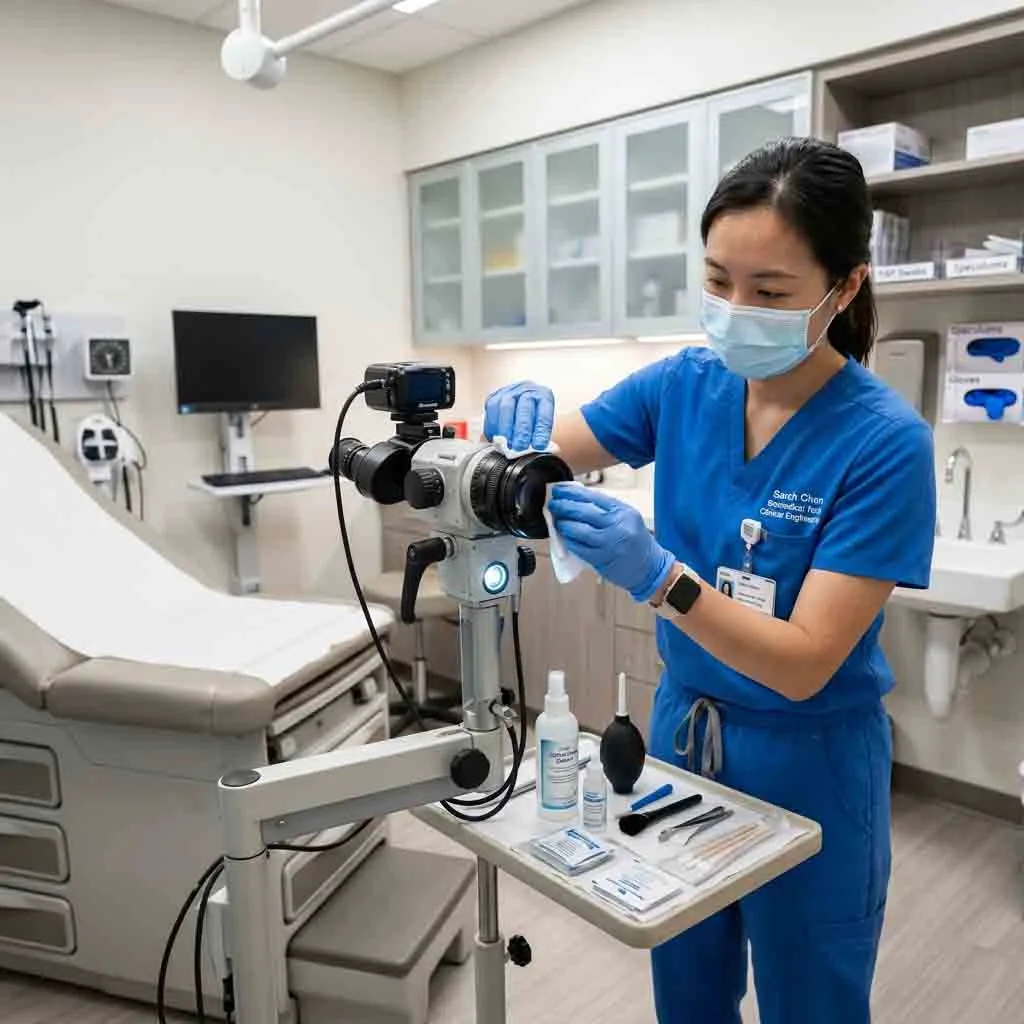
Use Approved Chemical Wipes: Wipe down all high-touch surfaces—handgrips, magnification drums, focus knobs, articulating arm handles, and the keyboard/mouse—using EPA-registered hospital disinfectants (e.g., CaviWipes, quaternary ammonium compounds, or 70% isopropyl alcohol).
-
Observe Contact Time: Disinfectants only kill pathogens if they remain visibly wet on the surface for the manufacturer’s required contact time (usually 1 to 3 minutes).
-
Avoid Spraying Liquids: Never spray liquid cleaners directly onto the colposcope head. Liquid can seep into the zooming mechanics, short-circuit the CMOS digital sensors, or cause fungal growth inside the optical housing. Always spray the liquid onto a cloth first, then wipe.
2. The Art of Cleaning Apochromatic Optical Lenses
The front objective lens is the most expensive and delicate component of an optical colposcope. These lenses feature microscopic, vacuum-deposited anti-reflective (AR) coatings designed to maximize light transmission and eliminate glare.
Plaintext
                     SAFE OPTICAL CLEANING PROTOCOL
+-------------------------------------------------------------------------+
| DO USE:                               | DO NOT USE:                     |
| - Photographic lens blower bulb       | - Compressed canned air (propellant)|
| - Lint-free optical lens tissue       | - Paper towels, Kleenex, or gauze|
| - 70% Isopropyl or Ether-Alcohol      | - Acetone, Ammonia, or Windex   |
| - Gentle outward spiral motion        | - Heavy pressure or dry scrubbing|
+-------------------------------------------------------------------------+
Why Gauze and Paper Towels Destroy Lenses
Surgical gauze and standard paper towels are made of wood pulp fibers. Scrubbing a glass lens with these materials is equivalent to using fine sandpaper. Over time, this strips the purple/green anti-reflective coating, leaving the lens permanently foggy and causing severe glare when the bright LED is turned on.
The Correct Step-by-Step Lens Cleaning Method
If a splash of saline, a fingerprint, or a fleck of silver nitrate lands on the objective lens:
-
Blow Away Dust: Use a rubber squeeze blower to remove loose dust particles. Wiping dust (which often contains microscopic silica sand) across the lens will scratch it.
-
Moisten the Tissue: Apply 1 or 2 drops of optical lens cleaner to a clean piece of optical lens paper (never apply liquid directly to the glass).
-
The Spiral Wipe: Place the moistened tissue in the direct center of the objective lens. With very light pressure, wipe in a continuous, expanding circular spiral from the center outward to the metal rim.
-
Discard the Tissue: Never reuse the same tissue for a second pass, as you will simply drag the picked-up oils back across the glass.
3. Mechanical Maintenance: Fixing the “Drifting Arm”
The most frequent mechanical complaint in gynecology clinics is a colposcope that refuses to stay still. When the doctor positions the optical head over the vaginal canal and lets go of the handles, the heavy head slowly drifts downward or swings sideways, ruining the focal plane right before a biopsy.
Modern colposcopes utilize articulated pantographic swing-arms fitted with internal mechanical counterbalance springs or gas struts. Drift occurs when this mechanical tension falls out of alignment.
Resolving Vertical (Up/Down) Drift
-
Ensure all accessories (cameras, cables, monitor screens) are attached to the colposcope. Balancing must be done with the machine fully weighted.
-
Locate the vertical tension adjustment knob (usually located on the main hinge of the suspension arm).
-
Pull the optical head down. If it immediately springs back up, the internal spring tension is too tight. Turn the adjustment knob toward the minus (-) sign.
-
Push the optical head up. If it immediately sinks toward the floor, the tension is too loose. Turn the adjustment knob toward the plus (+) sign.
-
The system is perfectly balanced when you can place the optical head at any vertical height and it remains frozen in mid-air.
Resolving Lateral (Side-to-Side) Swing
If the arm swings left or right independently, the heavy star-base is likely not sitting level on the floor.
-
Check the locking casters on the base.
-
Ensure the clinic floor is flat. If the floor is uneven, the heavy optical head will naturally swing downhill due to gravity. Lock the heavy-duty foot brakes on the wheels to stabilize the base.
4. Troubleshooting Illumination Issues
If a clinician complains that the cervix looks “dark,” “yellow,” or “flickering,” the diagnostic accuracy of the acetic acid test is severely compromised.
Legacy Halogen & Xenon Systems
If your clinic utilizes older halogen or xenon colposcopes, dim lighting usually stems from two issues:
-
Bulb Degradation: Halogen bulbs degrade rapidly, shifting from white light to a dim, muddy yellow after just 50 to 100 hours of use. If the light is yellow, replace the bulb immediately using surgical gloves (oils from bare fingers will cause the new glass bulb to shatter under high heat).
-
Fiber-Optic Cable Fractures: Older colposcopes generate light in a heavy box on the stand and transmit it to the head via a thick black fiber-optic cable. If nurses bend this cable too sharply or roll a heavy cart over it, the internal glass fibers shatter.
-
Test: Disconnect the cable and shine a flashlight into one end. Look at the other end. If you see dozens of black dots in the light circle, the cable is crushed and must be replaced.
-
The Modern Fix: Upgrading to Medical Cold LED
If you are constantly battling blown bulbs and broken cables, the most cost-effective long-term solution is to upgrade to a system utilizing Medical Cold LED illumination.
LEDs are mounted directly on the optical head (eliminating the fragile fiber-optic cable entirely). They deliver crisp 5500K daylight-white illumination that perfectly highlights green-filter vascular patterns, and they boast a lifespan exceeding 50,000 hours, effectively reducing illumination maintenance to zero.
5. Quick-Reference Troubleshooting Matrix for Clinics
Print this table and attach it to the colposcope stand for rapid biomedical or nursing reference:
| Symptom / Complaint | Probable Cause | Immediate Corrective Action |
| Blurry image at all magnifications | Dirty objective lens or out-of-focus eyepieces | Clean lens with optical tissue; Reset binocular diopter rings to “0”. |
| Image goes out of focus when zooming in | Loss of Parfocality (Diopters set incorrectly) | Zoom to max magnification, focus with main knob, zoom out, adjust eyepieces to clear image. |
| Optical head slowly sinks to the floor | Low vertical spring tension | Tighten the main Z-axis counterbalance tension knob on the swing arm. |
| Light is too yellow or very dim | Aging halogen bulb or broken fiber cable | Swap the bulb cartridge; Inspect fiber-optic cable for internal fractures. |
| Digital monitor shows “No Signal” | Disconnected HDMI/SDI cable or power loss | Trace the video cable from the camera head to the monitor; Check the power supply unit. |
| Heavy glare or “halo” around the image | Condensation/Fungus inside the lens | Do not open the sealed optical head. Call manufacturer for professional biomedical service. |
6. Why UMY Medical Colposcopes Are Built for Clinic Durability
When hospitals and international medical distributors procure capital equipment, they must evaluate the Total Cost of Ownership (TCO). A cheap colposcope that requires constant bulb replacements, suffers from drifting mechanical arms, and traps bacteria in poorly designed plastic joints will ultimately cost a facility thousands of dollars in downtime.
As a certified original equipment manufacturer (OEM), UMY Medical Equipment Co Ltd engineers colposcopes specifically to withstand the rigors of high-volume cervical cancer screening environments.
The UMY Medical Engineering Advantage:
-
Sealed, Antimicrobial Housings: UMY Medical colposcopes feature sleek, gap-free exterior housings coated in medical-grade antimicrobial paint. This allows clinical staff to perform rapid, aggressive chemical wipe-downs between patients without fear of liquid short-circuiting internal electronics.
-
Maintenance-Free LED Illumination: By integrating high-CRI Medical Cold LEDs directly into the optical and digital camera heads, UMY Medical completely eliminates the need for expensive halogen bulb inventories and fragile fiber-optic cables, driving illumination maintenance costs to zero.
-
Heavy-Duty Pantographic Mechanics: Designed with precision-machined internal gas struts, UMY Medical swing-arms provide reliable, drift-free “one-finger” positioning that retains its tension calibration for years, eliminating the constant need for biomedical readjustment.
-
ISO 13485 & CE Compliance: Manufactured under strict international medical device quality systems, UMY Medical ensures that every colposcope meets global electrical safety and hygiene standards, providing healthcare networks with dependable, low-maintenance diagnostic platforms.
Frequently Asked Questions (FAQ)
Q1: How often should our clinic schedule preventative maintenance for a colposcope?
A standard colposcope should undergo a formalized Preventative Maintenance (PM) check by a certified biomedical technician once every 12 months. This includes electrical safety testing (checking chassis grounding), calibrating arm tension, inspecting casters, and performing a deep optical cleaning.
Q2: Is it safe to use UV light wands to disinfect the colposcope?
While UVC light is an effective surface disinfectant, prolonged exposure to high-intensity UV light can degrade the rubber eyecups, dry out the plastic casing, and potentially damage the internal digital CMOS sensors over time. Standard chemical wiping (following the manufacturer’s IFU) remains the gold standard for non-critical device disinfection.
Q3: What should we do if fungus grows inside the optical lenses?
In highly humid, tropical climates, microscopic fungus can grow on the internal glass prisms if the colposcope is left uncovered in a damp room. If you see web-like structures inside the glass, do not attempt to dismantle the optical head yourself, as you will ruin the precise prism alignment. The unit must be sent to the manufacturer for professional disassembly, chemical fungicide cleaning, and re-collimation. Protect the scope by keeping it in an air-conditioned, dehumidified room when not in use.
Q4: The doctor complains of double vision (diplopia) when looking through the eyepieces. How do I fix this?
Double vision occurs when the distance between the two eyepieces does not match the physical distance between the doctor’s eyes. Instruct the clinician to grasp both ocular tubes and push them together or pull them apart (adjusting the Interpupillary Distance or IPD) until the two separate circles of light merge into one perfect, stereoscopic 3D circle.
Q5: Do we need to cover the colposcope with sterile drapes for a standard exam?
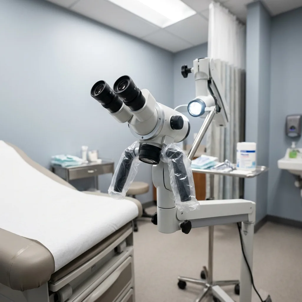
For routine colposcopic examinations and targeted punch biopsies, sterile draping is not required because the machine does not enter the sterile field. However, if the colposcope is rolled into an operating room for a surgical LEEP, cold knife conization, or laser ablation, it must be covered with a transparent, single-use sterile medical drape to comply with OR protocols.
Summary
A colposcope is the frontline defense in the global fight against cervical cancer. Protecting that diagnostic capability requires strict adherence to infection control protocols, gentle optical lens care, and proactive mechanical balancing. By implementing the standardized maintenance routines outlined in this guide, clinical teams can ensure their equipment remains safe, pristine, and ready for the next patient.