Anatomy of the Cervix: Why Magnification Matters
To understand what a physician looks for through a colposcope, one must understand the unique micro-anatomy of the uterine cervix.
The cervix connects the vaginal canal to the uterine cavity. Its surface consists of two distinct types of epithelial tissue that meet at a critical boundary:
Plaintext
                       THE CERVICAL TRANSFORMATION ZONE
+-------------------------------------------------------------------------+
| ECTOCERVIX (Outer Surface)  --> Stratified Squamous Epithelium (Smooth) |
|                                             |                           |
|                      [ SQUAMOCOLUMNAR JUNCTION (SCJ) ]                  |
|                                             |                           |
| ENDOCERVIX (Inner Canal)    --> Simple Columnar Epithelium (Glandular)  |
+-------------------------------------------------------------------------+
The Squamocolumnar Junction (SCJ)
-
The Ectocervix: The exterior surface visible during a standard speculum exam. It is covered by multi-layered, smooth, pink stratified squamous epithelium, similar to the lining of the vagina.
-
The Endocervix: The interior canal leading into the uterus. It is lined by a single layer of red, mucus-secreting columnar epithelium, which has a distinct grape-like, villous texture under magnification.
-
The Squamocolumnar Junction (SCJ): The precise microscopic border where the smooth squamous cells meet the columnar cells.
The Transformation Zone (TZ)
Throughout a woman’s reproductive life, hormonal shifts, puberty, and pregnancy cause the acidic environment of the vagina to stimulate the delicate columnar cells to transform into tough squamous cells. This natural process is called squamous metaplasia.
The dynamic area where this ongoing transformation takes place is known as the Transformation Zone (TZ).
Because the cells within the Transformation Zone are constantly dividing and transforming, they are uniquely vulnerable to infection by the Human Papillomavirus (HPV). More than 99% of all cervical precancers and invasive carcinomas originate directly within this microscopic zone.
Therefore, the primary clinical objective of any colposcopic examination is to completely visualize the entire Transformation Zone and the new Squamocolumnar Junction.
If the SCJ recedes deep inside the endocervical canal—which commonly occurs after menopause—the colposcopy is classified as “unsatisfactory” or “Type 3 TZ,” requiring specialized sampling of the inner canal.
2. The Step-by-Step Colposcopy Procedure
Undergoing a colposcopy is an outpatient procedure performed directly in a gynecologist’s consultation room. It does not require general anesthesia, intravenous lines, or surgical admission. The procedure typically takes between 10 and 20 minutes from start to finish.
Plaintext
                     THE CLINICAL COLPOSCOPY WORKFLOW
+-------------------------------------------------------------------------------+
| 1. Speculum Placement & Saline Wash   --> Clears mucus, checks baseline       |
| 2. White Light Inspection (4x - 8x)   --> Identifies gross morphology         |
| 3. Acetic Acid (3-5%) Application     --> Reveals acetowhite epithelium       |
| 4. Green Optical Filter Inspection    --> Examines abnormal vessels           |
| 5. Lugol's Iodine Application (Schiller)--> Identifies glycogen-depleted zones|
| 6. Directed Punch Biopsy & ECC        --> Collects tissue for pathology       |
| 7. Hemostasis (Monsel's Paste)        --> Controls localized bleeding         |
+-------------------------------------------------------------------------------+
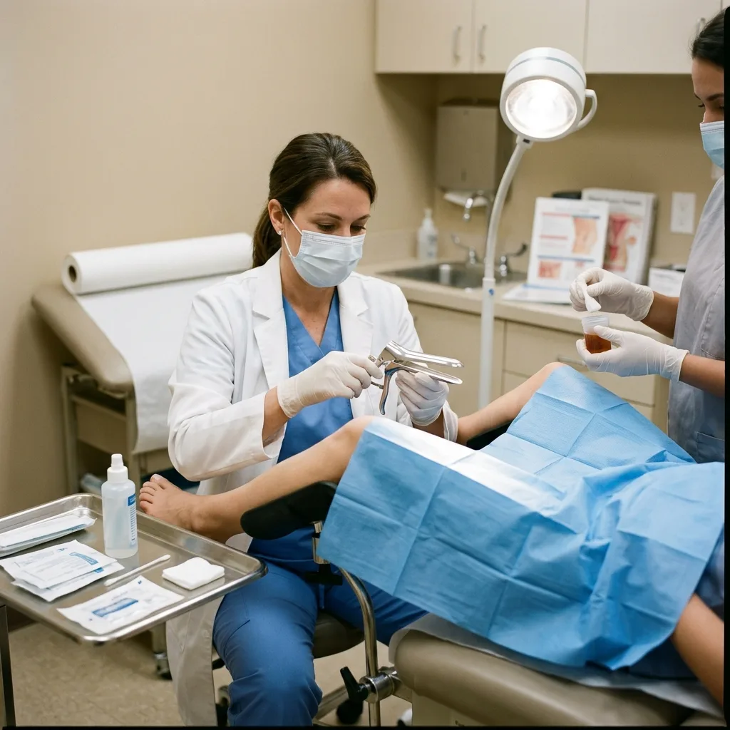
Step 1: Speculum Placement and Baseline Wash
The patient lies in the standard dorsal lithotomy position (feet in stirrups), exactly as during a routine pelvic exam.
-
The clinician inserts a sterile, lubricated bivalve speculum to gently hold the vaginal walls apart, exposing the cervix.
-
The colposcope is rolled up close to the examination table. The instrument never touches the patient; its optical lens head sits approximately 250 mm to 300 mm outside the vaginal opening.
-
The clinician washes the cervix with normal saline solution to remove excess cervical mucus and cellular debris. Under low magnification (4x to 8x), the baseline appearance of the cervix is documented, evaluating the natural color, gross erosion, nabothian cysts, or visible exophytic polyps.
Step 2: The Acetic Acid Test (The Vinegar Reaction)
The cornerstone of colposcopic diagnosis is the application of a 3% to 5% acetic acid (dilute medical vinegar) solution using a large cotton swab or gentle spray applicator.
Acetic acid acts as a temporary chemical dehydrating agent:
-
Normal Squamous Cells: Normal, healthy cervical cells contain small, mature nuclei surrounded by abundant clear cytoplasm. Acetic acid penetrates these cells easily, and the light from the colposcope passes through the translucent cells, reflecting off the vascular stroma underneath. The tissue continues to look pink.
-
Dysplastic (Precancerous) Cells: Cells infected with high-risk HPV that are undergoing malignant transformation exhibit an abnormally high nuclear-to-cytoplasmic ratio. Their enlarged, hyperchromatic nuclei and dense nuclear proteins coagulate upon contact with acetic acid.
-
The Acetowhite Effect: Because light cannot pass through this dense cloud of coagulated nuclear protein, it is reflected back to the viewer. The abnormal area turns an opaque, cloudy white against the surrounding pink tissue within 60 to 90 seconds.
The speed of appearance, density, thickness, and persistence of this acetowhite epithelium directly correlate with the severity of the cervical lesion.
Step 3: Green Filter Optical Inspection (Vascular Mapping)
Angiogenesis—the formation of abnormal new capillary networks—is a universal hallmark of growing precancerous and cancerous tissues. As cervical dysplasia deepens, rapid cellular proliferation creates disorganized micro-vessels.
Standard white light illumination can make subtle red capillaries blend into the surrounding pink tissue background. To overcome this, the clinician clicks a green optical filter into the light pathway of the colposcope.
Plaintext
                       OPTICAL PHYSICS OF THE GREEN FILTER
+-------------------------------------------------------------------------+
| White Light Source ---> Green Optical Glass Filter ---> Pure Green Light|
|                                                              |          |
|                                                              v          |
| Tissue Surface: Oxygenated Hemoglobin (Red Blood) STRONGLY ABSORBS GREEN|
| Visual Result:  Red capillaries appear pitch-black against a pale field.|
+-------------------------------------------------------------------------+
Because red blood contains hemoglobin, which absorbs green light wavelengths, red blood vessels appear pitch-black and razor-sharp against the pale cervical background.
Under medium to high magnification (12x to 20x), the doctor evaluates the architecture of these vessels, looking for three classic abnormal patterns:
-
Punctation: Dilated, looping capillary tips that reach the epithelial surface, appearing as fine or coarse red stippled dots.
-
Mosaicism: Capillaries arranged in geometric tile patterns or chicken-wire configurations surrounding blocks of acetowhite cells.
-
Atypical Vessels: Highly irregular, branching, coiled, or corkscrew-shaped vessels that abruptly terminate or change caliber. Atypical vessels are a primary indicator of invasive cervical carcinoma.
Step 4: The Lugol’s Iodine Test (Schiller’s Test)
To verify the borders of abnormal tissue before taking a biopsy, the doctor may apply Lugol’s iodine solution (an aqueous potassium iodide solution) to the cervix.
This test relies on the presence of glycogen:
-
Healthy, mature squamous cells are packed with glycogen, which avidly absorbs iodine, staining the healthy tissue a dark mahogany brown or black.
-
Precancerous and cancerous cells divide too rapidly to produce or store glycogen. Consequently, dysplastic tissue fails to take up the stain, standing out as bright mustard yellow or saffron yellow areas.
-
This striking boundary (known as an iodine-negative or Schiller-positive area) marks the exact tissue margins that must be sampled or excised.
3. Classifying Abnormal Findings: Understanding CIN
When a clinician looks through the colposcope, they use validated scoring systems (such as the Reid Colposcopic Index (RCI) or the Swede Score) to evaluate the tissue margins, color density, vessel patterns, and iodine staining.
These visual signs point toward a probable stage of Cervical Intraepithelial Neoplasia (CIN):
Plaintext
                      CIN PROGRESSION SPECTRUM
+-------------------------------------------------------------------------+
| NORMAL TISSUE  --> Organized layers, mature nuclei, full glycogen       |
| CIN 1 (Mild)   --> Dysplasia confined to lower 1/3 of epithelial layer  |
| CIN 2 (Moderate)--> Abnormal cells occupy lower 2/3 of epithelial layer |
| CIN 3 (Severe) --> Full-thickness cellular disorder (Carcinoma in Situ) |
| INVASIVE CANCER--> Malignant cells break through basement membrane      |
+-------------------------------------------------------------------------+
CIN 1 (Low-Grade Squamous Intraepithelial Lesion – LSIL)
-
What it means: Mild dysplasia. The cellular changes are confined strictly to the basal (lower) third of the cervical epithelium.
-
Colposcopic Appearance: Faint, translucent, slow-appearing acetowhite changes with feathered, irregular borders. Vascular patterns are either absent or show fine punctation.
-
Clinical Course: CIN 1 is considered a transient, acute manifestation of an active HPV infection. In over 70% to 80% of women, the immune system successfully clears the virus, and CIN 1 lesions regress to normal tissue on their own within 12 to 24 months without surgical intervention.
CIN 2 (Moderate Dysplasia)
-
What it means: Abnormal, hyperchromatic cells occupy between one-half and two-thirds of the epithelial thickness.
-
Colposcopic Appearance: Distinct, smooth-bordered, moderate-density acetowhite plaques that appear quickly after applying vinegar.
-
Clinical Course: CIN 2 sits on the biological borderline. While some cases in young women regress spontaneously, a substantial percentage progress to severe dysplasia. Most clinical guidelines recommend active management or close monitoring.
CIN 3 (Severe Dysplasia / Carcinoma In Situ)
-
What it means: High-grade, full-thickness cellular atypia. Abnormal cells extend through the entire thickness of the epithelial lining. However, the cells have not breached the basement membrane, meaning they have not become invasive cancer.
-
Colposcopic Appearance: Thick, dense, chalky, or oyster-shell white epithelium that turns white immediately upon acetic acid application. Coarse punctation, wide mosaic tile patterns, and peeling edges are common.
-
Clinical Course: CIN 3 is a true precancerous condition. Left untreated, a significant percentage of CIN 3 cases will eventually breach the basement membrane and turn into life-threatening invasive cervical cancer. Surgical excision is standard care.
4. Biopsy Procedures: Targeted Punch & Endocervical Curettage (ECC)
A colposcopic image provides an educated optical impression, but only a pathologist looking at a histological tissue slice under a laboratory microscope can render a definitive diagnosis. If abnormal tissue is identified during colposcopy, tissue samples must be taken.
Plaintext
                     CERVICAL TISSUE SAMPLING SITES
+-------------------------------------------------------------------------+
| [ Targeted Punch Biopsy ]  --> Samples visible acetowhite ectocervix    |
| [ Endocervical Curettage ] --> Scrapes invisible lining of inner canal  |
+-------------------------------------------------------------------------+
Targeted Punch Biopsy
Using elongated, spring-loaded biopsy forceps (such as Tischler, Kevorkian, or Baby Tischler forceps), the clinician takes one or more tiny pinch biopsies directly from the most severe-looking areas of the Transformation Zone.
-
The Sensation: The ectocervix has relatively few sensory pain fibers. Most patients feel a sharp pinch followed by a brief, mild-to-moderate uterine cramp (similar to a menstrual cramp).
-
Sample Size: Each tissue biopsy is roughly the size of a grain of rice (1 mm to 3 mm).
Endocervical Curettage (ECC)
If the Transformation Zone extends upward into the narrow endocervical canal, or if the initial Pap smear indicated abnormal glandular cells (AGC), the doctor must sample the tissue that cannot be seen with the colposcope.
-
A thin, spoon-shaped instrument called an endocervical curette is passed gently into the cervical os to scrape cells from the inner canal lining.
-
The Sensation: ECC typically causes a distinct, crampy sensation deep in the lower abdomen that lasts for 10 to 15 seconds.
-
Important Note: An ECC is strictly contraindicated during pregnancy, as it risks disrupting the amniotic sac or inducing premature labor.
Controlling Bleeding: Hemostasis
After the biopsies are collected, small bleeding points will be present on the cervix.
-
The clinician immediately applies a thick, mustard-colored paste called Monsel’s solution (ferric subsulfate) or a silver nitrate applicator stick to the biopsy sites.
-
Ferric subsulfate coagulates proteins, creating instant chemical clotting.
5. Post-Colposcopy Care and Recovery: What to Expect
Understanding normal post-procedure physical signs prevents unnecessary panic.
Plaintext
               POST-COLPOSCOPY RECOVERY TIMELINE
+-------------------------------------------------------------------------+
| FIRST 24 - 48 HOURS:                                                    |
| - Mild lower abdominal menstrual-like cramping (treat with Ibuprofen).  |
| - Discharge: Dark brown, black, or "coffee-ground" granular discharge   |
|   (This is dried Monsel's solution coming out - COMPLETELY NORMAL).     |
|                                                                         |
| FIRST 5 - 7 DAYS (Cervical Healing Window):                             |
| - NO sexual intercourse, NO tampon use, NO menstrual cups.              |
| - NO hot tubs, public swimming pools, or vaginal douching.              |
| - Use standard sanitary pads for light spotting.                        |
+-------------------------------------------------------------------------+
When to Contact the Clinic
While serious complications are exceedingly rare, patients must seek medical attention if they experience:
-
Heavy, bright-red vaginal bleeding soaking through more than one sanitary pad per hour.
-
Severe, worsening pelvic pain that does not respond to over-the-counter pain medication.
-
A high fever (above 38°C / 100.4°F) or chills.
-
A foul-smelling vaginal discharge (which may indicate a localized post-procedure infection).
6. Diagnostic Modality Comparison Matrix
The table below outlines how colposcopy fits alongside other standard cervical diagnostic tools:
| Modality | What It Tests | Turnaround Time | Primary Role | Invasive Level |
| Pap Smear (Cytology) | Loose cell morphology | 3 to 7 days | Mass primary screening | Non-invasive (Cell scrape) |
| HPV DNA Test | Viral presence (High-risk strains) | 2 to 5 days | Primary screening & risk strat | Non-invasive (Cell scrape) |
| Colposcopy | In-vivo tissue structure & vessels | Immediate (Real-time) | Visual triage of abnormal screens | Non-invasive (Optical view) |
| Punch Biopsy | Intact tissue histology | 5 to 10 days | Definitive clinical diagnosis | Minimally invasive (Tissue pinch) |
| LEEP / Conization | Large tissue excision | 7 to 14 days | Precancer treatment & diagnosis | Minor surgical intervention |
7. Understanding the Pathology Report and Next Steps
Approximately one to two weeks following the procedure, the pathology lab will complete its microscopic histological evaluation of the biopsy specimens.
The treatment plan is determined by the intersection of the patient’s age, HPV status, and biopsy histology:
Plaintext
                     CLINICAL MANAGEMENT PATHWAY
+-------------------------------------------------------------------------+
| BENIGN / CERVICITIS  --> Routine follow-up Pap smear in 12 months       |
| CIN 1 (Mild Dysplasia)--> Surveillance: Co-testing (Pap+HPV) in 1 year  |
| CIN 2 (Moderate)     --> Case-dependent: Observation vs. LEEP excision  |
| CIN 3 (Severe)       --> Treatment: Outpatient LEEP or Cone Biopsy      |
+-------------------------------------------------------------------------+
Management of High-Grade Lesions (CIN 2 and CIN 3)
When high-grade precancerous lesions are confirmed, preventive outpatient treatments are scheduled to eliminate the abnormal cells before they have any opportunity to invade deeper tissue layers:
-
Loop Electrosurgical Excision Procedure (LEEP): The most widely used modern method. Under local anesthesia, the gynecologist uses a thin, electrified wire loop to excise the entire abnormal Transformation Zone in a few seconds. The procedure simultaneously removes the disease and provides a clean tissue margin for the pathologist to verify clear borders.
-
Cold Knife Conization (CKC): Performed in an operating room under general anesthesia, the surgeon uses a scalpel to remove a cone-shaped wedge of the cervix. This is reserved for suspected invasive cancer, endocervical lesions, or complex cases where a LEEP cannot reach deep enough.
-
Ablative Therapies (Cryotherapy & Thermal Ablation): Instead of removing tissue, extreme cold (liquid nitrous oxide) or controlled heat destroys the abnormal epithelial cells, allowing healthy new tissue to regenerate. While effective, ablation destroys the tissue, leaving no specimen for the pathologist to confirm that cancer was not hiding underneath.
Frequently Asked Questions (FAQ)
Q1: Does a colposcopy hurt?
The visual examination with the colposcope is completely painless because the instrument remains outside the body. You will only feel the physical presence of the vaginal speculum, similar to a routine Pap smear. If a biopsy is performed, you will feel a momentary sharp pinch and a mild menstrual-like cramp lasting a few seconds.
Q2: Can I get a colposcopy if I am on my period?
It is strongly recommended to reschedule your colposcopy if you are experiencing heavy menstrual bleeding. Blood covers the surface of the cervix, preventing the acetic acid from reacting properly with the epithelial cells and obscuring the doctor’s view of the delicate vascular patterns. If you are only experiencing light spotting, contact your clinic—many doctors can perform the exam if bleeding is minimal.
Q3: Is colposcopy safe during pregnancy?
Yes, colposcopy is considered entirely safe during pregnancy. However, the goals of the exam change: the clinician inspects the cervix purely to rule out invasive cancer. If a suspicious lesion is found, a superficial punch biopsy can be taken safely. However, endocervical curettage (ECC) is never performed during pregnancy, and treatment for precancerous lesions (like LEEP) is almost always postponed until six to eight weeks postpartum.
Q4: Can I have sexual intercourse before my appointment?
You should abstain from vaginal sexual intercourse, tampon use, vaginal medications, and douching for at least 24 to 48 hours prior to your colposcopy. Sexual friction and foreign substances can cause microscopic surface trauma, inflame the epithelial cells, and interfere with the chemical staining reactions, making accurate diagnosis more difficult.
Q5: If my biopsy comes back as CIN 1, does that mean I will get cervical cancer?
No. CIN 1 represents a mild, early response to an HPV infection, not cancer. In more than 70% of healthy women, the immune system naturally suppresses or clears the virus, allowing the cervical cells to return to normal within one to two years. Unless CIN 1 persists for multiple years, it is usually managed with annual monitoring rather than aggressive surgical treatment.