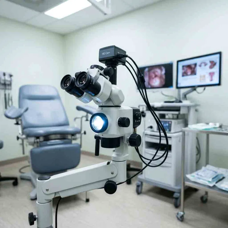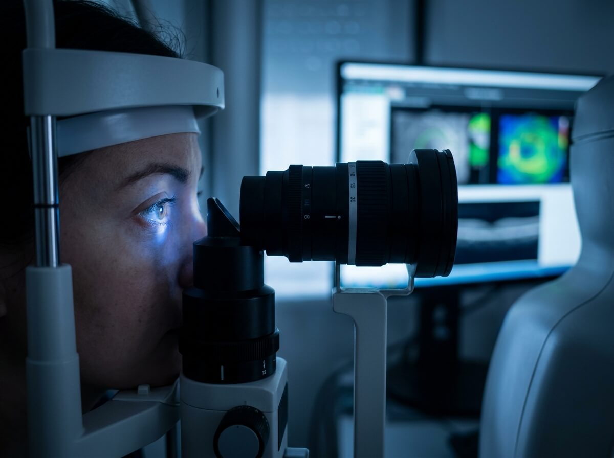
Ophthalmic practices rely on two distinct categories of technology: diagnostic instruments for early detection and optical measurement, and surgical systems for interventional procedures. While diagnostic units prioritize image resolution, non-invasive optical capture, and automated data processing, surgical platforms emphasize real-time fluidic control, microscopic visualization, precision cutting, and sterile field integrity.
For hospital purchasing managers, clinic owners, and B2B medical device distributors, understanding the technical specifications and operational differences between diagnostic and surgical devices is crucial for capital allocation and room planning.
+-----------------------------------------------------------------------------------+
|                     OPHTHALMIC EQUIPMENT FUNCTIONAL SPECTRUM                      |
+-----------------------------------------------------------------------------------+
| DIAGNOSTIC MODALITIES               | SURGICAL MODALITIES                         |
| - High-Resolution Optical Imaging   | - Real-Time Microscopic Visualization     |
| - Non-Invasive Structural Assessment| - High-Frequency Acoustic Emulsification  |
| - Automated Wavefront/IOP Metrics   | - Micro-Incision Vitreoretinal Fluidics   |
| - Pre-Operative Surgical Planning   | - Photocoagulation & Disruption Lasers    |
+-----------------------------------------------------------------------------------+
1. Key Operational Differences: Diagnostic vs. Surgical Equipment
| Feature / Metric | Diagnostic Ophthalmic Equipment | Surgical Ophthalmic Equipment |
| Primary Objective | Non-invasive tissue visualization & quantitative testing | Surgical tissue modification, resection, & restoration |
| Operator Interface | Automated auto-alignment, joystick, touch panel | Foot-pedal fluidic control, motorized micro-joysticks |
| Sterilization Need | Surface disinfection (alcohol wipe / chin-rest covers) | Full autoclave, EO gas, or disposable sterile packs |
| Data Requirements | DICOM export, EMR integration, normative databases | Real-time video recording, heads-up 3D display output |
| Power & Utility | Standard AC power (100 – 240 V) | High-power AC, pneumatic line / vacuum supply |
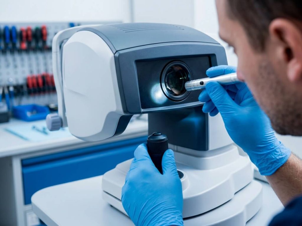
2. Deep Dive: High-Demand Surgical Ophthalmic Equipment
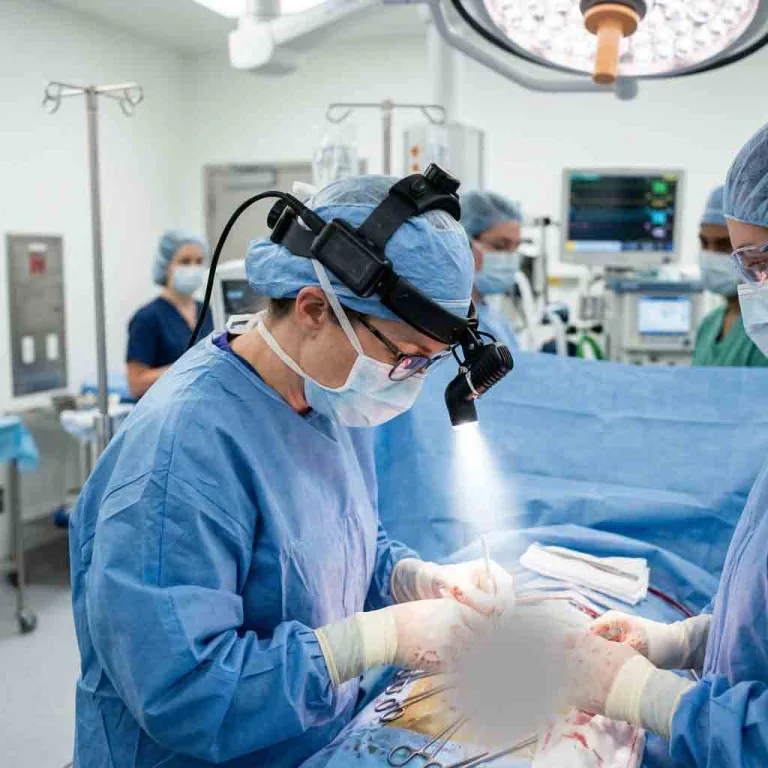
Surgical ophthalmic devices require strict adherence to safety standards, precise fluidics, and instant operator responsiveness during anterior and posterior segment surgeries.
Phacoemulsification Systems (Cataract Surgery)
-
Fluidic Pump Mechanisms:
-
Peristaltic Pumps: Provides flow-dominated control where vacuum builds upon occlusion, offering high safety and stability in anterior chamber depth.
-
Venturi Pumps: Delivers instant vacuum generation proportional to foot-pedal depression, enabling rapid nuclear fragment attraction.
-
-
Ultrasound Transducer Frequency: Operates typically between 28 kHz and 45 kHz to emulsify dense crystalline lenses with minimal thermal dissipate at the corneal incision site.
-
Torsional / Oscillatory Movement: Side-to-side ultrasonic motion reduces repulsive forces, enhancing cutting efficiency while protecting corneal endothelial cells.
Ophthalmic Surgical Microscopes
-
Red Reflex Optimization: Dual-beam stereo illumination systems maintain a bright, consistent red reflex essential for capsulorhexis and hydrodissection.
-
Optical Systems: Motorized zoom ratios (1:6 or higher), APO (apochromatic) optics, and integrated XY fluid-head positioning.
-
Integrated Intraoperative OCT (iOCT): Allows real-time visualization of retinal layers and corneal graft apposition directly within the surgeon’s eyepiece or 3D heads-up screen.
                  SURGICAL MICROSCOPE INTEGRATION
                                 |
     +---------------------------+---------------------------+
     |                                                       |
Optical & Illumination                         Digital & Assistant Modules
- Coaxial Red Reflex Illumination              - Integrated Intraoperative OCT (iOCT)
- Apochromatic (APO) Motorized Zoom            - Stereo Assistant Teaching Scope
- LED / Xenon High-Contrast Light              - 4K 3D Heads-Up Display Output
Vitrectomy Systems (Vitreoretinal Surgery)
-
Cut Rates: Modern pneumatic or ultra-speed cutters achieve rates up to 10,000 – 20,000 cuts per minute (CPM)}, minimizing traction on the peripheral retina.
-
Micro-Incision Vitrectomy Surgery (MIVS): Gauge sizes ranging from 23G, 25G, to ultra-fine 27G enable sutureless wound closure.
-
Integrated Intraocular Gas & Silicone Oil Injection: Automated pressure control for fluid-gas exchange during retinal detachment repairs.
Ophthalmic Lasers
-
Femtosecond Lasers: Used in FLACS (Femtosecond Laser-Assisted Cataract Surgery) and LASIK flap creation with sub-micron precision.
-
Nd:YAG Lasers (1064 nm): Photodisruption laser used for posterior capsulotomy and peripheral iridotomy.
-
Photocoagulation Green Lasers (532 nm) Solid-state lasers targeting retinal vascular pathology in diabetic retinopathy.
3. Direct Specification Comparison: Popular Equipment Categories
                        TONOMETRY TECHNOLOGY COMPARISON
                                     |
       +-----------------------------+-----------------------------+
       |                                                           |
Non-Contact Air-Puff Tonometer                Goldmann Applanation Tonometer (GAT)
- Method: Electro-optical air-puff sensing    - Method: Direct corneal applanation (7.35 mm²)
- Anesthesia: Not required                    - Anesthesia: Topical anesthetic & fluorescein
- Ideal for: High-speed primary screening     - Ideal for: Clinical gold-standard verification
Tonometry: Non-Contact Tonometer (NCT) vs. Goldmann Applanation Tonometer (GAT)
-
NCT (Diagnostic): Non-contact, rapid air-puff measurement suitable for general screening without topical anesthesia. Includes corneal thickness compensation algorithms.
-
GAT (Clinical Standard): Direct contact prism applanation mounted on a slit lamp. Requires topical anesthetic and fluorescein dye; remains the baseline standard for glaucoma management.
Retinal Assessment: Fundus Camera vs. Surgical Ophthalmoscope
-
Fundus Camera (Diagnostic): Captures high-resolution digital color photos and fluorescein angiograms of the retina for documentation and long-term disease tracking.
-
Indirect Ophthalmoscope (Diagnostic/Surgical): Wearable binocular system providing dynamic, wide-angle 3D visualization during peripheral retinal examinations and intraoperative scleral buckling.
4. Technical Specification & Selection Matrix for Procurement
| Device Category | Critical Specification | Preferred Benchmark | Primary Clinical Benefit |
| Phaco Unit | Max Cut Rate & Vacuum | ≥ 4000 CPM, 0 – 600 mmHg | Chamber stability, rapid lens extraction |
| Surgical Microscope | Working Distance & Zoom | 175 – 200 mm, Motorized 1:6 APO | High maneuvering space, crisp resolution |
| Vitrectomy Unit | MIVS Gauge Compatibility | 23G, 25G, 27G ultra-speed | Sutureless procedures, faster healing |
| YAG Laser | Pulse Energy & Spot Size | 0.3 – 10 mJ, 8 – 10 um | Precise capsulotomy without lens pitting |
| Slit Lamp Diagnostic | Magnification Steps | 5-Step (6x, 10x, 16x, 25x, 40x) | Detailed anterior & posterior examination |
5. B2B Buying Considerations: ROI, Sterilization & Installation
When evaluating diagnostic versus surgical equipment packages for commercial supply or institutional purchase, consider the following checklist:
-
Cleanroom & Sterilization Compatibility: Surgical handpieces and accessories must withstand repeated steam autoclaving (134°C) or low-temperature hydrogen peroxide gas plasma without insulation breakdown.
-
Consumable & Tubing Set Costs: Surgical phaco and vitrectomy units require ongoing purchases of single-use fluidic cassettes, pack tubing, and surgical tips. Evaluate the long-term cost per case.
-
Ergonomics & Footprint: Diagnostic rooms require compact designs for rapid patient turnover, whereas operating rooms require ceiling-mounted or mobile floor-stand systems with customizable foot-pedal mapping.
-
Power Backup Integration: Surgical suites require dedicated Uninterruptible Power Supply (UPS) systems capable of maintaining full system operation during power fluctuations.
6. Frequently Asked Questions (FAQ)
Q1: What is the main operational difference between Venturi and Peristaltic pumps in phaco machines?
Peristaltic pumps build vacuum only when the aspiration tip is occluded by nuclear material, giving surgeons precise flow control. Venturi pumps create instant vacuum in the cassette directly from airflow, sucking fragments to the tip faster regardless of occlusion.
Q2: Can a diagnostic OCT be used inside an operating room?
Standard diagnostic OCTs are tabletop units designed for outpatient clinics. Intraoperative OCT (iOCT) systems are specialized compact modules directly integrated into the optical path of a surgical microscope, allowing live scanning of tissue during surgery.
Q3: What sterilization method is recommended for surgical phaco handpieces?
Most OEM surgical phaco handpieces are rated for standard gravity or prevacuum autoclaving (132°C – 134°C). Always follow manufacturer guidelines to protect piezoelectric crystals from thermal shock.