1. Executive Overview: Setting Up a Modern Ophthalmic Diagnostic Clinic
Establishing or upgrading an eye care practice requires a balanced investment in diagnostic accuracy, patient throughput, and equipment reliability. Ophthalmic diagnostic devices serve as the foundation of vision care, allowing clinicians to detect refractive errors, anterior segment pathologies, retinal diseases, and glaucoma at early stages.
For hospital procurement officers, clinic administrators, and equipment distributors, selecting the right mix of primary screening instruments and advanced imaging modalities directly impacts operational efficiency and clinical outcomes.
+-----------------------------------------------------------------------------------+
|                        COMPLETE OPHTHALMIC CLINIC WORKFLOW                        |
+-----------------------------------------------------------------------------------+
|  1. Screening & Refraction   | Autorefractor / Keratometer, Digital Phoropter     |
|  2. Anterior Segment Exam    | Slit Lamp Biomicroscope, Corneal Topographer       |
|  3. Glaucoma & IOP Screening | Non-Contact Tonometer, Visual Field Analyzer       |
|  4. Posterior & Retina Exam  | Optical Coherence Tomography (OCT), Fundus Camera  |
+-----------------------------------------------------------------------------------+
2. Primary Refraction & Preliminary Screening Equipment
Preliminary screening tools measure vision acuity and objective refractive error, forming the initial phase of any eye examination.
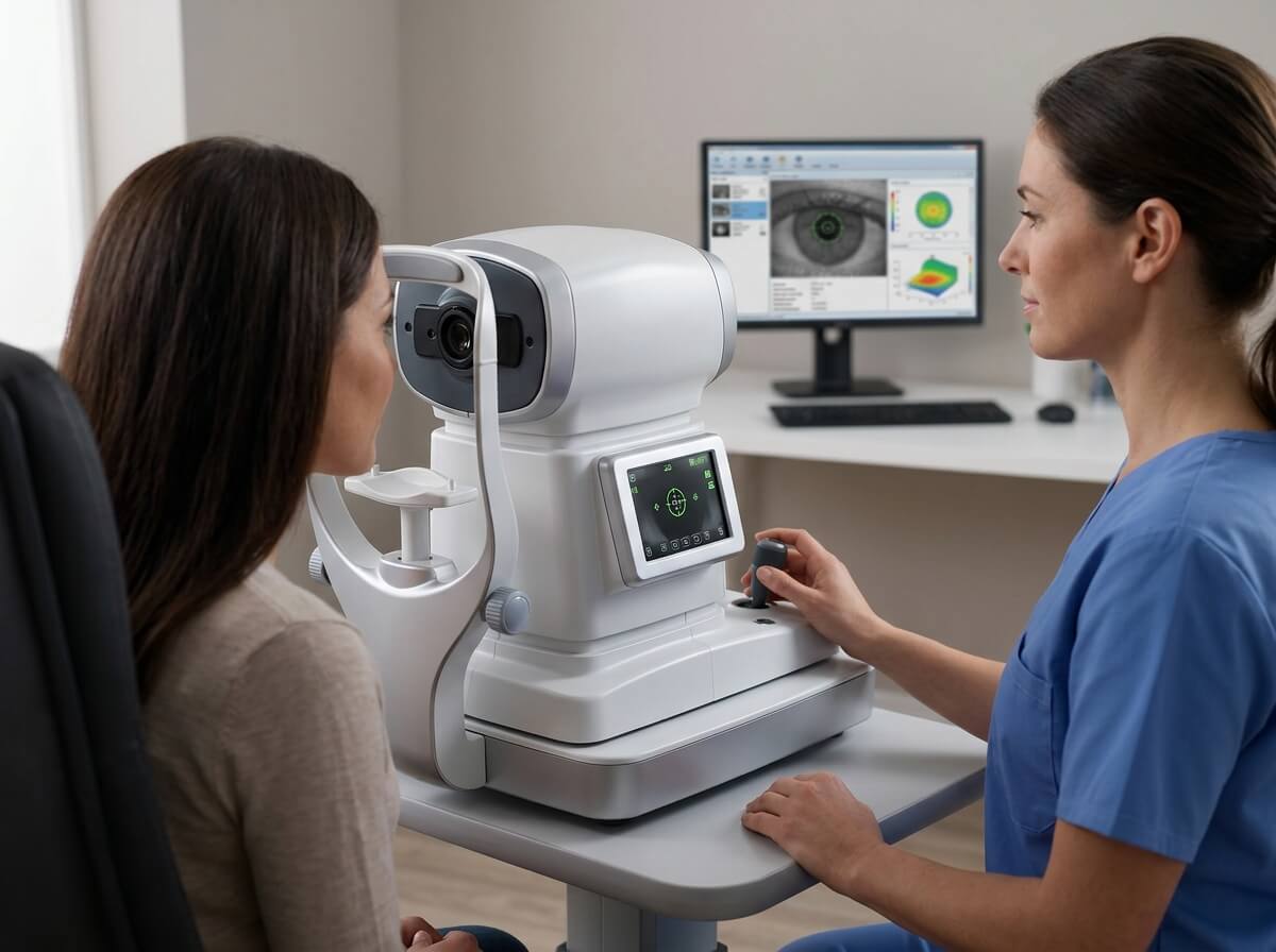
Autorefractors and Auto Keratometers
-
Primary Function: Measures objective refractive error (sphere, cylinder, axis) and corneal curvature (keratometry) within seconds.
-
Key Features to Evaluate: Dual CCD sensor technology, automatic pupil tracking, peripheral keratometry measurement, and minimum pupil size capability ($\le 2.0\text{ mm}$).
-
Target Applications: General optical shops, pediatric eye clinics, high-volume refractive surgical screening.
Manual and Digital Phoropters
-
Primary Function: Allows practitioners to refine subjective refraction, test binocular vision, and determine exact lens prescriptions.
-
Key Features to Evaluate: Wireless tablet integration, synchronized cross-cylinder lenses, silent lens interchange mechanisms, and seamless EMR connectivity.
-
Target Applications: Optometry practices, refractive surgery centers, outpatient ophthalmology clinics.
Automated Lensmeters (Lensometers)
-
Primary Function: Measures the power of existing single-vision, bifocal, and progressive spectacle or contact lenses.
-
Key Features to Evaluate: Shack-Hartmann wavefront sensors, UV/blue light transmittance testing, and automatic progressive lens detection.
3. Anterior Segment Examination & Topography Instruments
Diagnostic devices evaluating the cornea, anterior chamber, iris, and crystalline lens are crucial for diagnosing dry eye syndrome, cataracts, and corneal ectasias.
          ANTERIOR SEGMENT DIAGNOSTICS
       |
       +-----------------------------+-----------------------------+
       |                                                           |
Slit Lamp Biomicroscope                               Corneal Topographer / Tomographer
- Magnification: 6x to 40x                            - Placido Disc / Scheimpflug Imaging
- Illumination: LED / Halogen                         - Pachymetry & Elevation Mapping
- Dry Eye & Tear Film Analysis                        - Keratoconus Screening
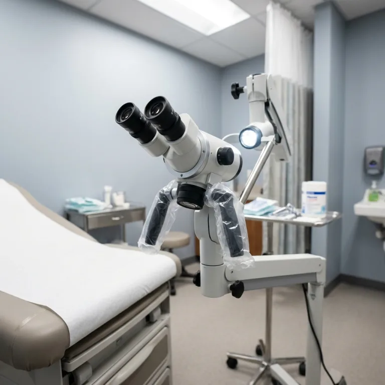
Slit Lamp Biomicroscopes
-
Tower Illumination (Haag-Streit Style): Positioned above the microscope head, providing superior light control for general anterior segment exams and posterior viewing with auxiliary lenses.
-
Integrated Digital Cameras: High-resolution CMOS cameras ($5.0\text{ MP}$ or higher) paired with beam splitters allow real-time image capture, video recording, and patient education.
-
Light Source: LED illumination provides uniform color temperature, cooler operation, and significantly longer lifespan compared to traditional halogen bulbs.
Corneal Topographers and Tomographers
-
Placido Disc Systems: Projects concentric light rings onto the cornea to map anterior surface curvature, ideal for contact lens fitting and astigmatism analysis.
-
Scheimpflug Imaging Systems: Captures 3D cross-sectional images of the entire anterior segment, providing elevation maps, corneal pachymetry, and keratoconus screening.
4. Posterior Segment Imaging & Retinal Diagnostics
Advanced posterior segment diagnostics enable detailed visualization of the retina, optic nerve head, macula, and retinal vasculature.
Optical Coherence Tomography (OCT)
-
Spectral-Domain OCT (SD-OCT): Offers scan speeds of 20,000 to 100,000 A-scans/second, yielding high-resolution cross-sectional images of the macula and retinal nerve fiber layer (RNFL).
-
Swept-Source OCT (SS-OCT): Uses longer wavelengths ($\sim 1050\text{ nm}$) for deeper tissue penetration, enabling clear imaging of the choroid and vitreous interface.
-
OCT Angiography (OCTA): Non-invasive visualization of microvascular networks without fluorescein dye injection, critical for managing diabetic retinopathy and wet AMD.
Digital Fundus Cameras
-
Mydriatic Fundus Cameras: Requires pupil dilation, offering wide-field fluorescein angiography (FA) and indocyanine green angiography (ICGA).
-
Non-Mydriatic Fundus Cameras: Enables fast, comfortable retinal photography through small, un-dilated pupils ($\ge 3.3\text{ mm}$), perfect for diabetic retinopathy screening networks.
-
Ultra-Widefield (UWF) Imaging: Captures up to $200^\circ$ of the retina in a single shot, detecting peripheral retinal tears and vascular abnormalities.
5. Glaucoma Screening & Intraocular Pressure (IOP) Devices
Early detection of elevated intraocular pressure and visual field loss is essential for preventing irreversible vision loss from glaucoma.
              GLAUCOMA DIAGNOSTIC SUITE
       |
       +-----------------------------+-----------------------------+
       |                                                           |
Tonometry (IOP Measurement)                   Perimetry (Visual Field Analysis)
- Non-Contact Air-Puff Tonometer              - Standard Automated Perimetry (SAP)
- Goldmann Applanation Tonometer (GAT)        - Threshold & Fast Screening Strategies
- Rebound Tonometer (Handheld)                - Progression Analysis Software
-
Non-Contact Tonometers (NCT): Delivers a gentle air puff to measure IOP without topical anesthesia, ideal for high-volume primary screening.
-
Goldmann Applanation Tonometers (GAT): Mounted directly on slit lamps; widely considered the clinical gold standard for precise IOP measurement.
-
Automated Visual Field Perimeters: Measures central and peripheral visual field sensitivity using threshold testing protocols (e.g., 24-2, 30-2, 10-2) to detect early glaucomatous field defects.
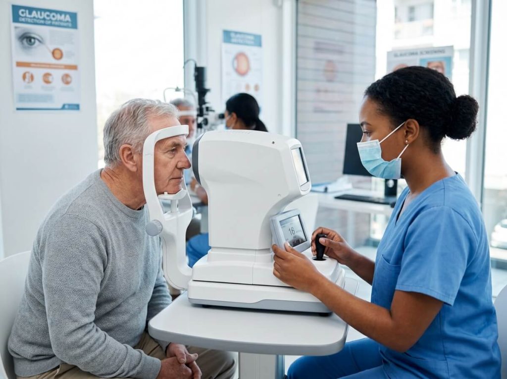
Optometrist performing glaucoma screening on an elderly patient using a desktop non-contact tonometer.
6. Essential Ophthalmic Equipment Comparison Matrix
| Equipment Category | Key Technology / Specs | Primary Diagnostic Target | Clinical Setting | Relative Cost Range |
| Autorefractor / Keratometer | Wavefront / Dual CCD, Auto-tracking | Refractive errors, corneal radius | Primary Screening, Optical Shops | Low to Moderate |
| Slit Lamp Biomicroscope | LED/Halogen, 3-5 step magnification | Anterior segment, cornea, lens | Universal (All Clinics) | Low to Moderate |
| Optical Coherence Tomography | SD-OCT / SS-OCT, OCTA | Retinal layers, macula, RNFL, optic nerve | Retina Clinics, Hospitals | High to Premium |
| Digital Fundus Camera | Non-mydriatic, 45° to 200° UWF | Diabetic retinopathy, macular degeneration | Screening Labs, Retina Practices | Moderate to High |
| Non-Contact Tonometer | Soft air puff, corneal thickness compensation | Intraocular pressure (IOP) | General Screening, Glaucoma | Moderate |
| Visual Field Analyzer | LED perimeter bowl, DICOM compatible | Glaucoma, neurological field loss | Glaucoma Clinics, Hospitals | Moderate to High |
7. B2B Procurement & Equipment Selection Checklist
When sourcing ophthalmic equipment from international manufacturers or distributors, evaluate these operational metrics:
-
EMR / PACS Connectivity: Ensure support for DICOM 3.0 standards and seamless data transfer to Electronic Medical Record (EMR) software.
-
Maintenance & Calibration Requirements: Check factory warranty terms, spare parts availability, and localized technical support service-level agreements (SLAs).
-
Power Voltage Adaptability: Verify dual-voltage compatibility ($100\text{–}240\text{ V AC}, 50/60\text{ Hz}$) for global export and distribution.
-
Certifications & Compliance: Confirm regulatory compliance such as CE Marking, ISO 13485 manufacturing quality standards, and FDA approval where applicable.
8. Frequently Asked Questions (FAQ)
Q1: What is the minimum equipment required to open a basic optometry or eye clinic?
A basic diagnostic room requires an autorefractor/keratometer, a digital or manual phoropter with a chart projector, a slit lamp biomicroscope, an automated lensmeter, and a tonometer (non-contact or rebound).
Q2: What is the difference between SD-OCT and SS-OCT?
Spectral-Domain OCT (SD-OCT) uses a spectrometer with wavelengths around $840\text{ nm}$, providing high-resolution retinal imaging. Swept-Source OCT (SS-OCT) uses a tunable laser at $1050\text{ nm}$, allowing deeper signal penetration through cataracts and into the choroid at faster scan rates.
Q3: Why is corneal pachymetry important in IOP measurement?
Corneal thickness affects applanation tonometer readings. Thicker corneas can artificially inflate measured IOP, while thinner corneas can lead to underestimation. Integrated pachymeters automatically calculate adjusted IOP values.