Executive Overview: The Evolution of Hemodynamic Assessment
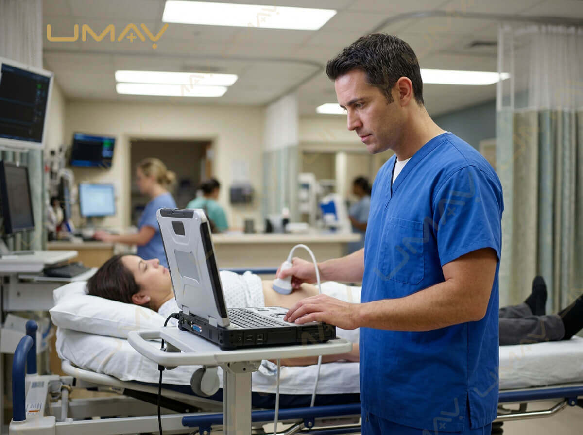
Color Doppler ultrasound represents one of the most critical advances in non-invasive medical diagnostics. By overlaying real-time color-coded blood flow dynamic maps onto conventional grayscale (B-mode) anatomical images, Color Doppler systems allow clinicians to evaluate cardiac structures, peripheral blood vessels, organ perfusion, and vascular lesions in real time without ionizing radiation or invasive catheterization.
For medical procurement specialists, clinic managers, and OEM distributors, understanding the physics, key operational modes, and performance limits of Color Doppler ultrasound equipment is essential. Selecting a system with high Doppler sensitivity, robust beamforming architecture, and effective clutter filtering directly impacts diagnostic confidence and long-term institutional value.
1. Fundamental Physics: Understanding the Doppler Shift
Color Doppler imaging relies on the Doppler Effect—the change in frequency of a wave reflected from a moving target relative to the stationary transducer source. In medical ultrasound, moving red blood cells (erythrocytes) within vascular structures act as the scattering targets.
2. Core Doppler Ultrasound Imaging Modes
A. Color Flow Mapping (CFM / Color Doppler)
Color Flow Mapping processes Doppler frequency shifts within a designated Region of Interest (ROI) and assigns color pixels based on flow direction and mean velocity:
-
Red Color (BART Convention): Indicates blood moving towards the transducer face (Positive Doppler Shift).
-
Blue Color: Indicates blood moving away from the transducer face (Negative Doppler Shift).
-
Color Turbulence / Mosaic Pattern: High-velocity or turbulent flows (e.g., downstream of a vascular stenosis) display yellow, green, or mosaic color mixing due to variance in flow directions.
B. Power Doppler Imaging (PDI) / Directional Power Doppler
Unlike standard Color Doppler, which measures flow velocity and direction, Power Doppler integrates the total area under the Doppler spectrum, displaying the total energy or amplitude of the reflected Doppler signal.
-
Key Advantage: Up to 3 to 5 times more sensitive to slow blood flow than standard CFM, making it immune to Doppler angle dependency (works even at $90^\circ$).
-
Primary Clinical Use: High-resolution evaluation of low-velocity microvascular perfusion, such as renal cortical flow, testicular torsion, thyroid nodules, and tendon inflammation (MSK).
C. Pulsed Wave Doppler (PW Doppler / Spectral Doppler)
Pulsed Wave Doppler transmits short acoustic pulses and uses a user-defined “sample volume gate” to measure exact blood flow velocities at a specific depth along the ultrasound line of sight.
-
Quantitative Analysis: Displays a real-time spectral waveform showing Peak Systolic Velocity (PSV), End Diastolic Velocity (EDV), Resistive Index (RI), and Pulsatility Index (PI).
-
Nyquist Limit & Aliasing: Because PW Doppler pulses at a specific Pulse Repetition Frequency (PRF), velocities exceeding the Nyquist limit ($\text{PRF} / 2$) suffer from spectrum wrapping (aliasing).
D. Continuous Wave Doppler (CW Doppler)
Continuous Wave Doppler utilizes two dedicated transducer crystals within a single probe: one continuously transmitting sound waves and the other continuously listening to reflections.
-
Key Advantage: Capable of accurately measuring extremely high blood flow velocities (exceeding 6 to 8 m/s) without aliasing.
-
Primary Clinical Use: Essential in Echocardiography for calculating severe aortic valve stenosis pressure gradients and regurgitant jet velocities.
3. Side-by-Side Comparison of Doppler Modes
| Doppler Mode | Measured Parameter | Angle Dependence | Aliasing Risk | Primary Clinical Target |
| Color Flow Mapping (CFM) | Mean Flow Velocity & Direction | High (Needs < 60°) | Yes (High velocities) | Carotid stenosis, DVT, Valvular regurgitation |
| Power Doppler (PDI) | Signal Amplitude / Energy | Minimal (Angle Independent) | No | Microvascular perfusion, Organ ischemia, MSK |
| Pulsed Wave (PW) | Exact Velocity Spectrum at Gate | Critical (Angle Correct) | Yes (Nyquist Limit) | Hemodynamic quantification (PSV/EDV/RI) |
| Continuous Wave (CW) | Peak Velocity along Beam Line | Moderate | No Aliasing | High-velocity Cardiac Valvular Jets (Cardiology) |
4. Key B2B Procurement & Hardware Performance Metrics
When evaluating Color Doppler equipment proposals from OEM manufacturers or international distributors, procurement officials should assess these crucial technical indicators:
-
Digital Beamformer Channel Count: Systems with 64, 128, or 256 physical processing channels deliver superior signal-to-noise ratios, reducing clutter artifacts in deep vascular exams.
-
Dynamic Range & Wall Filter Control: High dynamic range (> 120 dB) paired with intelligent adaptive wall filters removes low-frequency tissue motion artifacts while retaining low-velocity vein signals.
-
Auto-Trace Spectral Waveform Analysis: Integrated real-time AI auto-tracing calculates PSV, EDV, RI, and S/D ratios with a single touch, boosting outpatient patient turnover rates.
-
Transducer Bandwidth & Frequency Multi-Stepping: Broad-band linear probes (e.g., 5.0 to 14.0 MHz) allow clinicians to easily toggle between superficial thyroid scans and deeper femoral artery studies.
5. Frequently Asked Questions (FAQ)
Q1: What causes “Aliasing” in Color Doppler, and how can it be resolved?
Aliasing occurs when blood flow velocity exceeds the system’s Nyquist limit ($\text{PRF} / 2$), making forward flow appear as backward flow (color wrap-around). To resolve aliasing, operators should increase the Velocity Scale (PRF), lower the transducer baseline, or switch to a lower frequency probe.
Q2: Why is Power Doppler preferred over standard Color Doppler in rheumatology and MSK?
Power Doppler is significantly more sensitive to slow capillary and synovial blood flow, which are common indicators of joint inflammation and early arthritis. Additionally, because Power Doppler is angle-independent, it eliminates signal loss around curved tendon structures.
Q3: What is the difference between Single-Beam and Multi-Beamformer architecture in Doppler machines?
Multi-beamformers process multiple parallel acoustic receive lines per transmitted pulse. This significantly enhances frame rate (FPS) during Color Doppler imaging, preventing lag or stuttering during fast cardiac or arterial wall movements.
Conclusion: Maximizing Clinical Diagnostic Precision
Color Doppler ultrasound remains an indispensable diagnostic tool across modern clinical practice. Equipping your medical facility with a high-performance Color Doppler system ensures fast, accurate vascular assessment, superior patient care, and reliable long-term clinical versatility.