The Clinical Impact of Surgical Visualization
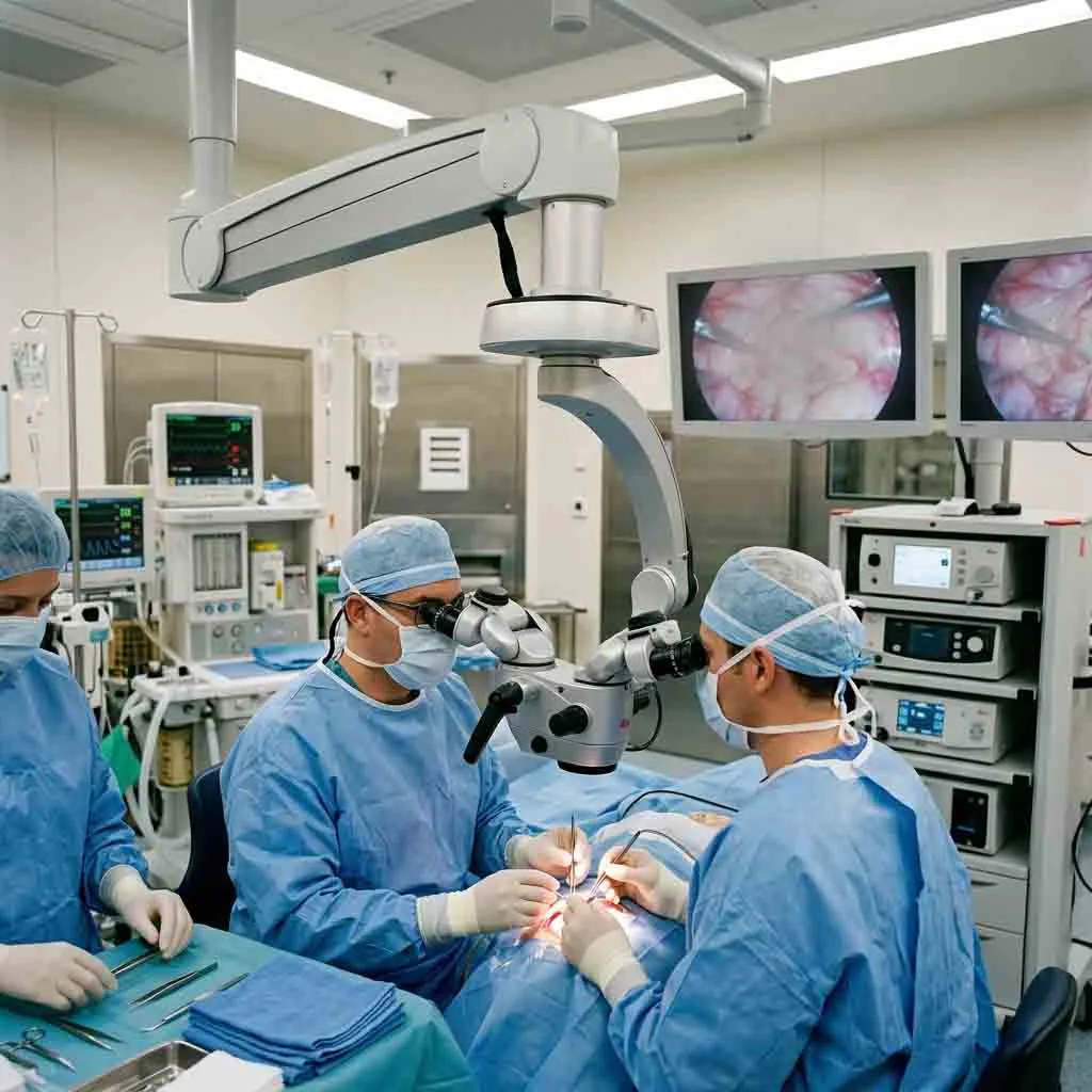
In microsurgery, the margin between procedural success and permanent functional deficit is measured in fractions of a millimeter. The human eye has a visual resolving limit of approximately 0.2 millimeters at close range. When navigating delicate anatomical structures—such as intracranial aneurysms, middle ear ossicles, retinal membranes, or peripheral nerves—unaided vision is insufficient.
Plaintext
                      CLINICAL MAGNIFICATION SPECTRUM
  +-------------------------------------------------------------------------+
  | DENTAL / ENT         --> 3x to 15x  --> Canal mapping, tympanoplasty    |
  | NEUROSURGERY / SPINE --> 4x to 20x  --> Tumor margins, vessel clipping  |
  | OPHTHALMIC / PLASTIC --> 6x to 25x+ --> Capsulorhexis, 1.0mm vessel tie |
  +-------------------------------------------------------------------------+
The introduction of specialized operating microscopes transformed these disciplines from exploratory, high-risk interventions into standardized, minimally invasive procedures. By combining high-resolution stereoscopic depth perception with intense, shadow-free coaxial illumination, the surgical microscope allows clinicians to distinguish subtle tissue planes, protect critical neurovascular bundles, and perform micro-suturing with sub-millimeter precision.
Because each surgical specialty deals with unique anatomical depths, tissue characteristics, and patient positioning, operating microscope systems are engineered with specialty-specific optical paths, focal ranges, and mechanical configurations.
2. Ophthalmology: Precision in Anterior and Posterior Segments
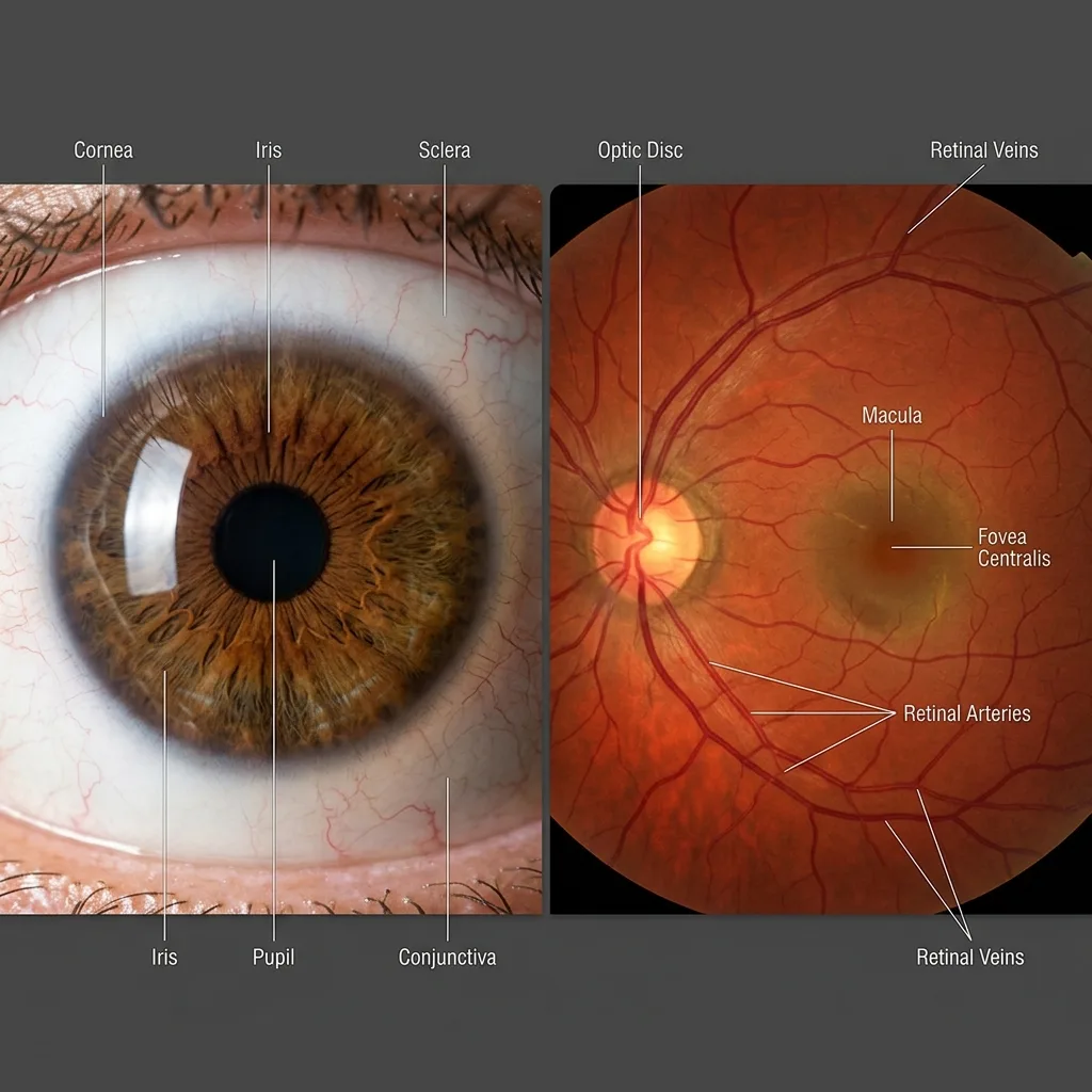
Ophthalmology was the first medical discipline to adopt operating microscopes routinely. Operating on the human eye requires exceptional optical clarity, sub-millimeter depth control, and specialized lighting systems capable of illuminating transparent and semi-transparent intraocular tissues.
Plaintext
                  OPHTHALMIC MICROSCOPE CONFIGURATION
+-------------------------------------------------------------------------+
| ANTERIOR SEGMENT (Cataract, Cornea)  --> Coaxial Red Reflex Illumination|
| POSTERIOR SEGMENT (Retina, Vitreous) --> Non-Contact Wide-Angle Viewing |
| FOOT PEDAL CONTROL                   --> Motorized X-Y, Zoom, and Focus |
+-------------------------------------------------------------------------+
A. Anterior Segment Surgery: Cataracts & Cornea
The most common ophthalmic application is cataract extraction via phacoemulsification and intraocular lens (IOL) implantation:
-
The Critical Role of the Red Reflex: During cataract surgery, the surgeon must perform a continuous curvilinear capsulorhexis—tearing a circular opening in the transparent anterior lens capsule. To see this transparent membrane, the microscope uses true coaxial illumination (light entering along the exact visual axis of the optical path). This light reflects off the vascularized retina at the back of the eye, creating a bright red or orange background glow known as the Red Reflex. Any tear, opacity, or capsule edge stands out in sharp silhouette against this red reflection.
-
Corneal Transplantation (Keratoplasty): In procedures such as Penetrating Keratoplasty (PK) or endothelial keratoplasty (DSEK/DMEK), high-magnification apochromatic optics allow the surgeon to place uniform 10-0 nylon corneal sutures and verify graft adherence without inducing irregular astigmatism.
B. Posterior Segment Surgery: Vitreoretinal Procedures
Operating on the posterior segment (vitreous body and retina) presents different optical challenges:
-
Wide-Angle Fundus Viewing: Viewing the retina requires integrating wide-angle viewing attachments (such as non-contact optical inverter systems) below the objective lens. These systems invert the aerial image generated by the cornea and lens, allowing vitreoretinal surgeons to visualize the peripheral retina out to the ora serrata.
-
Epiretinal Membrane (ERM) Peeling: Surgeons operate under high magnification (15x to 25x) using micro-forceps to peel pathologic cellular membranes that are only a few micrometers thick off the delicate internal limiting membrane (ILM) of the macula, directly treating macular holes and retinal traction.
-
Motorized Foot-Pedal Ergonomics: Because both of the ophthalmic surgeon’s hands are occupied holding micro-instruments inside the eye, ophthalmic operating microscopes feature multi-function foot consoles. The surgeon controls motorized focusing, continuous optical zoom, and motorized X-Y lateral movement using foot switches without interrupting the procedure.
3. Neurosurgery & Spine: Navigating Deep, Narrow Corridors
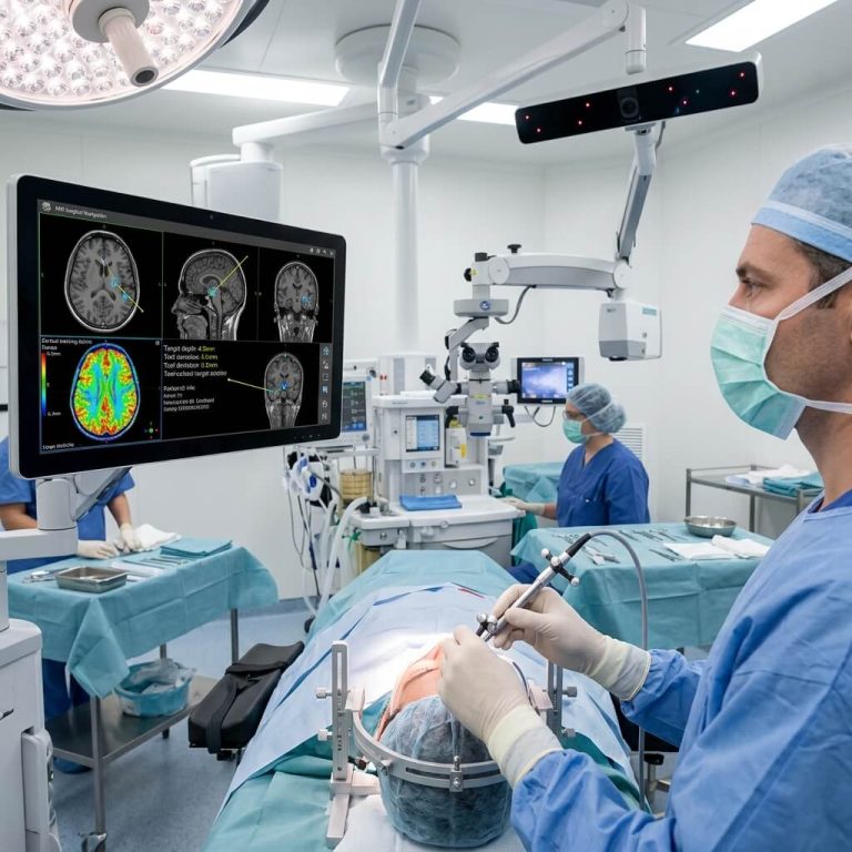
Neurosurgical operating microscopes represent the pinnacle of optical and mechanical engineering. Neurosurgeons must access lesions located deep within the cranial cavity or spinal canal through narrow, minimally invasive corridors while avoiding eloquent brain tissue.
Plaintext
                  NEUROSURGICAL MICROSCOPE WORKFLOW
+-------------------------------------------------------------------------+
| DEEP SURGICAL CORRIDOR  --> High-Intensity Xenon or High-Lux LED        |
| VARIABLE CAVITY DEPTH   --> Motorized Vario Objective (200 - 500 mm)    |
| ANGIOGRAPHY INTEGRATION --> Intraoperative Fluorescence (ICG / 5-ALA)   |
| CO-OBSERVATION          --> Face-to-Face Stereo Assistant Bridge        |
+-------------------------------------------------------------------------+
A. Vascular Neurosurgery: Aneurysms & AVMs
-
Cerebral Aneurysm Clipping: When treating intracranial aneurysms (such as those in the Circle of Willis), surgeons navigate narrow skull base fissures. The microscope delivers coaxial light deep into the fissure without casting shadows. The surgeon inspects the aneurysm neck, verifies that perforating micro-vessels are preserved, and applies titanium clips.
-
Arteriovenous Malformations (AVM): High-contrast apochromatic optics help the neurosurgeon distinguish between arterial feeding vessels, nidus vessels, and venous drainage channels, reducing the risk of catastrophic intraoperative hemorrhage.
-
Fluorescence Integration (ICG Angiography): Many modern neurosurgical microscopes incorporate near-infrared fluorescence filters. By injecting Indocyanine Green (ICG) dye intravenously, the surgeon can switch the microscope to near-infrared mode to confirm real-time blood flow through clipped aneurysms and bypass grafts on the microscope display.
B. Neuro-Oncology & Skull Base Tumors
-
Glioma & Meningioma Resection: Differentiating infiltrating tumor margins from healthy functional brain parenchyma requires maximum optical resolution and tissue contrast. Microscopes equipped with blue excitation filters facilitate 5-ALA (5-Aminolevulinic Acid) fluorescence-guided surgery, causing malignant glioma cells to glow bright red/violet under blue light.
-
Transsphenoidal Pituitary Surgery: Operating through the nasal cavity to reach the pituitary gland demands deep light penetration and a long working distance, allowing micro-curettes and aspirators to work without colliding with the optical head.
C. Minimally Invasive Spine Surgery (MISS)
-
Microdiscectomy & Decompression: When treating herniated intervertebral discs or spinal stenosis, the surgeon works through narrow tubular retractors (often 16 to 22 mm in diameter).
-
Motorized Variable Working Distance: Spine surgery requires continuous focal adjustments as the surgeon moves from the superficial muscle fascia down to the deep spinal dura. Microscopes featuring motorized variable focus (Vario objectives adjustable from 200 mm to 500 mm) allow instant focus adjustments via handgrip buttons without repositioning the suspension arm.
4. Otolaryngology (ENT): Micro-Anatomy of the Ear and Larynx
Otolaryngologists utilize surgical microscopes across two primary anatomical zones: the temporal bone (otology) and the vocal tract (laryngology).
Plaintext
                      ENT CLINICAL APPLICATIONS
                                  |
     +----------------------------+----------------------------+
     |                                                         |
     v                                                         v
[ OTOLOGY & NEUROTOLOGY ]                     [ MICROLARYNGEAL SURGERY ]
- Tympanoplasty (Eardrum repair)              - Vocal cord polyps and nodules
- Mastoidectomy (Cholesteatoma)               - Subglottic stenosis dilation
- Stapedectomy (0.4mm Piston Insertion)       - 400 mm Long Working Distance Lens
A. Otology & Neurotology (Middle and Inner Ear)
The middle ear houses the smallest bones in the human body: the malleus, incus, and stapes. Interventions here demand high magnification and tremor-free mechanical stability:
-
Tympanoplasty: Repairing a perforated tympanic membrane requires 8x to 16x magnification to place fascia or cartilage grafts beneath the eardrum margin.
-
Stapedectomy: In patients with otosclerosis, the stapes footplate becomes fixed, causing conductive hearing loss. Under high magnification, the ear surgeon removes the fixed stapes arch, uses a micro-drill or laser to create a microscopic fenestration (0.5 mm to 0.8 mm) in the footplate, and crimps a tiny titanium or Teflon piston prosthesis onto the incus bone.
-
Cholesteatoma Removal (Mastoidectomy): Removing destructive epidermal cysts from the mastoid cavity requires wide-field optical orientation to eradicate disease while avoiding the adjacent facial nerve (cranial nerve VII) and semicircular canals.
B. Microlaryngeal Surgery (Laryngology)
-
Vocal Cord Lesions: Operating on vocal cord nodules, polyps, cysts, and early glottic cancers requires a unique optical setup. The patient is placed under general anesthesia with a rigid metal laryngoscope positioned through the mouth down to the vocal folds.
-
Long Working Distance Requirements: Because the vocal cords sit deep within the neck at the end of a long rigid scope, the operating microscope must utilize an objective lens with a long working distance of 400 mm. A standard 200 mm or 250 mm lens cannot be used, as it would not leave enough clearance for the surgeon to manipulate long micro-laryngeal scissors, forceps, and laser fibers.
5. Dental & Endodontic Microsurgery
In dentistry, the operating microscope has elevated endodontics (root canal therapy) and restorative microsurgery from tactile guesswork to direct visual diagnosis.
Plaintext
                  ENDODONTIC MICROSCOPE BENEFITS
+-------------------------------------------------------------------------+
| CANAL ANATOMY    --> Identifies hidden MB2 canals and lateral branches  |
| RETREATMENT      --> Visualizes and retrieves separated endodontic files|
| APICOECTOMY      --> Inspects root apex for micro-fractures under 20x   |
| ERGONOMIC POSTURE--> Eliminates neck bending via 0-180° inclinable tube |
+-------------------------------------------------------------------------+
A. Locating Hidden and Calcified Canals
Traditional root canal therapy relies on radiographs and tactile feel. However, complex root anatomy often contains hidden channels:
-
The Mesiobuccal 2 (MB2) Canal: In maxillary first molars, the MB2 canal is present in over 70% to 90% of teeth, yet it is frequently missed under unaided vision due to calcification. Missed canals are a leading cause of endodontic treatment failure.
-
High magnification (10x to 20x) combined with intense coaxial illumination allows endodontists to trace subtle developmental grooves in the pulpal floor and unroof calcified secondary dentin to locate hidden canal orifices.
B. Instrument Retrieval and Perforation Repair
-
Separated Files: If a nickel-titanium root canal file breaks inside a curved canal, a surgical microscope provides the illumination and magnification needed to visualize the top of the broken metal fragment. The clinician can then apply ultrasonic tips to trough around the instrument and retrieve it without destroying the root.
-
Perforation Management: Accidental root perforations can be visualized directly and sealed hermetically using biocompatible materials like Mineral Trioxide Aggregate (MTA).
C. Surgical Endodontics (Apicoectomy)
When conventional root canal therapy cannot resolve a periapical infection, surgical endodontics is indicated:
-
The clinician reflects a gingival flap, creates a small cortical bone window, and resects the diseased apical 3 mm of the tooth root.
-
Under 15x to 20x magnification, the resected root face is inspected with a micro-mirror for micro-fractures, isthmuses, and unsealed canal fins. An ultrasonic retro-tip prepares a 3 mm cavity inside the root tip, which is then filled with retro-filling material to achieve a complete apical seal.
D. Ergonomic Preservation for Dental Clinicians
Dentists and endodontists suffer from high rates of chronic cervical spine and lumbar disc herniation caused by working in a hunched position over the oral cavity. An operating microscope equipped with an ergonomic 0–180 degree inclinable binocular tube allows the practitioner to sit in an upright, neutral position with their back and neck supported, looking straight ahead into the eyepieces while the optical head is angled down into the patient’s mouth.
6. Plastic, Reconstructive & Hand Surgery: Microvascular Anastomosis
Plastic and reconstructive surgery frequently involves transferring living tissue (skin, fat, muscle, or bone) from one part of the body to another to reconstruct defects caused by cancer resections or severe trauma.
Plaintext
                 MICROVASCULAR ANASTOMOSIS PROCEDURE
+-------------------------------------------------------------------------+
| Vessel Diameter: 0.8 mm to 2.5 mm                                       |
| Suture Material: 9-0 to 11-0 Nylon (Finer than human hair)              |
| Magnification:   12x to 25x High Resolution                             |
| Clinical Goal:   100% Patency with zero intimal inversion or thrombus   |
+-------------------------------------------------------------------------+
A. Free Tissue Transfer (Free Flaps)
-
Reconstruction Examples: Breast reconstruction using a Deep Inferior Epigastric Perforator (DIEP) flap, or jaw reconstruction using a vascularized fibula free flap following oral cancer resection.
-
Vascular Anastomosis: Once the flap is transferred to the recipient site, its arterial and venous pedicles must be connected to local recipient vessels to restore blood supply. These vessels are typically 1.0 mm to 2.5 mm in diameter.
-
Under 12x to 20x magnification, the reconstructive surgeon places interrupted sutures using 9-0 or 10-0 nylon thread on a microscopic needle. The microscope allows the surgeon to visualize the inner endothelial layer (intima) of the blood vessel clearly, ensuring that sutures pass through all vessel layers cleanly without catching the opposing wall or causing platelet aggregation that could trigger thrombosis and flap loss.
B. Digit and Limb Replantation
-
When severed fingers or limbs are replanted, tiny digital arteries, veins, and nerves must be repaired. Digital vessels in children can be smaller than 0.8 mm in diameter.
-
High-magnification apochromatic lenses ensure the surgeon can align and coapt tiny epineural nerve sheaths using 10-0 or 11-0 sutures, restoring sensory and motor function to the replanted digit.
7. Cross-Specialty Comparison Matrix
The table below summarizes the contrasting technical and optical requirements across the major surgical specialties:
| Clinical Specialty | Typical Procedures | Optimal Working Distance | Common Magnification | Essential Hardware / Optical Feature |
| Ophthalmology | Phacoemulsification, Vitrectomy, Keratoplasty | 175 mm – 200 mm | 6x – 25x | Coaxial Red Reflex system, foot-controlled motorized X-Y coupling |
| Neurosurgery | Aneurysm clipping, Glioma resection, Skull base | 200 mm – 500 mm (Variable) | 4x – 20x | Motorized Vario lens, deep cavity Xenon/LED, ICG fluorescence filters |
| Spine Surgery | Microdiscectomy, ACDF, Spinal decompression | 250 mm – 450 mm (Variable) | 3x – 12x | Long working distance for tubular retractors, flexible counterbalance arm |
| Otology (Ear) | Tympanoplasty, Stapedectomy, Mastoidectomy | 200 mm – 250 mm | 6x – 20x | High mechanical stability, assistant co-observation tube, laser interface |
| Laryngology (Vocal) | Vocal cord polyps, Laryngeal papillomas | 350 mm – 400 mm | 4x – 16x | Extended working distance to bypass rigid laryngoscope instruments |
| Dental / Endodontics | MB2 canal location, Apicoectomy, File removal | 200 mm – 300 mm | 3x – 20x | Compact footprint, 0–180° inclinable binocular tube, orange/green filters |
| Plastic / Reconstructive | DIEP flap, Replantation, Lymphatic anastomosis | 200 mm – 300 mm | 10x – 25x+ | Face-to-face dual binocular bridge, high depth of field for vessel coupling |
8. Specialty Illumination Accessories: Filters and Safety
Modern operating microscopes utilize specialized optical filters to optimize illumination for specific clinical tasks while protecting delicate tissues:
-
Retinal Protection Filter (Yellow Filter): In ophthalmic microscopes, prolonged exposure to intense white or blue light can cause phototoxic retinal damage. A yellow filter blocks harmful short-wavelength blue light during cataract procedures when full-intensity illumination is required.
-
Orange Filter (Dental Curing Protection): Dental composite resin materials cure when exposed to blue light wavelengths (typically 450–470 nm). Dental operating microscopes include a flip-in orange filter that blocks the curing spectrum, allowing the dentist to manipulate, shape, and place composite restorations under high magnification without premature hardening.
-
Green Filter (Red Free / Vascular Enhancement): Used in neurosurgery, ophthalmology, and ENT to increase the contrast of vascular networks. Blood vessels absorb green light strongly, causing capillaries and bleeding points to appear sharp and black against surrounding pale tissues.
Frequently Asked Questions (FAQ)
Q1: Why can’t a hospital use the same operating microscope for both eye surgery and neurosurgery?
While both specialties demand premium optics, their structural configurations conflict. Ophthalmic surgery requires a short focal working distance (around 175–200 mm), a specialized coaxial red reflex lighting system, and motorized X-Y foot controls. Neurosurgery requires deep-cavity illumination, a motorized variable focal range (200–500 mm) to reach deep cranial spaces, and specialized counterbalanced suspension arms that support heavy laser or camera attachments.
Q2: What is the purpose of an assistant scope (co-observation tube)?
In teaching hospitals and complex surgeries (such as neurosurgery and reconstructive free-flap procedures), two surgeons work simultaneously. An assistant scope uses a beam splitter to redirect 50% of the optical image to a secondary binocular tube. In “face-to-face” setups, the lead surgeon and assistant sit opposite each other, seeing the exact same surgical field in full stereoscopic 3D.
Q3: Why is a 400 mm working distance mandatory for vocal cord surgery?
During microlaryngeal surgery, a rigid hollow metal laryngoscope is inserted into the patient’s mouth and down into the throat to expose the vocal cords. The surgeon uses instruments that are 200 mm to 300 mm long. An objective lens with a working distance of 400 mm is required so the microscope head remains safely outside the patient’s oral space, leaving room to insert and manipulate long instruments.
Q4: Does high magnification always mean a better view during microsurgery?
No. There is an inverse physical relationship between magnification, field of view, and depth of field. As magnification increases, the field of view narrows and the depth of field becomes shallower, requiring frequent refocusing. Skilled microsurgeons use low magnification (3x to 6x) for anatomical orientation and tissue dissection, reserving high magnification (15x to 25x) specifically for micro-suturing, fine nerve coaptation, or micro-drilling.
Q5: Can dental operating microscopes record high-definition patient videos?
Yes. Modern dental and surgical microscopes incorporate integrated or beam-splitter-attached 4K digital video cameras. These allow clinicians to stream live procedures to ceiling monitors for dental assistant coordination, document clinical steps for patient communication, and archive footage for academic presentations and legal records.
Summary
The clinical versatility of the operating microscope has cemented its role as an indispensable piece of capital equipment across modern surgical departments. From providing the life-saving red reflex in cataract surgery to revealing hidden calcified canals in endodontics and enabling microvascular anastomosis in trauma reconstruction, specialized surgical optics directly elevate clinical success rates.